V体育ios版 - Effective delivery of large genes to the retina by dual AAV vectors
- PMID: 24150896
- PMCID: PMC3927955
- DOI: 10.1002/emmm.201302948
VSports在线直播 - Effective delivery of large genes to the retina by dual AAV vectors
Abstract
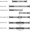
Retinal gene therapy with adeno-associated viral (AAV) vectors is safe and effective in humans VSports手机版. However, AAV's limited cargo capacity prevents its application to therapies of inherited retinal diseases due to mutations of genes over 5 kb, like Stargardt's disease (STGD) and Usher syndrome type IB (USH1B). Previous methods based on 'forced' packaging of large genes into AAV capsids may not be easily translated to the clinic due to the generation of genomes of heterogeneous size which raise safety concerns. Taking advantage of AAV's ability to concatemerize, we generated dual AAV vectors which reconstitute a large gene by either splicing (trans-splicing), homologous recombination (overlapping), or a combination of the two (hybrid). We found that dual trans-splicing and hybrid vectors transduce efficiently mouse and pig photoreceptors to levels that, albeit lower than those achieved with a single AAV, resulted in significant improvement of the retinal phenotype of mouse models of STGD and USH1B. Thus, dual AAV trans-splicing or hybrid vectors are an attractive strategy for gene therapy of retinal diseases that require delivery of large genes. .
Figures (V体育安卓版)


EGFP protein quantification by ELISA of retinas from C57BL/6 mice 1 month following subretinal injection of AAV2/8 vectors encoding for EGFP under the control of the PR-specific Rhodopsin (RHO) promoter. The scatter plot depicts EGFP protein levels from each retina; the mean value for each group is depicted and indicated with a solid line VSports注册入口. The number ( n) of eyes analyzed is depicted under the x axis. ** P ANOVA<0. 001. More details on the statistical analysis including specific statistical values can be found in the Statistical analysis paragraph of the Materials and methods section.
Fluorescence analysis of retinal cryosections from Large White pigs 1 month following subretinal injection of AAV2/8 vectors encoding for EGFP under the control of the PR-specific RHO promoter V体育官网入口. The pictures are representative of: n = 4 or n = 3 eyes injected with dual AAV TS or hybrid AK, respectively. The scale bar (100 μm) is depicted in the figure. NS, AAV normal size; TS, dual AAV trans-splicing; AK, dual AAV hybrid AK; DAPI, 4′,6′-diamidino-2-phenylindole staining; EGFP, native EGFP fluorescence; Merge, overlay of DAPI and EGFP pictures; RPE, retinal pigmented epithelium; PR, photoreceptors.

Representative Western blot analysis of C57BL/6 retinal lysates 1 month following the injection of dual AAV trans-splicing (TS) and hybrid AK (AK) vectors encoding for ABCA4 under the control of the PR-specific Rhodopsin promoter (RHO PROMOTER). The arrow points at full-length proteins, the molecular weight ladder is depicted on the left, 150 μg of protein were loaded in each lane. The number ( n) and percentage of ABCA4-positive retinas out of total retinas analyzed is depicted V体育2025版. AK, retinas injected with dual AAV hybrid AK vectors; TS, retinas injected with dual AAV TS vectors; neg, retinas injected with AAV vectors expressing EGFP, as negative controls; α-3xflag, Western blot with anti-3xflag antibody; α-Dysferlin, Western blot with anti-Dysferlin antibody, used as loading control.
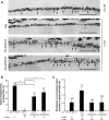
Representative pictures of immuno-electron microscopy analysis with anti-HA antibody of retinal sections from wild-type BALB/c (WT) and Abca4−/− mice injected with either dual AAV or with negative control vectors. The black dots represent the immuno-gold labelling of the ABCA4-HA protein. The scale bar (200 nm) is depicted in the figure VSports.
Representative pictures of transmission electron microscopy analysis showing lipofuscin granules content in the RPE of WT and Abca4−/− mice injected with either dual AAV or negative control vectors VSports app下载. The black arrows indicate lipofuscin granules. The scale bar (1. 6 μm) is depicted in the figure.
Quantification of the mean number of lipofuscin granules counted in at least 30 fields (25 μm2) for each sample. The number ( n) of eyes analyzed is depicted below each bar. The mean value is depicted above the corresponding bar. Values are represented as mean ± standard error of the mean (s. e. m. ). * P ANOVA < 0. 05; ** P ANOVA < 0. 001. More details on the statistical analysis including specific statistical values can be found in the Statistical analysis paragraph of the Materials and methods section V体育官网.
 "VSports"
Representative pictures of transmission electron microscopy analysis of retinal sections from wild-type BALB/c (WT) and Abca4−/− mice injected with dual AAV trans-splicing (TS-ABCA4) and hybrid AK vectors (AK-ABCA4) or with either AAV vectors expressing EGFP or 5′-or 3′-half of the dual hybrid AK vectors (neg), as negative controls V体育ios版. The dotted lines indicate the edges of RPE cells. The scale bar (3. 8 μm) is depicted in the figure.
Quantification of the mean RPE thickness counted in at least 30 fields for each sample. The number ( n) of eyes analyzed is depicted below each bar. Abca4−/− neg includes Abca4−/− mice injected with either AAV vectors expressing EGFP ( n = 2) or 5′-( n = 3) or 3′-( n = 4) half of the dual hybrid AK vectors (neg total n = 9). The mean value is depicted above the corresponding bar. Values are represented as mean ± standard error of the mean (s.e.m.). * P ANOVA < 0.05.
Recovery from light desensitization in 3 month-old Abca4−/− and BALB/c mice at 6 weeks post-injection. The relative b-wave is the ratio between the post-and the pre-desensitization b-wave amplitudes both evoked by 1 cd s/m2. The time refers to the minutes post-desensitization. The mean recovery (%) at 60 min is depicted.

Representative Western blot analysis of C57BL/6 eyecups 1 month following the injection of dual AAV trans-splicing (TS) and hybrid AK (AK) vectors encoding for MYO7A-HA under the control of the ubiquitous chicken beta-actin (CBA) promoter. The arrow indicates full-length proteins, the molecular weight ladder is depicted on the left, 100 μg of proteins were loaded in each lane. The number ( n) and percentage of MYO7A-positive eyecups out of total eyecups analyzed is depicted. AK, eyes injected with dual AAV hybrid AK vectors; TS, eyes injected with dual AAV TS vectors; neg, eyes injected with either 5′-or 3′-half of the dual AAV TS and hybrid AK vectors; α-HA, Western blot with anti-hemagglutinin (HA) antibody; α-Dysferlin, Western blot with anti-Dysferlin antibody, used as loading control.
Quantification of MYO7A levels expressed from dual AAV vectors in sh1−/− eyecups relative to endogenous Myo7a expressed in littermate sh1+/– eyecups. sh1−/− eyes were injected with dual AAV TS and hybrid AK vectors encoding MYO7A under the control of the CBA promoter and analyzed 1.5 months later. sh1+/– eyes were injected with AAV vectors expressing EGFP. The number ( n) of eyes analyzed is depicted below each bar. The quantification was performed by Western blot analysis using the anti-Myo7a antibody and measurements of MYO7A and Myo7a band intensities normalized to Dysferlin. The histograms show the expression of MYO7A protein as percentage relative to sh1+/– Myo7a; the mean value is depicted above the corresponding bars. Values are represented as mean ± standard error of the mean (s.e.m.).

Semi-thin retinal sections representative of both sh1+/+ and sh1+/– eyes ( sh1+/+) injected with AAV vectors expressing EGFP and of sh1−/− eyes injected with dual AAV trans-splicing (TS-MYO7A), hybrid AK (AK-MYO7A) or the 5′-or 3′-half vectors (neg), as negative controls. The arrows point at correctly localized melanosomes, the scale bar (10 μm) is depicted in the figure.
Quantification of melanosome localization in the RPE villi of sh1 mice 2–3 months following subretinal delivery of dual AAV vectors. The n of eyes analyzed is depicted below each bar. The quantification is depicted as the mean number of apical melanosomes/100 μm, the mean value is depicted above the corresponding bar. sh1−/− neg includes sh1−/− eyes injected with AAV vectors expressing either 5′-( n = 1) or 3′-( n = 2) half of the dual TS vectors, or 5′-half ( n = 2) of the dual hybrid AK vectors (neg total n = 5). Values are represented as mean ± standard error of the mean (s.e.m.). * P ANOVA < 0.05, ** P ANOVA < 0.001.
Quantification of the number of rhodopsin gold particles at the PR connecting cilium of sh1 mice 2–3 months following subretinal delivery of dual AAV vectors. The n of eyes and connecting cilia analyzed is depicted below each bar. sh1−/− neg includes sh1−/− eyes injected with AAV vectors expressing either 5′-half of the dual TS vectors ( n = 3) or 5′-half ( n = 2) of the dual hybrid AK vectors (neg total n = 5). The quantification is depicted as the mean number of gold particles per length of connecting cilium, the mean value is depicted above the corresponding bar. Values are represented as mean ± standard error of the mean (s.e.m.). More details on the statistical analysis including specific statistical values can be found in the Statistical analysis paragraph of the Materials and methods section.
References
-
- Allikmets R. A photoreceptor cell-specific ATP-binding transporter gene (ABCR) is mutated in recessive Stargardt macular dystrophy. Nat Genet. 1997;17:122. - PubMed
-
- Auricchio A. Fighting blindness with adeno-associated virus serotype 8. Hum Gene Ther. 2011;22:1169–1170. - V体育官网 - PubMed
-
- Auricchio A, Hildinger M, O'Connor E, Gao GP, Wilson JM. Isolation of highly infectious and pure adeno-associated virus type 2 vectors with a single-step gravity-flow column. Hum Gene Ther. 2001;12:71–76. - PubMed
Publication types
"VSports在线直播" MeSH terms
- V体育ios版 - Actions
- VSports手机版 - Actions
- VSports注册入口 - Actions
- "VSports" Actions
- Actions (V体育安卓版)
- Actions (VSports)
- Actions (VSports手机版)
- "V体育2025版" Actions
Substances (V体育平台登录)
- "V体育ios版" Actions
- Actions (V体育ios版)
- "V体育官网" Actions
- VSports手机版 - Actions
- Actions (VSports最新版本)
- Actions (V体育2025版)
"VSports最新版本" Supplementary concepts
- "VSports手机版" Actions
"VSports手机版" Grants and funding
LinkOut - more resources
"V体育安卓版" Full Text Sources
"V体育官网入口" Other Literature Sources

