<b dropzone="FvHhpnRv"></b> <b draggable="U9cLT"><bdo draggable="m0eSp"></bdo></b><area dropzone="5Vw1mYkh"></area> Skip to main page content
U.S. flag

An official website of the United States government

Dot gov

The . gov means it’s official. Federal government websites often end in . gov or . mil. Before sharing sensitive information, make sure you’re on a federal government site. VSports app下载.

Https

The site is secure. The https:// ensures that you are connecting to the official website and that any information you provide is encrypted and transmitted securely V体育官网. .

. 2013 Nov;14(11):1146-54.
doi: 10.1038/ni.2731. Epub 2013 Oct 6.

VSports app下载 - Steady-state production of IL-4 modulates immunity in mouse strains and is determined by lineage diversity of iNKT cells

Affiliations

Steady-state production of IL-4 modulates immunity in mouse strains and is determined by lineage diversity of iNKT cells

You Jeong Lee et al. Nat Immunol. 2013 Nov.

Erratum in

  • Nat Immunol. 2014 Mar;15(3):305

Abstract

Invariant natural killer T cells (iNKT cells) can produce copious amounts of interleukin 4 (IL-4) early during infection. However, indirect evidence suggests they may produce this immunomodulatory cytokine in the steady state. Through intracellular staining for transcription factors, we have defined three subsets of iNKT cells (NKT1, NKT2 and NKT17) that produced distinct cytokines; these represented diverse lineages and not developmental stages, as previously thought VSports手机版. These subsets exhibited substantial interstrain variation in numbers. In several mouse strains, including BALB/c, NKT2 cells were abundant and were stimulated by self ligands to produce IL-4. In those strains, steady-state IL-4 conditioned CD8(+) T cells to become 'memory-like', increased serum concentrations of immunoglobulin E (IgE) and caused dendritic cells to produce chemokines. Thus, iNKT cell-derived IL-4 altered immunological properties under normal steady-state conditions. .

"V体育安卓版" PubMed Disclaimer

Conflict of interest statement

Competing financial interests

The authors declare no competing financial interests.

Figures (V体育平台登录)

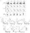
Figure 1
Figure 1. BALB/c iNKT cells produce IL-4 in the steady state
(a) Flow cytometric analysis shows hCD2 expression in conventional CD4 SP thymocytes (top row) and CD1d tetramer binding iNKT cells from thymus, spleen and liver (bottom three rows) of 7 week-old B6 and BALB/c KN2+/− mice. (b) Percentages and numbers of hCD2+ iNKT cells in thymus, spleen and liver of 7–8 week old B6-KN2 (N=4~10) and BALB/c-KN2 (N=4~13) mice. Horizontal bars indicate mean values. Unpaired two tailed t-tests were used to compare B6 and BALB/c mice. ***p<0.0001, **p=0.0095 (c) Representative FACS data of T-bet and Eomes expression in CD8 SP thymocytes of indicated mouse strains.
Figure 2
Figure 2. PLZF, ROR-γt and T-bet differentiate NKT1, NKT2 and NKT17 cells
(a) Thymic iNKT cells from 7 week-old B6 and BALB/c mice were stained for intracellular PLZF, T-bet and ROR-γt. We designated 3 distinct populations as NKT1, NKT2 and NKT17 cells. (b) NK1.1 and CD44 expression on each iNKT subset is shown. Historical stages are indicated by S1, S2, and S3. (c) Thymocytes of BALB/c KN2+/− mice were depleted of CD8 and CD24 positive cells by MACS, stimulated with PMA and ionomycin for 4 hours and stained for intracellular cytokines and hCD2. (d) Frequencies and numbers of each iNKT subset in thymi of 7–8 week-old B6 (N=11) and BALB/c (N=9) mice were compared. Horizontal bars indicate mean values. Unpaired two tailed t-tests were used to compare B6 and BALB/c mice. ***p<0.001.
Figure 3
Figure 3. IL4 producing NKT2 cells do not give rise to NKT1 cells
Thymocytes from B6 and BALB/c T-betGFP KN2+/− mice were enriched iNKT cells by depleting CD8 and CD24 positive cells and sorted hCD2+ or hCD2 NKT2 cells. Cells were labeled with VCT, injected into congenic host mice and analyzed four days later for T-betGFP expression from donor cells. Pooled data of 3 independent experiments are shown.
Figure 4
Figure 4. Steady state IL-4 is produced by NKT2 cells, and is enhanced in the absence of T-bet
(a) hCD2 staining histograms of each iNKT cell subset in B6 and BALB/c mice in the steady state are shown. In the panels on the right, hCD2 expresion on NKT2 cells of B6 and BALB/c mice are overlaid. Data are representative of at least three independent experiments. (b and c) Representative profiles of iNKT cell transcription factor expression are shown (left) with statistical analysis (right) for six week old mice. One-way ANOVA was used to compare B6 WT (N=10), B6 T-bet+/− (N=15), and B6 T-bet−/ − (N=4). ***p<0.001, *p<0.05. Horizontal bars indicate mean values. (d) Representative FACS profiles of T-bet and Eomes staining in CD8 SP thymocytes are shown.
Figure 5
Figure 5. Interstrain comparison of iNKT subsets: NKT2 cell production of IL-4 is associated with memory-like CD8 T cells
(a) Representative FACS profiles of thymic iNKT cells and CD8 SP thymocytes in 7–8 week-old indicated strains. (b) Percentage of iNKT cells among total thymocytes (left panel) and percentage of each subset (NKT1, NKT2 and NKT17) among total thymic iNKT cells (right panels), N=3~9. (c) Percentages of thymic CD8 SP thymocytes that express Eomes as detected by intracellular staining. Horizontal bars indicate mean values. (d) Correlation of Eomes expression in CD8 SP thymocytes with the frequency of NKT2 cells among total thymocytes (left) or IL-4 mRNA in NKT2 cells (right). Arbituary units represent the IL-4 message level in sorted NKT2 cells, normalized to GAPDH, and multiplied by the frequency of NKT2 cells among total thymocytes. Linear regression was used to calculate goodness of fit (R2).
Figure 6
Figure 6. IL-4 producing iNKT cells are stimulated by self ligands
(a) BM derived DCs pulsed with solvent (gray), βGlcCer (thick) or αGalCer (thin) were injected into B6 Nur77GFP mice and splenic iNKT cells were analyzed 16 hours later. Data are representative of two independent analyses with a total of 4 mice per group. (b) Thymocytes from B6 and BALB/c Nur77GFP mice were analyzed for expression of GFP in each iNKT subset. Data are representative of 3 independent experiments. (c) GFP expression in BALB/c Nur77GFP KN2+/− mice shows IL-4 producing NKT2 cells (hCD2+) express a higher level of GFP than hCD2 negative NKT2 cells. Representative data from 3 independent experiments is shown. (d) Gene expression of Nur77 was compared to IL-4 in sorted NKT2 cells (mRNA arbitrary units normalized to Gapdh). Linear regression was used to calculate goodness of fit(R2). (e) hCD2 expression is dependent on sustained TCR signaling. Thymic iNKT cells of BALB/c KN2+/− mice were MACS enriched by depleting CD8+ and CD24+ cells, labeled with VCT and injected into WT BALB/c (N=5) or BALB/c CD1d KO (N=5) mice. Six days later, hCD2 expression in NKT2 cells was analyzed. MFI, mean fluorescence intensity. An unpaired two tailed t-test was used to compare hCD2 MFI in WT or CD1d KO hosts. ***p=0.0004. (f) iNKT subsets have distinct TCR repertoires. TCR Vβ7+ usage is shown among NKT1, NKT2 and NKT17 cells in B6 mice (N=6). One-way ANOVA was used to compare the frequency of NKT1, NKT2 and NKT17 cells. ***p<0.001, *p<0.05.
Figure 7
Figure 7. Steady state IL-4 produced by iNKT cells influences B cells and thymic DCs
(a) Serum IgE concentrations measured by ELISA in indicated mice are shown. Unpaired two tailed t-tests were used to compare indicated two groups. N=5~16, *** p<0.001, **p<0.01, *p<0.05 (b) Thymic CD8α+ or SIRPα+− DCs were sorted as described in experimental procedures and analyzed for the expression of CCL17 and CCL22 by real time RT-PCR. One-way ANOVA was used to compare CCL17 and CCL22 levels in SIRPα+ DCs. Pooled data from four independent experiments using 6 to 12 mice total in each group. *** p<0.001, ** p<0.01, *p<0.05. Horizontal bars indicate mean values. Thy, thymocytes.

Comment in

V体育2025版 - References

    1. Lee YJ, Jameson SC, Hogquist KA. Alternative memory in the CD8 T cell lineage. Trends Immunol. 2011;32:50–56. - PMC (V体育官网) - PubMed
    1. Berg LJ. Signalling through TEC kinases regulates conventional versus innate CD8(+) T-cell development. Nat Rev Immunol. 2007;7:479–485. - V体育官网入口 - PubMed
    1. Weinreich MA, Odumade OA, Jameson SC, Hogquist KA. T cells expressing the transcription factor PLZF regulate the development of memory-like CD8+ T cells. Nat Immunol. 2010;11:709–716. - PMC - PubMed
    1. Alonzo ES, et al. Development of promyelocytic zinc finger and ThPOK-expressing innate gamma delta T cells is controlled by strength of TCR signaling and Id3. J Immunol. 2010;184:1268–1279. - PMC - PubMed
    1. Verykokakis M, Boos MD, Bendelac A, Kee BL. SAP protein-dependent natural killer T-like cells regulate the development of CD8(+) T cells with innate lymphocyte characteristics. Immunity. 2010;33:203–215. - PMC - PubMed

Publication types

MeSH terms

"VSports" Substances