V体育安卓版 - Paneth cells as a site of origin for intestinal inflammation
- PMID: 24089213
- PMCID: PMC3862182
- DOI: 10.1038/nature12599 (VSports手机版)
Paneth cells as a site of origin for intestinal inflammation
Abstract
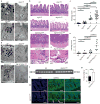
The recognition of autophagy related 16-like 1 (ATG16L1) as a genetic risk factor has exposed the critical role of autophagy in Crohn's disease. Homozygosity for the highly prevalent ATG16L1 risk allele, or murine hypomorphic (HM) activity, causes Paneth cell dysfunction. As Atg16l1(HM) mice do not develop spontaneous intestinal inflammation, the mechanism(s) by which ATG16L1 contributes to disease remains obscure. Deletion of the unfolded protein response (UPR) transcription factor X-box binding protein-1 (Xbp1) in intestinal epithelial cells, the human orthologue of which harbours rare inflammatory bowel disease risk variants, results in endoplasmic reticulum (ER) stress, Paneth cell impairment and spontaneous enteritis. Unresolved ER stress is a common feature of inflammatory bowel disease epithelium, and several genetic risk factors of Crohn's disease affect Paneth cells. Here we show that impairment in either UPR (Xbp1(ΔIEC)) or autophagy function (Atg16l1(ΔIEC) or Atg7(ΔIEC)) in intestinal epithelial cells results in each other's compensatory engagement, and severe spontaneous Crohn's-disease-like transmural ileitis if both mechanisms are compromised. Xbp1(ΔIEC) mice show autophagosome formation in hypomorphic Paneth cells, which is linked to ER stress via protein kinase RNA-like endoplasmic reticulum kinase (PERK), elongation initiation factor 2α (eIF2α) and activating transcription factor 4 (ATF4) VSports手机版. Ileitis is dependent on commensal microbiota and derives from increased intestinal epithelial cell death, inositol requiring enzyme 1α (IRE1α)-regulated NF-κB activation and tumour-necrosis factor signalling, which are synergistically increased when autophagy is deficient. ATG16L1 restrains IRE1α activity, and augmentation of autophagy in intestinal epithelial cells ameliorates ER stress-induced intestinal inflammation and eases NF-κB overactivation and intestinal epithelial cell death. ER stress, autophagy induction and spontaneous ileitis emerge from Paneth-cell-specific deletion of Xbp1. Genetically and environmentally controlled UPR function within Paneth cells may therefore set the threshold for the development of intestinal inflammation upon hypomorphic ATG16L1 function and implicate ileal Crohn's disease as a specific disorder of Paneth cells. .
Conflict of interest statement
The authors declare no competing financial interests.
Figures


References
-
- Jostins L, et al. Host-microbe interactions have shaped the genetic architecture of inflammatory bowel disease. Nature. 2012;491:119–124. - "V体育安卓版" PMC - PubMed
-
- Cadwell K, et al. Virus-plus-susceptibility gene interaction determines Crohn’s disease gene Atg16L1 phenotypes in intestine. Cell. 2010;141:1135–1145. - PMC (VSports注册入口) - PubMed
-
- Kaser A, et al. XBP1 links ER stress to intestinal inflammation and confers genetic risk for human inflammatory bowel disease. Cell. 2008;134:743–756. - V体育ios版 - PMC - PubMed
-
- Treton X, et al. Altered endoplasmic reticulum stress affects translation in inactive colon tissue from patients with ulcerative colitis. Gastroenterology. 2011;141:1024–1035. - "V体育2025版" PubMed
Publication types
- "VSports最新版本" Actions
- VSports - Actions
MeSH terms
- Actions (V体育官网入口)
- "V体育官网入口" Actions
- "V体育2025版" Actions
- V体育安卓版 - Actions
- V体育平台登录 - Actions
- V体育安卓版 - Actions
- Actions (V体育2025版)
- V体育官网 - Actions
Substances
- VSports最新版本 - Actions
- Actions (V体育官网)
- "VSports在线直播" Actions
- VSports注册入口 - Actions
- "VSports注册入口" Actions
"V体育官网" Grants and funding
- R37 DK044319/DK/NIDDK NIH HHS/United States
- R01 DK088199/DK/NIDDK NIH HHS/United States
- R01 DK051362/DK/NIDDK NIH HHS/United States
- V体育平台登录 - DK0034854/DK/NIDDK NIH HHS/United States
- "V体育2025版" R01 DK053056/DK/NIDDK NIH HHS/United States
- G0601840/MRC_/Medical Research Council/United Kingdom
- DK051362/DK/NIDDK NIH HHS/United States
- VSports - 260961/ERC_/European Research Council/International
- V体育官网 - G1002610/MRC_/Medical Research Council/United Kingdom
- R01 DK044319/DK/NIDDK NIH HHS/United States
- "VSports" 100140/WT_/Wellcome Trust/United Kingdom
- DK044319/DK/NIDDK NIH HHS/United States
- P30 DK034854/DK/NIDDK NIH HHS/United States
- DK088199/DK/NIDDK NIH HHS/United States
- P 21530/FWF_/Austrian Science Fund FWF/Austria
- DK053056/DK/NIDDK NIH HHS/United States
- R56 DK053056/DK/NIDDK NIH HHS/United States (V体育2025版)
LinkOut - more resources (VSports app下载)
Full Text Sources (V体育ios版)
Other Literature Sources
Medical
Molecular Biology Databases
Research Materials (V体育官网)

