Skip to main page content
U.S. flag

An official website of the United States government

Dot gov

The . gov means it’s official. Federal government websites often end in . gov or . mil VSports app下载. Before sharing sensitive information, make sure you’re on a federal government site. .

Https

The site is secure. The https:// ensures that you are connecting to the official website and that any information you provide is encrypted and transmitted securely V体育官网. .

. 2013;9(9):e1003614.
doi: 10.1371/journal.ppat.1003614. Epub 2013 Sep 5.

Vibrio cholerae evades neutrophil extracellular traps by the activity of two extracellular nucleases

Affiliations

Vibrio cholerae evades neutrophil extracellular traps by the activity of two extracellular nucleases

Andrea Seper et al. PLoS Pathog. 2013.

VSports - Abstract

The Gram negative bacterium Vibrio cholerae is the causative agent of the secretory diarrheal disease cholera, which has traditionally been classified as a noninflammatory disease. However, several recent reports suggest that a V. cholerae infection induces an inflammatory response in the gastrointestinal tract indicated by recruitment of innate immune cells and increase of inflammatory cytokines. In this study, we describe a colonization defect of a double extracellular nuclease V. cholerae mutant in immunocompetent mice, which is not evident in neutropenic mice. Intrigued by this observation, we investigated the impact of neutrophils, as a central part of the innate immune system, on the pathogen V. cholerae in more detail VSports手机版. Our results demonstrate that V. cholerae induces formation of neutrophil extracellular traps (NETs) upon contact with neutrophils, while V. cholerae in return induces the two extracellular nucleases upon presence of NETs. We show that the V. cholerae wild type rapidly degrades the DNA component of the NETs by the combined activity of the two extracellular nucleases Dns and Xds. In contrast, NETs exhibit prolonged stability in presence of the double nuclease mutant. Finally, we demonstrate that Dns and Xds mediate evasion of V. cholerae from NETs and lower the susceptibility for extracellular killing in the presence of NETs. This report provides a first comprehensive characterization of the interplay between neutrophils and V. cholerae along with new evidence that the innate immune response impacts the colonization of V. cholerae in vivo. A limitation of this study is an inability for technical and physiological reasons to visualize intact NETs in the intestinal lumen of infected mice, but we can hypothesize that extracellular nuclease production by V. cholerae may enhance survival fitness of the pathogen through NET degradation. .


VSports在线直播 - Conflict of interest statement

The authors have declared that no competing interests exist.

Figures
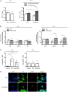
Figure 1
Figure 1. Intestinal colonization, inflammatory gene expression and neutrophil infiltration.
A. Shown are the recovered CFU per colon from immunocompetent (open bars) or neutropenic (gray bars) C57BL/6 mice infected with V. cholerae WT or ΔdnsΔxds mutant. Eight to ten weeks old streptomycin-treated mice were inoculated with the respective V. cholerae strain (7×10⁹ to 1010 CFU per mouse). At 24 h or 72 h post infection, the colons were collected, homogenized in LB medium and plated for CFU counting. Shown are medians of the recovered CFU for each data set (n = 5 for 24 h and n≥7 for 72 h). The error bars indicate the interquartile range. Significant differences between the data sets are marked by asterisks (P<0.05; Mann-Whitney U Test). B. Shown is the induction of inflammatory gene expression upon V. cholerae infection. Eight to ten weeks old streptomycin-treated C57BL/6 mice were inoculated with V. cholerae WT, ΔdnsΔxds mutant or left uninfected for mock-inoculated controls. At 24 h or 72 h post infection, the proximal 1 cm of the ascending colon was collected, RNA was extracted, reverse transcribed to cDNA and used as template for qRT-PCR analysis of the indicated genes. Gene expression was normalized to the housekeeping gene 36B4. Shown are median gene expression levels compared to mock-inoculated controls (indicated by the dashed line at 1) for each data set (n = 5 for 24 h and n≥7 for 72 h). The error bars indicate the interquartile range. The dotted line indicates a 2-fold upregulation compared to the mock-inoculated control mice. Significant differences between the data sets are marked by asterisks (P<0.05; Mann-Whitney U Test). C. Infiltration of the epithelium with neutrophils was quantified by randomly counting fifty high-power fields (HPF, 400× magnification) in tissue sections of ceca of mice colonized for 24 h or 72 h with V. cholerae WT, ΔdnsΔxds mutant or mock-inoculated controls (n = 5 for 24 h and n≥7 for 72 h). The results are expressed as the median number of neutrophils per field. The error bars indicate the interquartile range. Significant differences between the data sets are marked by asterisks (P<0.05; Kruskal-Wallis test followed by post-hoc Dunn's multiple comparisons). D. Shown is a representative immunofluorescence staining of neutrophils in ceca from mice colonized for 24 h with V. cholerae WT or ΔdnsΔxds mutant. Neutrophils were visualized by indirect immunofluorescence using primary antibodies against histone H1 and neutrophil myeloperoxidase (MPO) and DNA was visualized with DAPI (blue channel). Alexa Fluor 488- and 568-conjugated secondary antibodies were used for visualization of histone H1 (green channel) and MPO (red channel), respectively. Pictures were taken with a Nikon C1 confocal microscope at 60× magnification. Maximum intensity projections from Z-stacks are shown.
Figure 2
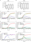
Figure 2. V. cholerae stimulates ROS production and NET formation in human neutrophils.
A–B. ROS production by human neutrophils incubated with the indicated V. cholerae strains and MOI was measured by a luminometric assay. The y-axis shows the area under the curve representing the ROS production over 6 h. Shown are medians of at least six measurements out of three independent donors. The error bars represent the interquartile range. The ROS dynamics are available as supporting figure S2B and C. C–H. DNA release of neutrophils incubated with PMA or the respective V. cholerae strain and MOI. Untreated neutrophils served as an unstimulated control. Staining of DNA by the cell impermeant fluorescent DNA dye Sytox green was measured in 10 min intervals. Values are presented as percentage of DNA fluorescence compared with the Triton ×100 lysis control (100%) indicating NET formation, respectively. DNAse I was added after 6 h for the indicated data sets (+DNAse I). Shown are medians of at least six measurements out of three (C–F) or two (G and H) independent donors.
Figure 3
Figure 3. The two extracellular nucleases of V. cholerae are able to degrade NETs.
A. DNA release of neutrophils was stimulated with PMA (indicated by the arrow “PMA”) and followed by incubation with the respective V. cholerae strain (MOI 40, time point of addition is indicated by the arrow “Vch”). Staining of DNA by the cell impermeant fluorescent DNA dye Sytox green was measured in 10 min intervals. Values are presented as percentage of DNA fluorescence compared with the Triton ×100 lysis control (100%) indicating NET formation, respectively. Shown are medians of at least six measurements out of three independent donors. B. Human neutrophils were stimulated with V. cholerae WT or ΔdnsΔxds mutant (MOI 4) in presence of the cell impermeant fluorescent DNA dye Sytox green and monitored by live cell imaging. Shown are images of the indicated time points. The complete movies are available as movie S1 (V. cholerae WT) and S2 (ΔdnsΔxds mutant) in the supporting information.
Figure 4
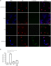
Figure 4. Visualization of NET formation by V. cholerae.
Human neutrophils were stimulated for 6 h either with PMA, V. cholerae WT or ΔdnsΔxds mutant (MOI 40) or left untreated (unstimulated). A. Shown are representative immunofluorescent micrographs of DNA (DAPI, blue) and neutrophil elastase (NE, Cy3 conjugated, red) stained samples as well as V. cholerae expressing GFP (green). B. Microscopic evaluation of NET formation by human neutrophils stimulated with PMA, V. cholerae WT or ΔdnsΔxds mutant or left untreated (unstim). The percentage of cells undergoing NET formation was determined using ImageJ as explained in the Materials and Methods section. Shown are medians with the interquartile range. At least nine images from two independent donors were analyzed for each data set. Significant differences between the data sets are marked by asterisks (P<0.05; Kruskal-Wallis test followed by post-hoc Dunn's multiple comparisons).
Figure 5
Figure 5. The two extracellular nucleases of V. cholerae facilitate survival upon contact with NETs.
A. Induction of dns and xds upon presence of NETs (gray bars) or extracellular DNA (open bars). NET formation of human neutrophils was stimulated by PMA followed by incubation with WT V. cholerae (MOI 4). After incubation with NETs or extracellular DNA the bacterial RNA was extracted, reverse transcribed to cDNA and used as template for qRT-PCR analysis of the indicated genes. Expression of the analyzed genes was compared to the control condition using the same procedure but without the presence of NETs or extracellular DNA (indicated by the dashed line at 1) and normalized to the 16S rRNA. The dotted line indicates a 2-fold upregulation compared to the control conditions. The data are presented as medians with the interquartile range (n = 6). B. Quantitative analysis of bacterial entrapment by activated human neutrophils. NET formation was stimulated with PMA followed by incubation with the indicated V. cholerae strains (MOI 40). After incubation the plates were centrifuged, the supernatants were removed and the wells were carefully washed with fresh medium to remove not entrapped bacteria. Shown is the percentage of entrapped bacteria compared with the total number of bacteria in the well (100%). The data are presented as medians with the interquartile range (n = 6). Significant differences between the data sets are marked by asterisks (P<0.05; Mann-Whitney U Test). C. Quantitative analysis of bacterial killing after coincubation of PMA stimulated neutrophils with the respective V. cholerae strain (MOI 40). Data are presented as median percentage of killed bacteria compared with the respective control treated with DNAse I and cytochalasin D to avoid extracellular and phagocytic killing. The data are presented as medians with the interquartile range (n = 6). Significant differences between the data sets are marked by asterisks (P<0.05; Mann-Whitney U Test).

VSports - References

    1. WHO (2012) Cholera annual report 2011. Weekly epidemiological record 87: 289–304. - V体育安卓版 - PubMed
    1. (CDC) CfDCaP (2010) Cholera outbreak — Haiti, October 2010. MMWR Morb Mortal Wkly Rep 59: 1411. - PubMed
    1. Yildiz FH, Visick KL (2009) Vibrio biofilms: so much the same yet so different. Trends Microbiol 17: 109–118. - PMC - PubMed
    1. Colwell RR (2000) Viable but nonculturable bacteria: a survival strategy. J Infect Chemother 6: 121–125. - VSports注册入口 - PubMed
    1. Harris JB, LaRocque RC, Qadri F, Ryan ET, Calderwood SB (2012) Cholera. Lancet 379: 2466–2476. - VSports - PMC - PubMed

Publication types (V体育官网)

MeSH terms

"V体育2025版" LinkOut - more resources