<time lang="AIrgv"></time><small id="6uOEEk"></small>
VSports app下载 - Skip to main page content
U.S. flag

An official website of the United States government

Dot gov

The VSports app下载. gov means it’s official. Federal government websites often end in . gov or . mil. Before sharing sensitive information, make sure you’re on a federal government site. .

Https

The site is secure. The https:// ensures that you are connecting to the official website and that any information you provide is encrypted and transmitted securely V体育官网. .

. 2013 Aug 26;210(9):1871-88.
doi: 10.1084/jem.20122762. Epub 2013 Aug 19.

Lung dendritic cells induce migration of protective T cells to the gastrointestinal tract

Affiliations

Lung dendritic cells induce migration of protective T cells to the gastrointestinal tract

Darren Ruane et al. J Exp Med. .

Abstract

Developing efficacious vaccines against enteric diseases is a global challenge that requires a better understanding of cellular recruitment dynamics at the mucosal surfaces. The current paradigm of T cell homing to the gastrointestinal (GI) tract involves the induction of α4β7 and CCR9 by Peyer's patch and mesenteric lymph node (MLN) dendritic cells (DCs) in a retinoic acid-dependent manner VSports手机版. This paradigm, however, cannot be reconciled with reports of GI T cell responses after intranasal (i. n. ) delivery of antigens that do not directly target the GI lymphoid tissue. To explore alternative pathways of cellular migration, we have investigated the ability of DCs from mucosal and nonmucosal tissues to recruit lymphocytes to the GI tract. Unexpectedly, we found that lung DCs, like CD103(+) MLN DCs, up-regulate the gut-homing integrin α4β7 in vitro and in vivo, and induce T cell migration to the GI tract in vivo. Consistent with a role for this pathway in generating mucosal immune responses, lung DC targeting by i. n. immunization induced protective immunity against enteric challenge with a highly pathogenic strain of Salmonella. The present report demonstrates novel functional evidence of mucosal cross talk mediated by DCs, which has the potential to inform the design of novel vaccines against mucosal pathogens. .


Figures (V体育ios版)
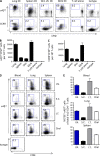
Figure 1.
Figure 1.
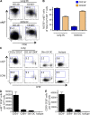
Lung DCs induce integrin α4β7 and CCR9. (A–C) CD11c+ DCs isolated from the lung, spleen, skin-draining LNs, or MLNs were pulsed with OVA and cultured with CFSE-labeled OT-II T cells at a ratio of 1:2 for 5–7 d. (A) T cell expression of α4β7 (top) and CCR9 (bottom) was measured by flow cytometry and plotted against CFSE dilution. (B and C) Quantification of the number of proliferated (CFSElo) α4β7+ (B) or CCR9+ (C) cells showing cumulative data from three independent experiments. (D and E) CFSE-labeled CD45.1+Vα2+CD4+OT-II cells were transferred into naive CD45.2+ recipients. 2 h after transfer, mice were immunized with OVA protein + polyICLC or cholera toxin via the indicated route, and the percentage of CFSEloCD45.1+Vα2+CD4+ cells expressing α4β7 was determined in blood, lung, and spleen 4 d after immunization. (D) Representative flow cytometry data showing CFSE dilution versus α4β7 expression. (E) Cumulative data from three independent experiments showing frequency of α4β7+CFSElo cells among total Vα2+CD4+ cells.
Figure 2.
Figure 2.
Lung CD103+ and CD24+11b+ DCs induce integrin α4β7 and CCR9 in vitro. (A and B) MHCII+CD11chiCD11b+CD103 and MHCII+CD11chiCD11bCD103+ DCs were flow-sorted from the lung or MLN, pulsed with OVA, and cultured with CFSE-labeled OT-II T cells at a ratio of 1:2 for 5–7 d. (A) T cell expression of α4β7 induced by lung DCs (top) and MLN DCs (bottom) was measured by flow cytometry and plotted against CFSE dilution. (B) Quantification of the number of proliferated (CFSElo) α4β7+ cells showing cumulative data from three independent experiments. (C–E) Lung-derived MHCII+CD11chiCD103CD11b+CD24+ and MHCII+CD11chiCD103CD11b+CD64+ cells were flow-sorted. Additionally, MHCII+CD11chi DCs were isolated from skin-draining LNs. These DCs were pulsed with OVA and cultured with CFSE-labeled OT-II T cells at a ratio of 1:2 for 5–7 d (C) T cell expression of α4β7 (top) and CCR9 (bottom) was measured by flow cytometry and plotted against CFSE dilution. (D and E) Quantification of the number of proliferated (CFSElo), α4β7+ (D) or CCR9+ (E) cells showing cumulative data from three independent experiments.
Figure 3.
Figure 3.
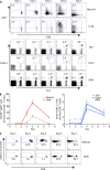
After i.n. immunization, integrin α4β7 is induced in the mediastinal LN- and lung-resident cells, followed by the appearance of migratory cells in the GI tissues. CD45.1+Vα2+CD4+OT-II cells were transferred into naive CD45.2+ recipients. 2 h after transfer, mice were immunized with OVA protein + polyICLC via the i.n. route, and the transferred CD45.1+Vα2+CD4+ cells in mediastinal LN (med LN), lung, SILP, colon, and MLN were examined on days 1, 2, 3, 4, and 7 after immunization. (A) Representative flow cytometry data showing CFSE dilution versus α4β7 expression in the med LN and lung at the respective time points (top two rows) and the frequency of adoptively transferred Vα2+CD45.1+CD4+ T cells in the SILP, colon and MLN (bottom three rows). (B) Cumulative data showing frequency of α4β7+CFSElo cells among total Vα2+CD4+ cells (left) and the frequency of Vα2+CD45.1+ cells among total CD4+ T cells (right). (C) Proliferation (CFSE dilution) of the adoptively transferred CD45.1+CD4+Vα2+ T cells is compared between the med LN (top) and MLN (bottom) of the recipient mice on days 1, 2, 3, 4, and 7.
Figure 4.
Figure 4.
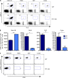
Administration of FTY-720 leads to accumulation of transferred CD45.2+Vα2+CD4+ OT-II cells in the mediastinal LNs, but not in the MLNs, after i.n. vaccination. CFSE-labeled CD45.1+Vα2+CD4+OT-II cells were adoptively transferred to naive CD45.2+ recipients that were immunized after 2 h with OVA protein and polyICLC, delivered i.n. On days 0–3, the recipient mice were administered FTY-720 (1 µg/g mouse). The mice were sacrificed on day 4, and the frequency of transferred CD45.1+Vα2+CD4+ T cells was quantified in the med LN, MLN, SILP, and colon. (A) Representative flow cytometry plot comparing the frequency of adoptively transferred CD45.1+ CD4+ Vα2+ OT-II cells between WT mice (top) and FTY-720 administered mice (bottom). (B) Quantification of the number of transferred CD45.1+CD4+Vα2+ OT-II cells in the med LN, MLN, SILP, and colon showing cumulative data from three independent experiments. *, P < 0.05; **, P < 0.01; ***, P < 0.001. (C) Proliferation of the adoptively transferred CD45.1+CD4+Vα2+ T cells is compared between WT and FTY-720 administered mice in the indicated tissues.
Figure 5.
Figure 5.
i.n. immunization–induced migration of T cells to the GI tract is integrin α4β7-dependent and is more efficient than s.c. immunization–induced T cell migration to the GI tract. (A and B) 2 h after adoptive transfer of CFSE-labeled CD45.1+Vα2+CD4+ OT-II cells to naive CD45.2+ mice, the recipient mice were immunized i.n. or s.c. with OVA protein and polyICLC. On day 7 after immunization, the frequency of CD45.1+Vα2+CD4+ T cells was determined in the SILP, colon, and MLN. Representative flow cytometry plots (A) and cumulative data from three independent experiments (B) are shown. (C and D) After 2 h of adoptive transfer of CFSE-labeled CD45.1+Vα2+CD4+ OT-II cells to naive CD45.2+ mice, the recipient mice were immunized i.n. with OVA protein and polyICLC. On days 0 and 3, mice received 100 µg of anti-α4β7 antibody delivered i.p. with the control mice receiving 100 µl of PBS i.p. On day 7 after immunization, the frequency of CD45.1+Vα2+CD4+ T cells was determined in the colon, IEL, SILP, MLN, blood, spleen, med LN, and lung. Representative flow cytometry plots (C) and cumulative data from three independent experiments comparing anti-α4β7 and PBS administered mice (D) are shown.
Figure 6.
Figure 6.
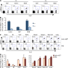
Induction of integrin α4β7 by lung DCs requires RA and TGF-β signaling. (A) Demonstration of ALDH activity in lung-derived DCs using flow cytometry. The MFI of a fluorescent ALDH substrate was quantified for lung DCs in the presence (top two curves) or absence (middle two curves) of the ALDH inhibitor-DEAB. MLN CD103+ DCs and splenic CD8α+ DCs served as the positive and negative controls, respectively (bottom two curves). (B and C) CD11c+ DCs isolated from the lung and MLN were pulsed with OVA and cultured with CFSE-labeled OT-II T cells at a ratio of 1:2 for 5–7 d. RAR-β inhibitor, LE540 (1 µM), or DMSO control is added to the DC/OT-II cultures. (B) Representative flow cytometry plots comparing the expression of α4β7 on CD4+OT-II cells with and without RAR-β antagonist LE-540. (C) Quantification of the number of α4β7+Vα2+CD4+ T cells induced by the respective DCs, in the absence (black bars) or presence (white bars) of RAR-β antagonist LE-540. (D) WT CD45.1+Vα2+CD4+OT-II cells, dnRAR-OT-II cells, and TGF-βRdef OT-II cells (described in the Materials and methods) were transferred into CD45.2+ recipient mice and OVA/polyICLC was administered i.n. Cumulative data from three experiments comparing the percentage of α4β7+CFSEloVα2+CD4+ T cells between the WT OT-II, dnRAR OT-II, and TGF-βRdef OT-II recipients is shown. (E) After the transfer of CD45.1+Vα2+CD4+OT-II cells, mice were immunized with OVA alone, with OVA + lipopolysaccharide (LPS), with OVA + polyICLC, or with polyICLC alone. Cumulative data from three experiments comparing the percentage of α4β7+CFSEloVα2+CD4+ T cells between these mouse groups.
Figure 7.
Figure 7.
After i.n. immunization, induction of integrin α4β7 is mediated by DCs. DT was administered to CD11c-DTR chimeras (CD11c-DTR bone marrow into WT mice), or zDC-DTR chimeras (zDC-DTR bone marrow into WT mice) (described in the Materials and methods). 24 h later, we transferred CFSE-labeled CD45.1+Vα2+CD4+ OT-II cells to CD11c-DTR chimeras (A and B) or to zDC-DTR chimeras (C and D). Mice administered PBS served as the respective controls. Representative flow cytometry plots (A and C) and cumulative data from three experiments each (B and D), showing the in vivo induction of integrin α4β7 on CFSEloCD45.1+Vα2+CD4+ OT-II cells in CD11c-DTR and zDC-DTR mice, respectively.
Figure 8.
Figure 8.
Ablation of CD11b+ cells attenuates the induction of α4β7 on transferred OT-II cells after i.n. immunization. Two doses of DT were administered to CD11b-DTR chimeras (CD11b-DTR bone marrow into WT mice; described in the Materials and methods) on days 0 and 1. On day 3 after DT, CFSE-labeled CD45.1+Vα2+CD4+ OT-II cells were transferred and the mice were immunized i.n. with OVA/polyICLC. PBS-administered CD11b-DTR mice served as controls. Representative flow cytometry plots (A) and cumulative data from three independent experiments showing the induction of integrin α4β7 on adoptively transferred CD45.1+Vα2+CD4+ OT-II cells in PBS or DT-administered animals (B). (C) Representative flow cytometry plots comparing the frequency of MHCII+CD11c+-gated CD11c+CD11b+ cells in the lung (top) and med LN (bottom) of CD11b-DTR bone marrow chimeric mice administered PBS or DT. (D) Quantification of CD11b and CD11b+ DC in the lung and med LN of CD11b-DTR bone marrow chimeric administered PBS or DT, showing cumulative data from three independent experiments.
Figure 9.
Figure 9.
Ablation of CD103+ DCs does not impact the induction of α4β7 on transferred OT-II cells after i.n. immunization. (A and B) DT or PBS was administration to langerin-DTR mice (described in the Materials and methods). 24 h later CFSE-labeled CD45.1+Vα2+CD4+ OT-II cells were transferred and the mice were immunized with OVA/polyICLC. Representative flow cytometry plots (A) and cumulative data from three experiments (B) are shown, comparing the expression of α4β7 on CFSElo CD45.1+Vα2+CD4+ OT-II cells. (C and D) CD45.1+Vα2+CD4+ OT-II cells were transferred to WT or batf3−/− mice, followed by immunized with OVA/polyICLC, delivered i.n. Representative flow cytometry plots (C) and cumulative data from three experiments (D) are shown, comparing the expression of α4β7 on CFSElo CD45.1+Vα2+CD4+ OT-II cells.
Figure 10.
Figure 10.
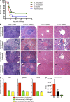
i.n. immunization protects against enteric challenge with highly pathogenic Salmonella. (A–C) C56Bl/6 mice were immunized with OVA/polyICLC delivered i.n. or s.c. in a prime-boost fashion or p.o. with OVA/CT, and challenged orally 7 d after the boost with Salmonella-OVA. Unimmunized mice challenged with Salmonella-OVA served as controls. Data from three independent experiments is presented here. (A) Kaplan-Meier curves comparing the survival of unimmunized (red) mice or mice immunized i.n. (green), s.c. (black), or p.o. (blue) against oral challenge with Salmonella-OVA (106PFU). (B) Hematoxylin and eosin sections examining the MLN (i, v, ix, and xiii), spleen (ii, vi, x, and xiv), and liver (iii, vii, xi, and xv), all at 200× magnification (bar, 100 µm), and liver (iv, viii, xii, and xvi) at 400× magnification (bar, 50 µm) from a WT (unimmunized, unchallenged) mouse (i–iv), an unimmunized, Salmonella-challenged mouse (v–viii), a representative i.n.-immunized, Salmonella-challenged mouse #1 (ix–xii), and a representative i.n.-immunized, Salmonella-challenged mouse #2 (xiii–xvi). (C) Cumulative data from three experiments showing the inflammation score from the WT (unimmunized, unchallenged), unimmunized, and Salmonella-challenged groups. (D) C56Bl/6 mice were immunized with heat and paraformaldehyde-inactivated S. typhimurium delivered i.n. or s.c. in a prime-boost fashion, and challenged orally 7 d after the boost with Salmonella-OVA. Unimmunized mice challenged with Salmonella-OVA served as controls. CFU of S. typhimurium per gram of splenic tissue is compared between the WT, s.c.-immunized, and i.n.-immunized mice.

V体育官网 - Comment in

References

    1. Bennett C.L., Clausen B.E. 2007. DC ablation in mice: promises, pitfalls, and challenges. Trends Immunol. 28:525–531 10.1016/j.it.2007.08.011 - DOI - PubMed
    1. Bennett C.L., van Rijn E., Jung S., Inaba K., Steinman R.M., Kapsenberg M.L., Clausen B.E. 2005. Inducible ablation of mouse Langerhans cells diminishes but fails to abrogate contact hypersensitivity. J. Cell Biol. 169:569–576 10.1083/jcb.200501071 - DOI - PMC - PubMed
    1. Berlin C., Bargatze R.F., Campbell J.J., von Andrian U.H., Szabo M.C., Hasslen S.R., Nelson R.D., Berg E.L., Erlandsen S.L., Butcher E.C. 1995. alpha 4 integrins mediate lymphocyte attachment and rolling under physiologic flow. Cell. 80:413–422 10.1016/0092-8674(95)90491-3 - DOI - PubMed
    1. Bhat P.V. 1998. Retinal dehydrogenase gene expression in stomach and small intestine of rats during postnatal development and in vitamin A deficiency. FEBS Lett. 426:260–262 10.1016/S0014-5793(98)00355-X - VSports最新版本 - DOI - PubMed
    1. Bienenstock J., McDermott M., Befus D., O’Neill M. 1978. A common mucosal immunologic system involving the bronchus, breast and bowel. Adv. Exp. Med. Biol. 107:53–59 10.1007/978-1-4684-3369-2_7 - DOI - PubMed

MeSH terms

"VSports手机版" Substances