Skip to main page content
U.S. flag

An official website of the United States government

Dot gov

The . gov means it’s official. Federal government websites often end in VSports app下载. gov or . mil. Before sharing sensitive information, make sure you’re on a federal government site. .

Https

The site is secure V体育官网. The https:// ensures that you are connecting to the official website and that any information you provide is encrypted and transmitted securely. .

. 2013 Sep;19(9):1132-40.
doi: 10.1038/nm.3265. Epub 2013 Aug 18.

Activation of the Nlrp3 inflammasome in infiltrating macrophages by endocannabinoids mediates beta cell loss in type 2 diabetes

Affiliations

VSports注册入口 - Activation of the Nlrp3 inflammasome in infiltrating macrophages by endocannabinoids mediates beta cell loss in type 2 diabetes

Tony Jourdan et al. Nat Med. 2013 Sep.

V体育官网 - Abstract

Type 2 diabetes mellitus (T2DM) progresses from compensated insulin resistance to beta cell failure resulting in uncompensated hyperglycemia, a process replicated in the Zucker diabetic fatty (ZDF) rat. The Nlrp3 inflammasome has been implicated in obesity-induced insulin resistance and beta cell failure VSports手机版. Endocannabinoids contribute to insulin resistance through activation of peripheral CB1 receptors (CB₁Rs) and also promote beta cell failure. Here we show that beta cell failure in adult ZDF rats is not associated with CB₁R signaling in beta cells, but rather in M1 macrophages infiltrating into pancreatic islets, and that this leads to activation of the Nlrp3-ASC inflammasome in the macrophages. These effects are replicated in vitro by incubating wild-type human or rodent macrophages, but not macrophages from CB₁R-deficient (Cnr1(-/-)) or Nlrp3(-/-) mice, with the endocannabinoid anandamide. Peripheral CB₁R blockade, in vivo depletion of macrophages or macrophage-specific knockdown of CB₁R reverses or prevents these changes and restores normoglycemia and glucose-induced insulin secretion. These findings implicate endocannabinoids and inflammasome activation in beta cell failure and identify macrophage-expressed CB₁R as a therapeutic target in T2DM. .


Figures

Figure 1
Figure 1
Effects of peripheral CB1R blockade on body weight, adiposity, hepatic lipogenesis and glycemic control in ZDF rats. Eight-week-old male ZDF rats treated for 28 d by oral gavage with 3 mg per kg body weight per day JD5037 or vehicle, (a) Effects of vehicle (gray columns and squares) and JD5037 (black columns and triangles) treatment of ZDF rats on body weight, adiposity and food intake compared to lean controls (white columns and open circles), n-20 pergroup. (b–d) Effects of vehicle (gray) and JD5037 (black) treatment of the same ZDF rats compared to lean controls (white) on hepatic triglycerides (FG), plasma alanine aminotransferase (ALT) and hepatic Fas and Scd1 (b; n = 20 per group); on fasting blood glucose, HbA1c plasma insulin and C-peptide levels (c; n = 20 per group), and on the glucose-infusion rate (GIR), the percentage of suppression of hepatic glucose production (HGP) and the rate of glucose disappearance (Rd) during a euglycemic-hyperinsuiinemic clamp (d; n = 5 per group). Data are expressed as means means ± s.e.m. from 20 rats pergroup; * P < 0.05, ** P < 0.01, *** p < 0.001.
Figure 2
Figure 2
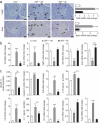
The effect of chronic JD5037 treatment on beta cell survival and function in ZDF rats, (a–d) Treatments, as described in Figure 1, were started in 8-week-old diabetic rats (a–c) or 6-week-old prediabetic ZDF rats (d), (a) Immunohistochemical identification of insulin, TUNEL-positive cells and CB1R protein in islets from lean rats and ZDF rats treated with vehicle or JD5037 for 4 weeks. Bar graphs show Ins1 and Cnr1 mRNA levels and anandamide (AEA) content of isolated islets or the percentage of TUNEL-positive cells; n = 15 rats per group. Arrows mark antigen-positive cells; significant difference from lean control, * P < 0.05, *** P < 0.001, (b) Glucose-induced insulin and C-peptide release in vivo (left and middle) or in isolated islets (right) from lean, vehicle-treated ZDF or JD5037-treated ZDF rats. In the left and middle graphs, the yellow segment indicates the glucose-induced increase in plasma insulin or C-peptide, respectively, over baseline insulin levels shown in white, gray or black; n = 10 rats or 8 islet preparations per group; * P < 0.05, ** P < 0.01, *** P < 0.001 relative to basel ine plasma insulin level or value in corresponding group of islets on 0,5 gl–1 glucose, (c) Slc2a2 and Gck expression in islets from lean, vehicle-treated or JD5037-treated ZDF rats; n = 10 preparations per group; Pvalues as in a. (d) Baseline blood glucose, insulin and C-peptide levels in 6-week-old prediabetic ZDF rats and their biweekly change during 12 weeks of daily treatment with vehicle (gray columns and squares) or JD5037 (black columns and triangles). Mean ± s.e.m. values in age-matched lean controls are shown by white columns and open circles; n = 10 rats per group during weeks 6–18. Significant difference from corresponding value in lean (* P < 0.05, ** P < 0.005 or *** p < 0.001) or vehicle-treated (### P < 0.001) ZDF rats. Scale bars, 50 μm.
Figure 3
Figure 3
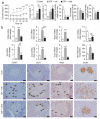
Macrophage content and Mrp3 expression in islets of lean and diabetic rats, (a) Immunohistochemical stains for CD68+ and Nlrp3 in islets of lean, ZDF and JD5037-treated ZDF rats; arrows mark antigen-positive cells. Scale bars, 50 μm. CD68 and Nlrp3 mRNA levels measured in isolated islets are shown on the right, (b) Pro- and anti-inflammatory gene expression in the same islets as in a. (c) Expression of Il18, Il1b, Il1r, Il1rn and Txnip, as well as p65-NFκB, IL-18 and IL-1β protein and caspase-1 activity, in the same isets. Eight-week-old ZDF rats treated for 28 d with vehicle or JD5037 13 mg per kg body weight per day), with age-matched lean controls; n = 20 rats per group; * P < 0.05, ** P < 0.01, *** P < 0.001. Scale bars, 50 μm. Data are expressed as means + s.e.m.
Figure 4
Figure 4
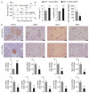
Effects of macrophage depletion on glycemic control and proinflammatory signaling in islets of ZDF rats, (a) Blood glucose, insulin and C-peptide levels and insulin resistance analyzed by insulin clamps in lean littermate controls (white columns or circles) and In ZDF rats treated with empty liposomes (rah, gray col umns or squares) or clodronate-containing liposomes (clodr, black columns or triangles) Glycemic control was monitored for 20 d. (b) Top, mRNA expression of Cd68 (as a marker of macrophages), Cnr1, Nlrp3, Ins1, Tnf, Ccl2, Txnip and anandamide content of islets isolated from pancreata of lean littermate rats (white columns) and ZDF rats treated with empty liposomes (veh, gray columns) or clodronate (clodr, black columns). Co umns and bars represent means ± s.e.m. from eight rats per group; ** P < 0.01, *** p < 0.001 relative to lean group, Bottom, representative immunohistochemistry stains (top) for CD68, CB1R, Nlrp3 and insulin protein expression in pancreatic sections from lean control rats and ZDF rats treated with empty liposomes or clodronate. Arrows mark antigen-positive cells. Scale bars, 50 mμ.
Figure 5
Figure 5
Effects of macrophage-selective siRNA knockdown of CB1R in ZDF rats, (a) Baseline blood glucose in 8-week-old ZDF rats during 10 d of treatment with scrambled (Scrb) GeRPs (gray columns and circles) or CB1R GeRPs (black columns and squares) and plasma insulin, C-peptide and pancreatic AEA levels at end of treatment, n = 6–8 rats per group; * P < 0.05, ** P < 0.01, *** P < 0.001. (b)Top, immunohistochemistry stains for islet insulin, macrophages and CB1R and Nlrp3 protein (with arrows marking antigen-positive cells). Scale bars, 50 μm. Bottom, mRNA expression of Ins1, Cd68. Cnr1, Nlrp3, Il18, Il1b, Tnf, Ccl2 and Pycard in islets isolated from ZDF rats treated with scrambled (gray) or CB1R GeRPs (black); n = 6 pergroup. * P < 0.05, ** P < 0.01, *** p < .001. Data are expressed as means ± s.e.m.
Figure 6
Figure 6
Proinflammatory gene and protein expression in human macrophages and rat and human isolated islets treated with AEA, IL-1β or high giucose. (a) Relative effects of AEA (1 μM; light gray), JD5037 (100 nM; dafk gray) or their combination (biack) versus a vehicle control (veh; white) on CNR1, CNR2, PYXARD and NLRP3 and on IL-1β and IL-18 secretion in human peripheral macrophages. Data are expressed as means ± s.e.m. from cells from six to eight different donors. MDM, monocyte-derived macrophages, (b) Effects of AEA (1 μM; light gray), high glucose HG; blue (33 mM) and IL-1β (30 ng ml−1; red) versus a vehicle control (veh; white) on the secretion of IL-1β, MCP-l, IL-6 and TNF-α by islets from lean and ZDF rats, (c) Effects of vehicle, AEA, high glucose and IL-1β on IL-1β and MCP-l secretion by human islets. Data in b and c are expressed as means ± s.e.m. from six to eight preparations, each containing ten islets. (d) Schematic illustration of the mechanism of CB1R-mediated beta cell loss in the diabetic islet, This figure was prepared using a template on the Servier medical art website {http://www.servier.frirservier-medical-art).

Comment in

References

    1. Defronzo RA. Banting Lecture. From the triumvirate to the ominous octet: a new paradigm for the treatment of type 2 diabetes mellitus. Diabetes. 2009;58:773–795. - PMC - PubMed
    1. Shoelson SE, Lee J, Goldline AB. Inflammation and insulin resistance. J. Clin. invest. 2006;116:1793–1801. - PMC - PubMed
    1. Ehses JA, et al. Increased number ot islet-associated macrophages in type 2 diabetes. Diabetes. 2007;56:2356–2370. - PubMed
    1. Masters SL, et al. Activation of the NLRP3 inflammasome by islet amyloid polypeptide provides a mechanism for enhanced IL-1β in type 2 diabetes. Nat. Immunol. 2010;11:897–904. - PMC - PubMed
    1. Youm YH, et al. Elimination of the NLRP3-ASC inflammasorne protects against chronic obesity-induced pancreatic damage. Endocrinology. 2011;152:4039–4045. - V体育ios版 - PMC - PubMed

Publication types

VSports在线直播 - MeSH terms