"VSports手机版" Skip to main page content
U.S. flag

An official website of the United States government

Dot gov

The . gov means it’s official. Federal government websites often end in . gov or . mil. Before sharing sensitive information, make sure you’re on a federal government site. VSports app下载.

Https

The site is secure. The https:// ensures that you are connecting to the official website and that any information you provide is encrypted and transmitted securely. V体育官网.

. 2013 Aug 27;110(35):14330-5.
doi: 10.1073/pnas.1311998110. Epub 2013 Aug 12.

Human circulating influenza-CD4+ ICOS1+IL-21+ T cells expand after vaccination, exert helper function, and predict antibody responses

Affiliations

Human circulating influenza-CD4+ ICOS1+IL-21+ T cells expand after vaccination, exert helper function, and predict antibody responses

Fabiana Spensieri et al. Proc Natl Acad Sci U S A. .

Abstract

Protection against influenza is mediated by neutralizing antibodies, and their induction at high and sustained titers is key for successful vaccination. Optimal B cells activation requires delivery of help from CD4(+) T lymphocytes. In lymph nodes and tonsils, T-follicular helper cells have been identified as the T cells subset specialized in helping B lymphocytes, with interleukin-21 (IL-21) and inducible costimulatory molecule (ICOS1) playing a central role for this function. We followed the expansion of antigen-specific IL-21(+) CD4(+) T cells upon influenza vaccination in adults. We show that, after an overnight in vitro stimulation, influenza-specific IL-21(+) CD4(+) T cells can be measured in human blood, accumulate in the CXCR5(-)ICOS1(+) population, and increase in frequency after vaccination. The expansion of influenza-specific ICOS1(+)IL-21(+) CD4(+) T cells associates with and predicts the rise of functionally active antibodies to avian H5N1. We also show that blood-derived CXCR5(-)ICOS1(+) CD4(+) T cells exert helper function in vitro and support the differentiation of influenza specific B cells in an ICOS1- and IL-21-dependent manner. We propose that the expansion of antigen-specific ICOS1(+)IL-21(+) CD4(+) T cells in blood is an early marker of vaccine immunogenicity and an important immune parameter for the evaluation of novel vaccination strategies. VSports手机版.

Keywords: CD4 help; humoral response; predictivity. V体育安卓版.


Conflict of interest statement

Conflict of interest statement: The authors are employees of Novartis Vaccines and Diagnostics, Srl.

Figures

Fig. 1.
Fig. 1.
HI antibody response to vaccination. HI antibody responses to the (A) H5N1 and (B) H3N2 vaccine strains at baseline (Pre), 21 d after the first dose (Post 1), and 21 d after the second dose (Post 2) of respective vaccine. The lines crossing the box plots represent median titers, and whiskers indicate the minimum and maximum values (***P < 0.0001 by Tukey–Kramer test).
Fig. 2.
Fig. 2.
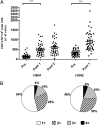
Expansion and cytokine profile of H5N1- and H3N2-specific CD4+ T cells after vaccination. Total cytokine positive (CTK+) CD4+ T cells in PBMCs of vaccines after overnight in vitro stimulation with H5N1 (Left) or H3N2 (Right). (A) Number of CTK+ CD4+ in PBMCs collected before (Pre), 3 wk after the first vaccination (Post 1), and 3 wk after the second vaccination (Post 2). Data are expressed as number of CTK+ CD4+/106 CD4+ T cells ± SEM (n = 44). (B) Cytokine profile of influenza-specific total CD4+ T cells 3 wk after the first vaccination (Post 1). The pie charts show the relative proportions of CD4+ T cells producing one (white), two (striped), three (gray), or four (black) cytokines. Shown is the average distribution in the whole database (n = 44; *P < 0.05, **P < 0.01, and ***P < 0.001, Tukey–Kramer test).
Fig. 3.
Fig. 3.
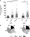
Expansion and cytokine profile of IL-21+ H5N1- and H3N2-specific CD4+ T cells after vaccination. IL-21+ CD4+ T cells in PBMCs of vaccines after overnight in vitro stimulation with H5N1 (Left) or H3N2 (Right). (A) Numbers of IL-21+ CD4+ after overnight stimulation with H5N1 or H3N2 before (Pre), 3 wk after the first vaccination (Post 1), and 3 wk after the second vaccination (Post 2). Data are expressed as number of antigen-specific IL-21+ CD4+/106 CD4+ T cells ± SEM (n = 44). (B) The pie charts show the relative Post 1 proportions of IL-21+ CD4+ T cells synthesizing IL-21 alone (white) or in combination with one (striped), two (gray), or three (black) additional cytokines. Shown is the average distribution in the whole database (n = 44; *P < 0.05, **P < 0.01, and ***P < 0.001, Tukey–Kramer test).
Fig. 4.
Fig. 4.
Distribution of blood antigen-specific IL-21+ CD4+ T cells according to the expression of ICOS1 and CXCR5. Shown are the relative proportions of IL-21+ CD4+ T cells expressing CXCR5ICOS1+ (gray), CXCR5+ICOS1+ (white), CXCR5ICOS1 (striped), and CXCR5+ICOS1 (black) after overnight stimulation with H5N1 (Left) or H3N2 (Right). Shown is the average distribution in 10 subjects from the A/T group, analyzed 3 wk after the first vaccination.
Fig. 5.
Fig. 5.
Frequency of H5N1-specific ICOS1+IL-21+ CD4+ T cells at Post 1 significantly correlates with and predicts HI titers at Post 2. Associations between paired values of HI titers to H5N1 (Post 2) and fold increase of H5N1-specific CD4+ at (AC) Post 1 vs. Pre or (DF) at Post 2 vs. Pre. The association was calculated with fold increase in (A and D) H5N1 ICOS1+IL-21+ CD4+ T cells, (B and E) H5N1 ICOS1IL-21+ CD4+ T cells, and (C and F) H5N1 total CTK+ CD4+ T cells. Fisher two-tailed exact test was used. Horizontal dashed lines indicate the value of HI titer of 80, the accepted threshold of protective antibodies. Vertical dashed lines indicate the value of threefold increase in H5N1 CD4+ T cells (n = 44).
Fig. 6.
Fig. 6.
Influenza-specific CXCR5ICOS1+ CD4+ T cells provide cognate help for antigen-specific antibody production in vitro. (AF) Immunoglobulin content in day 10 supernatant from three independent experiments. Shown are total IgG (A, C, and E) and total IgM (B, D, and F). (G) H3N2-specific IgG, (H) H3N2-specific IgM, and (I) DT (CRM-197)-specific IgG content measured in the coculture supernatants of the experiment shown in E and F. The last column of I shows the content in DT (CRM)-specific IgG in the supernatants from B cells stimulated with CpG and IL-2 in the absence of CD4+ T cells. Values are reported as median fluorescence intensity (MFI).

VSports - References

    1. Haining WN, Pulendran B. Identifying gnostic predictors of the vaccine response. Curr Opin Immunol. 2012;24(3):332–336. - PMC (VSports在线直播) - PubMed
    1. Galli G, et al. Adjuvanted H5N1 vaccine induces early CD4+ T cell response that predicts long-term persistence of protective antibody levels. Proc Natl Acad Sci USA. 2009;106(10):3877–3882. - PMC (V体育官网) - PubMed
    1. Breitfeld D, et al. Follicular B helper T cells express CXC chemokine receptor 5, localize to B cell follicles, and support immunoglobulin production. J Exp Med. 2000;192(11):1545–1552. - PMC - PubMed
    1. Vogelzang A, et al. A fundamental role for interleukin-21 in the generation of T follicular helper cells. Immunity. 2008;29(1):127–137. - PubMed
    1. Fazilleau N, Mark L, McHeyzer-Williams LJ, McHeyzer-Williams MG. Follicular helper T cells: Lineage and location. Immunity. 2009;30(3):324–335. - PMC (V体育2025版) - PubMed

Publication types (V体育ios版)

Substances