Skip to main page content
U.S. flag

An official website of the United States government

Dot gov

The . gov means it’s official VSports app下载. Federal government websites often end in . gov or . mil. Before sharing sensitive information, make sure you’re on a federal government site. .

Https

The site is secure. The https:// ensures that you are connecting to the official website and that any information you provide is encrypted and transmitted securely. V体育官网.

. 2013 Oct;149(1):55-64.
doi: 10.1016/j.clim.2013.06.005. Epub 2013 Jun 18.

Medulloblastoma expresses CD1d and can be targeted for immunotherapy with NKT cells

Affiliations

Medulloblastoma expresses CD1d and can be targeted for immunotherapy with NKT cells

"VSports在线直播" Daofeng Liu et al. Clin Immunol. 2013 Oct.

Abstract

Medulloblastoma (MB) is the most common malignant brain tumor of childhood. Current therapies are toxic and not always curative that necessitates development of targeted immunotherapy. However, little is known about immunobiology of this tumor. In this study, we show that MB cells in 9 of 20 primary tumors express CD1d, an antigen-presenting molecule for Natural Killer T cells (NKTs). Quantitative RT-PCR analysis of 61 primary tumors revealed an elevated level of CD1d mRNA expression in a molecular subgroup characterized by an overactivation of Sonic Hedgehog (SHH) oncogene compared with Group 4. CD1d-positive MB cells cross-presented glycolipid antigens to activate NKT-cell cytotoxicity. Intracranial injection of NKTs resulted in regression of orthotopic MB xenografts in NOD/SCID mice. Importantly, the numbers and function of peripheral blood type-I NKTs were preserved in MB patients VSports手机版. Therefore, CD1d is expressed on tumor cells in a subset of MB patients and represents a novel target for immunotherapy. .

Keywords: Brain tumors; CD1d; Cellular immunotherapy; Medulloblastoma; NKT cells V体育安卓版. .


Figures

Figure 1
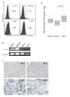
Figure 1. CD1d expression in medulloblastoma cells and tissues
(A) CD1d expression in four medulloblastoma cell lines was examined by flow cytometry: staining with isotype control (tinted) or anti-hCD1d (open). (B) CD1d mRNA expression in four medulloblastoma cell lines was analyzed by RT-PCR using GADPH as an amplification control. (C) CD1d expression in primary tumors was assessed by IHC staining. Representative sections shown are from two CD1d-positive (upper pannel) and two CD1d-negative (lower pannel) of 20 analyzed tumors. Original magnification, X10. (D) CD1d mRNA expression was analyzed in 61 primary tumors by Taqman qRT-PCR and quantified using delta Ct method relative to the expression of HPRT1. DAOY and CHLA-255 cells were used as CD1d positive and negative controls, respectively.
Figure 2
Figure 2. NKT cell cytokine response to CD1d-positive MB cells
Resting NKT cells (14 days after stimulation) were co-cultured with indicated CD1d+ MB cells pulsed overnight with α-GalCer (100 ng/mL) or vehicle control. Cytokines (A) IFN-γ and (B) IL-4) were measured in the culture supernatants after 24 h using CBA multiplex assay. (C) Using the same settings as above, IFN-γ production was measured in the presence of anti-hCD1d blocking mAb 51.1 or isotype control. Data are M ± SD from one of three experiments with triplicates.
Figure 3
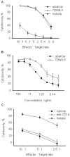
Figure 3. NKT cell cytotoxicity against CD1d positive MB cells
(A) DAOY/luc cells were pulsed with 100 ng/mL α-GalCer, 7WD8-5, or vehicle control overnight and co-cultured with resting NKT cells for 4 h at indicated E:T ratios. (B) DAOY/luc cells were pulsed with different concentrations of α-GalCer or 7WD8-5 overnight and co-cultured with resting NKT cells for 4 h at the fixed E:T ratio (2:1). NKT cell cytotoxicity was assessed by the loss of the target cell luminescence. Data were collected from six replicate wells per condition. (C) After pulsing overnight with β-GlcCer (2 μg/mL) or vehicle, DAOY/luc cells were blocked with anti-CD1d mAb 51.1 or isotype control for 30 min before adding NKT cells at the indicated E:T ratios. Data are M ± SD from one of three experiments with triplicates.
Figure 4
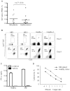
Figure 4. Normal NKT cell number and function in MB patients
Frequency of NKT cells in freshly isolated PBMC of MB patients and healthy donors was assessed by FACS using staining for CD3 and NKT iTCRα (6B11). (B) NKT cells were expanded from PBMCs of MB patients or healthy donors in culture with α-GalCer and IL-2. (C) NKT cells expanded from PBMC of a MB patient or a healthy donor were stimulated by α-GalCer-pulsed DAOY cells and the concentration of IFNγ was measured in the culture supernatants after 24 h using CBA multiplex assay. (D) The cytotoxicity of expanded NKT cells was tested against DAOY/luc cells pulsed with 100 ng/mL α-GalCer. Data are from a representative of three experiments.
Figure 5
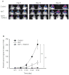
Figure 5. Intracranial transfer of NKT cells is effective against established tumor in an orthotopic MB model in NOD/SCID mice
A) DAOY/luc cells (2.5 × 10⁵) were intracranially injected into mouse brain at the position of bragma as described in methods. NKT cells alone or with 7DW8-5 (100 ng/mouse) were injected intracranially at day 5. Shown are representative bioluminescent images at indicated time intervals. (B) Tumor progression in indicated groups as measured by BL signal intensity. Data are M ± SD from 5 mice per group, one of three experiments.

References

    1. Packer RJ, Gajjar A, Vezina G, Rorke-Adams L, Burger PC, Robertson PL, et al. Phase III study of craniospinal radiation therapy followed by adjuvant chemotherapy for newly diagnosed average-risk medulloblastoma. J Clin Oncol. 2006;24:4202–4208. - PubMed
    1. Kim W, Choy W, Dye J, Nagasawa D, Safaee M, Fong B, et al. The tumor biology and molecular characteristics of medulloblastoma identifying prognostic factors associated with survival outcomes and prognosis. J Clin Neurosci. 2011;18:886–890. - PubMed
    1. Sadighi Z, Vats T, Khatua S. Childhood Medulloblastoma: The Paradigm Shift in Molecular Stratification and Treatment Profile. J Child Neurol. 2012;10:1302–1307. - PubMed (V体育官网入口)
    1. Louis DN, Ohgaki H, Wiestler OD, Cavenee WK, Burger PC, Jouvet A, et al. The 2007 WHO classification of tumours of the central nervous system. Acta Neuropathol. 2007;114:97–109. - PMC - PubMed
    1. Thompson MC, Fuller C, Hogg TL, Dalton J, Finkelstein D, Lau CC, et al. Genomics identifies medulloblastoma subgroups that are enriched for specific genetic alterations. J Clin Oncol. 2006;24:1924–1931. - PubMed

Publication types

MeSH terms (V体育官网入口)