Skip to main page content
U.S. flag

An official website of the United States government

Dot gov

The . gov means it’s official. Federal government websites often end in . gov or . mil. Before sharing sensitive information, make sure you’re on a federal government site. VSports app下载.

Https

The site is secure. The https:// ensures that you are connecting to the official website and that any information you provide is encrypted and transmitted securely V体育官网. .

. 2013 Sep;20(9):1230-40.
doi: 10.1038/cdd.2013.82. Epub 2013 Jul 5.

Milk fat globule-EGF factor 8 mediates the enhancement of apoptotic cell clearance by glucocorticoids

Affiliations

"VSports手机版" Milk fat globule-EGF factor 8 mediates the enhancement of apoptotic cell clearance by glucocorticoids

K Lauber et al. Cell Death Differ. 2013 Sep.

"V体育2025版" Abstract

The phagocytic clearance of apoptotic cells is essential to prevent chronic inflammation and autoimmunity. The phosphatidylserine-binding protein milk fat globule-EGF factor 8 (MFG-E8) is a major opsonin for apoptotic cells, and MFG-E8(-/-) mice spontaneously develop a lupus-like disease. Similar to human systemic lupus erythematosus (SLE), the murine disease is associated with an impaired clearance of apoptotic cells. SLE is routinely treated with glucocorticoids (GCs), whose anti-inflammatory effects are consentaneously attributed to the transrepression of pro-inflammatory cytokines. Here, we show that the GC-mediated transactivation of MFG-E8 expression and the concomitantly enhanced elimination of apoptotic cells constitute a novel aspect in this context. Patients with chronic inflammation receiving high-dose prednisone therapy displayed substantially increased MFG-E8 mRNA levels in circulating monocytes. MFG-E8 induction was dependent on the GC receptor and several GC response elements within the MFG-E8 promoter. Most intriguingly, the inhibition of MFG-E8 induction by RNA interference or genetic knockout strongly reduced or completely abolished the phagocytosis-enhancing effect of GCs in vitro and in vivo VSports手机版. Thus, MFG-E8-dependent promotion of apoptotic cell clearance is a novel anti-inflammatory facet of GC treatment and renders MFG-E8 a prospective target for future therapeutic interventions in SLE. .


Figures
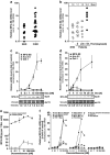
Figure 1
Figure 1
Enhanced expression by certain myeloid cells of MFG-E8 in response to treatment with glucocorticoids. (a) MFG-E8 mRNA expression was examined in peripheral blood monocytes from normal healthy donors (NHD, n=13) and patients with chronic inflammatory rheumatic diseases (CIRD, n=29) by qRT-PCR. The relative mRNA levels were normalized on 18S rRNA and calibrated on the mean value of the NHD population. (b) Results from (a) plotted against the weekly dose of prednisone (Pred) the patients were receiving. n=13 for NHD, n=15 for CIRD patients with 0 mg/week Pred, n=7 for CIRD patients with ≤35 mg/week Pred and n=7 for CIRD patients with >35 mg/week Pred. (c) PMA-differentiated THP-1 macrophages were treated with dexamethasone (Dex) for 24 h. MFG-E8 expression was examined on mRNA level by qRT-PCR and protein level by non-reducing SDS-PAGE with subsequent immunoblot analysis (loading control vinculin). The relative mRNA levels were normalized on 18S rRNA and calibrated on the untreated control population (means±S.D. from triplicates from one representative out of two experiments). Relative mRNA levels of Annexin A1 (Anx A1) and developmental endothelial locus-1 (Del-1) were measured as controls. (d) THP-1 macrophages were treated with 1 μM Dex for 0–24 h. MFG-E8 expression was examined on mRNA and protein level as in (c) (means±S.D. from triplicates from one representative out of two experiments). (e) MFG-E8 protein levels were quantified in culture supernatants and cellular lysates of dexamethasone-treated THP-1 macrophages treated as in (c) by ELISA. Means±s.d. of triplicates are depicted. (f) Primary human monocytes, monocytic cells lines, monocyte-derived primary human macrophages, macrophages differentiated from THP-1 or U937 cells, human foreskin fibroblasts, murine NIH3T3 fibroblasts and PHA-stimulated human lymphoblasts were treated with dexamethasone (Dex) for 24 h and MFG-E8 expression was examined on mRNA level by qRT-PCR as in (c). Mean values of duplicates from one representative out of 3 experiments are shown
Figure 2
Figure 2
Dexamethasone treatment dose- and time-dependently leads to enhanced uptake of apoptotic cells by macrophages. (a) PMA-differentiated THP-1 macrophages were treated with or without 1 μM Dex for 24 h and subsequently were fed with different ratios of apoptotic THP-1 cells, 6 μm microspheres or secondary necrotic fragments. After 2 h of incubation, phagocytosis was assessed flow cytometrically and is given as percentage of macrophages with ingested material (means±S.D. from quadruplicates from one representative out of three experiments). (b) THP-1 macrophages were treated with Dex for 24 h and subsequently applied to a phagocytosis assay with a ratio of 1 : 1 apoptotic THP-1 cells, 6 μm microspheres or secondary necrotic fragments, respectively. Phagocytosis was assessed as in (a) (means±S.D. from quadruplicates from one representative out of three experiments). (c) THP-1 macrophages were treated with 1 μM Dex for 0–24 h. Uptake of apoptotic THP-1 cells (ratio 1 : 1) was measured as in (a) (means±S.D. from quadruplicates from one representative out of three experiments). (d) Primary human macrophages differentiated with M-CSF from peripheral blood monocytes were treated with or without 1 μM Dex for 24 h and applied to a phagocytosis assay with autologous apoptotic neutrophils as in (a). Means±S.D. from quadruplicates from one representative out of two experiments are shown. (e) Primary human monocyte-derived macrophages were treated with Dex for 24 h and phagocytosis of autologous apoptotic neutrophils (ratio 1 : 2) was determined as in (b) (means±S.D. from quadruplicates from one representative out of two experiments)
Figure 3
Figure 3
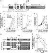
Different GREs in the promoter region of human MFG-E8 are required for dexamethasone-dependent MFG-E8 expression. (a) Schematic map of putative GREs within the minus 1000-bp region of the human MFG-E8 promoter. Positions of GREs and restriction sites are numbered according to NM_00592. (b) THP-1 macrophages were treated with Dex in the presence or absence of 10 μM RU486 for 24 h. MFG-E8 mRNA levels were quantified by qRT-PCR as in Figure 1c (means of duplicates from one representative out of two experiments). (c) THP-1 macrophages were treated with Dex in the presence or absence of 10 μg/ml of cycloheximide (CHX) for 24 h. MFG-E8 mRNA levels were assessed as in Figure 1c (mean values of duplicates from one representative out of two experiments). (d) Schematic map of the MFG-E8 promoter constructs employed in (e). (e) Different regions of the human MFG-E8 promoter cloned into pGL3 Basic (see (d)) were lipofected into THP-1 macrophages for 5 h and subsequently cells were treated with Dex for 16 h. Luciferase activities were measured in cellular lysates and calibrated on the corresponding untreated control populations (mean values±S.D. of triplicates from one representative out of three experiments). (f) THP-1 macrophages were treated with or without 1 μM Dex for 2 h before RNA synthesis inhibition with 100 ng/ml actinomycin D. The decay of MFG-E8 mRNA was measured by qRT-PCR. Half-lives (t1/2) of MFG-E8 mRNA were determined and are depicted in the plot (means of duplicates from one representative out of two experiments)
Figure 4
Figure 4
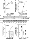
Inhibition of MFG-E8 expression by RNA interference or genetic knockout results in reduced phagocytosis promotion by dexamethasone treatment. (a) THP-1 monocytes were electroporated with an MFG-E8-specific siRNA oligonucleotide or the corresponding scrambled sequence before being differentiated into macrophages and treatment with Dex for 24 h. Phagocytosis of apoptotic prey cells was measured as in Figure 2b (means±S.D. from quadruplicates from one representative out of two experiments). (b) MFG-E8 knockdown, macrophage differentiation and Dex treatment of THP-1 cells were carried out as in (a) and MFG-E8 mRNA expression was assessed by qRT-PCR. Relative mRNA levels were normalized on 18S rRNA and calibrated on the untreated scramble-transfected control population (means of duplicates from one representative out of two experiments). (c) MFG-E8 knockdown, macrophage differentiation and Dex treatment of THP-1 cells were carried out as in (a) and MFG-E8 protein expression was detected by immunoblot analysis as in Figure 1c (one representative out of two experiments). (d) Thioglycollate-elicited peritoneal macrophages were prepared from wild-type and MFG-E8-knockout mice. In vitro macrophages were treated with 500 nM Dex for 24 h before quantitation of MFG-E8 mRNA by qRT-PCR (means of duplicates). In MFG-E8−/− cells, no MFG-E8 mRNA was detected. The inlay shows examples of the primary amplification curves. (e) Macrophages were prepared and treated as in (d). Subsequently, phagocytosis of apoptotic third-party thymocytes was measured and is depicted as phagocytic index (percentage of macrophages with ingested material × mean of internalized prey cell fluorescence) normalized on the corresponding untreated control population (n=8). P-values were calculated by heteroskedastic, unpaired, two-tailed Student's t-test analysis
Figure 5
Figure 5
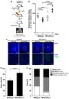
Dex-induced acceleration of irradiation-induced thymus atrophy is dependent on MFG-E8. (a) Schematic of the experimental setup. (b) C57BL/6 and MFG-E8−/− mice were treated with 1mg/kg/week Dex i.p. (for C57BL/6 n=6, for MFG-E8−/− n=4) or vehicle (for C57BL/6 n=6, for MFG-E8−/− n=5) for 1 week (Dex was applied in seven daily doses). Then, mice were irradiated with 0.5 Gy, killed after 8 h and thymi were excised. Thymus atrophy was measured as percentage of thymus weight in comparison with non-irradiated controls. Open and closed circles represent the values obtained for individual animals and bars indicate the means. P-values were calculated by heteroskedastic, unpaired, two-tailed Student's t-test analysis. (c) Paraffin sections of explanted thymi of Dex-treated, irradiated C57BL/6 and MFG-E8−/− mice were subjected to TUNEL–FITC staining for dying cells with DNA strand breaks and DAPI for total nuclei. Pictures were taken at × 2 (upper panel) or × 10 magnification (lower panel), respectively. (d) Explanted thymi of Dex-treated, irradiated C57BL/6 and MFG-E8−/− mice were mechanically disrupted, and flushed-out, collected cells were analyzed by FSC/SSC flow cytometry. The percentage of dead cells was determined from the percentage of cells with low FSC and high SSC. Means+S.E. are shown and P-value was calculated by heteroskedastic, unpaired, two-tailed Student's t-test analysis. (e) Collected cells from mechanically disrupted and flushed-out thymi of Dex-treated, irradiated C57BL/6 and MFG-E8−/− mice as prepared in (d) were subjected to 6-parameter FACS analyses. Different types of dead cells were classified as described in Materials and Methods. P-values were calculated by heteroskedastic, unpaired, two-tailed Student's t-test analysis

References

    1. Wickman G, Julian L, Olson MF. How apoptotic cells aid in the removal of their own cold dead bodies. Cell Death Differ. 2012;19:735–742. - PMC - PubMed
    1. Ravichandran KS. Beginnings of a good apoptotic meal: the find-me and eat-me signaling pathways. Immunity. 2011;35:445–455. - PMC - PubMed
    1. Poon IK, Hulett MD, Parish CR. Molecular mechanisms of late apoptotic/necrotic cell clearance. Cell Death Differ. 2010;17:381–397. - VSports注册入口 - PubMed
    1. Munoz LE, Lauber K, Schiller M, Manfredi AA, Herrmann M. The role of defective clearance of apoptotic cells in systemic autoimmunity. Nat Rev Rheumatol. 2010;6:280–289. - PubMed (V体育ios版)
    1. Gaipl US, Munoz LE, Grossmayer G, Lauber K, Franz S, Sarter K, et al. Clearance deficiency and systemic lupus erythematosus (SLE) J Autoimmun. 2007;28:114–121. - PubMed

Publication types

"VSports最新版本" MeSH terms