Skip to main page content
U.S. flag

An official website of the United States government

Dot gov

The . gov means it’s official. Federal government websites often end in . gov or . mil. Before sharing sensitive information, make sure you’re on a federal government site. VSports app下载.

Https

The site is secure. The https:// ensures that you are connecting to the official website and that any information you provide is encrypted and transmitted securely V体育官网. .

. 2013 Aug 2;288(31):22809-20.
doi: 10.1074/jbc.M113.482091. Epub 2013 Jun 21.

Parkinson disease protein DJ-1 binds metals and protects against metal-induced cytotoxicity

Affiliations

Parkinson disease protein DJ-1 binds metals and protects against metal-induced cytotoxicity

Benny Björkblom (VSports最新版本) et al. J Biol Chem. .

Abstract

The progressive loss of motor control due to reduction of dopamine-producing neurons in the substantia nigra pars compacta and decreased striatal dopamine levels are the classically described features of Parkinson disease (PD). Neuronal damage also progresses to other regions of the brain, and additional non-motor dysfunctions are common. Accumulation of environmental toxins, such as pesticides and metals, are suggested risk factors for the development of typical late onset PD, although genetic factors seem to be substantial in early onset cases. Mutations of DJ-1 are known to cause a form of recessive early onset Parkinson disease, highlighting an important functional role for DJ-1 in early disease prevention. This study identifies human DJ-1 as a metal-binding protein able to evidently bind copper as well as toxic mercury ions in vitro. The study further characterizes the cytoprotective function of DJ-1 and PD-mutated variants of DJ-1 with respect to induced metal cytotoxicity. The results show that expression of DJ-1 enhances the cells' protective mechanisms against induced metal toxicity and that this protection is lost for DJ-1 PD mutations A104T and D149A. The study also shows that oxidation site-mutated DJ-1 C106A retains its ability to protect cells. We also show that concomitant addition of dopamine exposure sensitizes cells to metal-induced cytotoxicity. We also confirm that redox-active dopamine adducts enhance metal-catalyzed oxidation of intracellular proteins in vivo by use of live cell imaging of redox-sensitive S3roGFP. The study indicates that even a small genetic alteration can sensitize cells to metal-induced cell death, a finding that may revive the interest in exogenous factors in the etiology of PD VSports手机版. .

Keywords: Cell Death; Cytotoxicity; Dj-1; Metals; Oxidative Stress; Parkinson Disease V体育安卓版. .


Figures (VSports最新版本)

FIGURE 1.
FIGURE 1.
Human DJ-1 purification and metal binding analysis. A, purified recombinant human DJ-1 analyzed by SDS-PAGE and Coomassie G-250 stained for detection of total protein. High purity nontagged human DJ-1 used for XRF indicated at arrow. B, combined XRF spectra of recombinant human DJ-1 exposed to metal-containing buffer (colored solid line), metal-containing buffer only (dotted line), and backsoak buffer without added metal salt (black solid line). Prominent DJ-1 metal binding peaks for Cu(I), Cu(II), and Hg(II) were detected as well as a weaker signal for Mn(II). Asterisk marks the internal titanium peak originating from the XRF instrument.
FIGURE 2.
FIGURE 2.
Metal exposure and analysis of cell survival. Cell survival data compared WT and DJ-1−/− MEF cells exposed to increasing concentrations of metals for 24 h. All values were compared with nontreated control set to 100% survival and plotted against the logarithmic scale for the used metal concentrations. A, CuCl, WT LC50 = 70 μm, DJ-1−/− LC50 = 30 μm. B, HgCl2, WT LC50 = 15 μm, DJ-1−/− LC50 = 4 μm. Statistically significant change in cell survival compared with DJ-1−/− cells is indicated with *, p < 0.05.
FIGURE 3.
FIGURE 3.
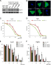
Characterization and comparison of WT or PD-mutated DJ-1 re-expression of cells to metal stresses. A, Western blot of wild type, DJ-1−/− MEF cells, and stable DJ-1 re-expression of MEF cells expressing human DJ-1 variants as indicated. Equal protein expression was detected using protein-specific antibodies raised against human DJ-1 and for actin as loading control. B, immunocytochemical staining of DJ-1−/− MEF cells or DJ-1 re-expression of cells expressing human DJ-1 variants as indicated. Green, DJ-1; blue, nucleus. Analyzed DJ-1 variants show equal subcellular localization, predominantly soluble and cytoplasmic. Scale bars, 10 μm. −1° = minus primary antibody. C and D, cell survival data comparing empty vector transfected DJ-1−/− cells or stably re-expressing human DJ-1 WT cells exposed to increasing concentrations of metal stresses for 24 h. C, CuCl, WT LC50 = 65 μm, DJ-1−/− LC50 = 32 μm. D, HgCl2, WT LC50 = 7 μm, DJ-1−/− LC50 = 3 μm. E and F, cell survival data comparing DJ-1−/− or re-expression of cells expressing human DJ-1 WT, PD-mutated human DJ-1 A104T, D149A, or the oxidation site-mutated human DJ-1 C106A. Cells were exposed to increasing concentrations of CuCl (E) and HgCl2 (F) metal stresses for 24 h before cell survival analysis. Statistically significant increase in cell survival was compared with DJ-1−/− cells indicated with *, p < 0.05.
FIGURE 4.
FIGURE 4.
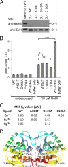
Point-mutated DJ-1 metal binding analysis. A, Western blot (WB) analysis, using anti-His6- and anti-DJ-1-specific antibodies of purified recombinant human DJ-1 WT and point-mutated DJ-1 A104T, DJ-1 D149A, DJ-1 C106A, and DJ-1 WT-His6 tag as positive control. B, AAS analysis of total amount of copper bound to recombinant human DJ-1 proteins, as shown in A, after 24 h exposure time. Statistically significant changes in total copper binding compared with WT DJ-1 are indicated with asterisks, **, p < 0.01; ***, p < 0.001. C, initial binding affinities for metal binding to DJ-1 recombinant human DJ-1 proteins, as shown in A. Calculated average MST dissociation constants (KD) for recombinant human DJ-1 WT and point-mutated variants using Cu(I), Cu(II), and Hg(II) as ligands. Lack of detected binding indicated with − sign. D, crystal structure model of dimeric human DJ-1 with the studied mutation sites indicated with *, and potential metal binding areas indicated within squares (dotted line). Protein structure model was adopted from Protein Data Bank code 2R1V.
FIGURE 5.
FIGURE 5.
Comparison of WT- or DJ-1-deficient cell survival in response to dopamine, MG132, or combined dopamine and metal-induced stress. A, cell survival data comparing DJ-1−/− and WT DJ-1-expressing MEF cells exposed to increasing concentrations of dopamine. The highest no observed effect concentration of dopamine observed at 33 μm in both DJ-1 knock-out and DJ-1 WT-expressing cells. WT LC50 = 185 μm, DJ-1−/− LC50 = 95 μm. Statistically significant change in cell survival indicated with *, p < 0.05. B, no change is cell survival was observed upon treatment with proteasome inhibitor MG132. C, table of LC50 values calculated from combined dopamine and metal-induced cell toxicity data shown in D–M. D–M, cell survival data comparing DJ-1−/− or cells stably re-expressing human DJ-1 WT, A104T, D149A, or C106A, exposed to 0, 6.6, or 33 μm dopamine in combination with increasing concentrations of copper (D–H) or mercury (I–M) stresses for a total of 18 h. Dopamine dose-dependent reduction of cell survival was observed in re-expressing DJ-1 WT and DJ-1 C106A cells, as well as in DJ-1−/−, DJ-1 A104T, and D149A re-expression cell lines exposed to copper or mercury.
FIGURE 6.
FIGURE 6.
Real time analysis of oxidative stress levels by use of CLSM live cell imaging of redox-sensitive S3roGFP. A, CLSM live cell imaging experiment showing redox-sensitive S3roGFP 408/488 nm ratios in transfected MEF DJ-1 WT cells. The cells were exposed to 0.5 mm H2O2 to mimic enhanced oxidative stress conditions 5 min after initiating image acquisition. After 35 min of imaging acquisition, the cells were reduced by treatment with 2 mm DTT. A clear change in the 408/488 nm ratio was observed upon oxidation and reduction of the imaged cells. B, CLSM images of S3roGFP-expressing cells treated as shown in A. The observed redox-dependent change in 408/488 nm ratio is visualized by pseudo-coloring according to included ratio scale bar 0–1.2 (blue, reduced; oxidized, red). Image size scale bar, 20 μm. C, CLSM live cell imaging experiment showing S3roGFP 408/488 nm ratios in DJ-1−/− and DJ-1 WT-expressing cells exposed to 100 μm dopamine, arrow. Mean value, ± S.E. for five cells per cell type are shown. D, S3roGFP 408/488 nm ratios in DJ-1 WT-expressing cells exposed to 100 μm CuCl, with or without addition of 33 μm dopamine. Control indicates nonexposed cells. Mean value, ± S.E. for 10 cells per treatment are shown. E, S3roGFP 408/488 nm ratios in DJ-1−/− cells treated as in D. Mean value, ± S.E. for 10 cells per treatment are shown. F, S3roGFP 408/488 nm ratios in DJ-1 WT-expressing cells exposed to 5 μm HgCl2, with or without addition of 33 μm dopamine. Mean value, ± S.E. for 12–17 cells per treatment are shown. G, S3roGFP 408/488 nm ratios in DJ-1−/− cells treated as in F. Mean value, ± S.E. for 13–19 cells per treatment are shown. Cells that died during the analysis were excluded from the shown quantified data.

References (VSports)

    1. Hirsch E., Graybiel A. M., Agid Y. A. (1988) Melanized dopaminergic neurons are differentially susceptible to degeneration in Parkinson's disease. Nature 334, 345–348 - PubMed (V体育2025版)
    1. Marsden C. D. (1996) in Oxford Textbook of Medicine (Weatherall D. J., Ledingham J. G., Warrell D. A., eds) 3rd Ed., pp. 3998–4022, Oxford University Press, Oxford
    1. Braak H., Del Tredici K., Rüb U., de Vos R. A., Jansen Steur E. N., Braak E. (2003) Staging of brain pathology related to sporadic Parkinson's disease. Neurobiol. Aging 24, 197–211 - PubMed
    1. Poewe W. (2007) in Parkinson's Disease and Movement Disorders (Jankovic J., Tolosa E., eds) 5th Ed., pp. 67–76, Lippincott Williams & Wilkins, Philadelphia
    1. Wang J. D., Huang C. C., Hwang Y. H., Chiang J. R., Lin J. M., Chen J. S. (1989) Manganese-induced parkinsonism: an outbreak due to an unrepaired ventilation control system in a ferromanganese smelter. Br. J. Ind. Med. 46, 856–859 - PMC - PubMed

Publication types (VSports最新版本)

MeSH terms