Skip to main page content
U.S. flag

An official website of the United States government

Dot gov

The . gov means it’s official. Federal government websites often end in . gov or . mil VSports app下载. Before sharing sensitive information, make sure you’re on a federal government site. .

Https

The site is secure. The https:// ensures that you are connecting to the official website and that any information you provide is encrypted and transmitted securely V体育官网. .

. 2013 Aug;33(16):3242-58.
doi: 10.1128/MCB.01674-12. Epub 2013 Jun 17.

C/EBPγ suppresses senescence and inflammatory gene expression by heterodimerizing with C/EBPβ

Affiliations

VSports手机版 - C/EBPγ suppresses senescence and inflammatory gene expression by heterodimerizing with C/EBPβ

Christopher J Huggins et al. Mol Cell Biol. 2013 Aug.

Abstract

C/EBPβ is an important regulator of oncogene-induced senescence (OIS). Here, we show that C/EBPγ, a heterodimeric partner of C/EBPβ whose biological functions are not well understood, inhibits cellular senescence VSports手机版. Cebpg(-/-) mouse embryonic fibroblasts (MEFs) proliferated poorly, entered senescence prematurely, and expressed a proinflammatory gene signature, including elevated levels of senescence-associated secretory phenotype (SASP) genes whose induction by oncogenic stress requires C/EBPβ. The senescence-suppressing activity of C/EBPγ required its ability to heterodimerize with C/EBPβ. Covalently linked C/EBPβ homodimers (β∼β) inhibited the proliferation and tumorigenicity of Ras(V12)-transformed NIH 3T3 cells, activated SASP gene expression, and recruited the CBP coactivator in a Ras-dependent manner, whereas γ∼β heterodimers lacked these capabilities and efficiently rescued proliferation of Cebpg(-/-) MEFs. C/EBPβ depletion partially restored growth of C/EBPγ-deficient cells, indicating that the increased levels of C/EBPβ homodimers in Cebpg(-/-) MEFs inhibit proliferation. The proliferative functions of C/EBPγ are not restricted to fibroblasts, as hematopoietic progenitors from Cebpg(-/-) bone marrow also displayed impaired growth. Furthermore, high CEBPG expression correlated with poorer clinical prognoses in several human cancers, and C/EBPγ depletion decreased proliferation and induced senescence in lung tumor cells. Our findings demonstrate that C/EBPγ neutralizes the cytostatic activity of C/EBPβ through heterodimerization, which prevents senescence and suppresses basal transcription of SASP genes. .


Figures
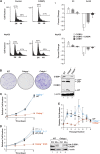
Fig 1
Fig 1
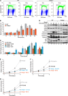
CEBPγ is required for efficient cellular proliferation. (A) Expression of C/EBPβ but not C/EBPγ increases G1 arrest in HepG2 cells and L929 fibroblasts. Cells were transfected with CD20 expression vector and C/EBPβ, C/EBPγ, or C/EBPβ and C/EBPγ, harvested after 24 h, and stained with propidium iodide. CD20-positive cells were analyzed for DNA content. Graphs (left panels) show the effects of C/EBPβ overexpression. Average percent changes are shown on the right; data are mean values ± SEM from five experiments. (B) WT, Cebpg−/−, and Cebpb−/− fibroblasts (MEFs) were isolated from E13.5 embryos and analyzed for colony growth. C/EBPβ and C/EBPγ protein levels were determined (right). Bands corresponding to the three C/EBPβ translational isoforms (LAP*, LAP, and LIP) are indicated. (C) Proliferation of WT, Cebpg+/, and Cebpg−/− MEFs was measured over 6 days. The data are mean values ± SEM from at least 10 independent MEF preparations, each assayed in triplicate. (D) Proliferation rates for WT and Cebpg−/− MEFs, without or with ectopic CEBPγ. The data are mean values ± SEM from at least 5 MEF isolates, assayed in triplicate. Right, C/EBPγ Western blot. Recombinant C/EBPγ is HA tagged and therefore migrates more slowly. (E) Long-term proliferative capacity of WT, Cebpg+/, and Cebpg−/− MEFs. Cells were propagated on a 3T3 protocol over 8 passages. The data are mean values ± SEM from 4 isolates of each genotype assayed in triplicate.
Fig 2
Fig 2
C/EBPγ is necessary for normal cell cycle progression. (A) WT and Cebpg−/− MEFs were synchronized by serum starvation, stimulated with serum, and labeled with BrdU. The percentage of cells in S phase after 18 h is shown. (B) Time course of cell cycle entry in WT and Cebpg−/− MEFs. Cells were serum starved, restimulated, and pulse-labeled with BrdU at the indicated times. Data are averages ± SEM from two experiments. (C) Expression of C/EBPγ or HPV oncoprotein E7 partially rescues delayed S-phase entry in Cebpg−/− MEFs. C/EBPγ and E7 were expressed in WT or Cebpg−/− MEFs, and the cells were analyzed as described above. Data are averages ± SEM from two experiments. (D) Cyclins D1, A, and E are reduced in Cebpg−/− MEFs. Lysates from serum-stimulated MEFs analyzed by Western blotting for CDKs (Cdk1/2/3/6), cyclins (A/B1/D1/E), and CDK inhibitors (p21/p27). (E to G) E7 and, to a lesser extent, cyclin D1 and c-Myc rescue growth of Cebpg−/− MEFs. WT and Cebpg−/− MEFs were transduced with vectors for E7 (E), cyclin D1 (F), or c-Myc (G) and proliferation was measured. Data are averages ± SEM from at least two experiments performed in triplicate.
Fig 3
Fig 3
Increased senescence in Cebpg−/− MEFs. (A) Morphology of WT and Cebpg−/− cells 6 days after being plated. (B) Increased SA–β-Gal expression in Cebpg−/− MEFs. The frequency of SA–β-Gal-positive cells is shown. Data are the mean values ± SEM from 2 experiments. (C) Expression of cell cycle inhibitors in WT and Cebpg−/− MEFs. Left, p16Ink4a and p19Arf protein levels at passages 2 to 4. A lysate from p16−/−/p19−/− MEFs is shown as a negative control (lane 1). Right, p15Ink4b and p18Ink4c protein levels in WT, Cebpb−/−, and Cebpg−/− MEFs at passage 2. The apparent increase in p18Ink4c expression in Cebpg−/− cells was not consistently observed.
Fig 4
Fig 4
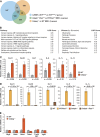
Cebpg−/− MEFs display a proinflammatory gene signature and enhanced expression of SASP genes. (A) Analysis of genes positively regulated by C/EBPβ and negatively regulated by C/EBPγ. Gene lists were generated from expression array comparisons: Cebpg−/− versus WT MEFs (increased expression; >+1.5-fold; P < 0.05), Cebpb−/−/RasV12 versus WT/RasV12 MEFs (decreased expression; <−1.5-fold; P = 0.05), and C/EBPβ-3T3RasV12 cells versus 3T3RasV12 cells (increased expression; >+1.5-fold; P < 0.05). NIH 3T3 data are from Basu et al. (8). Venn diagram depicts the intersections of these groups. The triple intersect represents genes that are positively regulated by C/EBPβ and inhibited by C/EBPγ. (B) GeneGo pathway and disease associations of the tri-intersect gene set. (C) Expression of representative SASP genes in WT and Cebpg−/− MEFs. mRNA levels were determined by qPCR; data are mean values ± SEM from 2 triplicate measurements from 2 independent MEF isolates. (D) SASP gene expression is induced by oncogenic RasV12 in WT but not Cebpb−/− MEFs. RNA isolated from control and RasV12-expressing MEFs was analyzed for SASP gene expression by qPCR. The data are mean values ± SEM from 2 triplicate measurements from 2 samples each. (E) Effect of C/EBPγ deficiency on expression of C/EBPβ target genes involved in tumor suppression or cell proliferation (8). The indicated genes were analyzed in WT and Cebpg−/− MEFs. Data are the means ± SEM from two experiments, assayed in triplicate.
Fig 5
Fig 5
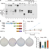
C/EBPγ stimulates cell proliferation by heterodimerizing with C/EBPβ. (A) EMSA was performed using a C/EBP consensus site probe and nuclear extracts from WT and Cebpg−/− MEFs (lanes 1 to 7). Antibody supershifts were performed to identify specific dimeric complexes. EMSAs were compared to those generated from cells overexpressing the indicated C/EBP proteins (lanes 8 to 14). The identities of C/EBP complexes are indicated. UT, untransfected cells; NRS, normal rabbit serum. (B) Domain structures of CEBPβ (LAP and LIP), C/EBPγ, and C/EBPγ-GLZ. LAP is the major C/EBPβ isoform and is expressed from the second in-frame AUG codon; LIP is a truncated translational isoform (34). C/EBPγ-GLZ contains the GCN4 leucine zipper. TAD, transactivation domain; REG, regulatory domain; BR, basic region; LZ, leucine zipper. (C) Proliferation assays of Cebpg−/− MEFs expressing C/EBPγ, LIP, or C/EBPγ-GLZ. The data are mean values ± SEM from at least 4 experiments assayed in triplicate. (D) Colony assays. The cells described in the legend to panel C were plated at a density of 5 × 10⁴ cells/dish and stained after 2 weeks. (E) The same populations were analyzed for senescent (SA–β-Gal-positive) cells (±standard deviation [SD]) from a representative experiment (>400 cells analyzed).
Fig 6
Fig 6
Opposing effects of C/EBPβ (LAP) homodimers and C/EBPγ:C/EBPβ heterodimers on cell proliferation. (A) Diagram of β∼β, γ∼β, and γ∼γ tethered dimers. Proteins are HA tagged, and internal in-frame AUG codons were deleted. (B) DNA binding of tethered dimers. Proteins were expressed in 293T cells ± RasV12 and cell extracts were subjected to EMSA. Protein levels were determined by HA Western blot. (C) Effects of linked dimers on proliferation of MEFs. Cebpb−/− or Cebpb−/−/RasV12 MEFs were infected with retroviruses encoding the indicated proteins, and cell proliferation was assayed. Data are the mean values ± SEM from 2 experiments assayed in triplicate. Right, C/EBP protein levels in transduced Cebpb−/−/RasV12 MEFs. The blot was probed with an HA antibody. The low levels of monomeric C/EBPβ and C/EBPγ in cells expressing tethered dimers result from partial proteolytic cleavage.
Fig 7
Fig 7
C/EBPβ homodimers inhibit growth and RasV12-induced transformation in NIH 3T3 cells. (A) Proliferation rates were determined for cells expressing the indicated C/EBP proteins. Right, C/EBP protein levels. The cells were plated for focus assays (B) or soft agar colony growth assays (C). Data are the mean values ± SEM from 3 experiments, each assayed in duplicate. (D) SASP gene expression in 3T3RasV12 cells expressing C/EBPβ, C/EBPγ, or linked dimers. Data are the mean values ± SEM from 2 measurements (performed in triplicate) from 2 independent samples.
Fig 8
Fig 8
C/EBPβ homodimers recruit the CBP coactivator. (A) C/EBPβ homodimers but not γ∼β heterodimers bind to CBP in a RasV12-dependent manner. C/EBP proteins were coexpressed with a Flag-tagged C-terminal fragment of CBP containing the TAZ2 domain (CBP1680-2441) in 293T cells, without or with RasV12. Cell lysates were immunoprecipitated with Flag antibody and analyzed for C/EBPβ. (B) Association of C/EBPβ, C/EBPγ, and CBP with SASP gene promoters in MEFs. ChIP assays were performed using control and RasV12-expressing cells; qPCR was performed with primers spanning C/EBP binding sites in the Il6, Il1a, and Il1b promoters (see materials and methods in the supplemental material). Binding was normalized to nonspecific (NS) antibody values. Data are from a representative experiment assayed in triplicate. (C) Increased binding of C/EBPβ to SASP gene promoters in Cebpg−/− MEFs. ChIP was performed as described for panel B. (D) RasV12-induced recruitment of CBP to SASP gene promoters requires C/EBPβ. CBP ChIP was performed on WT, Cebpb−/−, and Cebpg−/− MEFs. For each gene, the values are normalized to CBP binding in WT control cells (without RasV12). RasV12-expressing Cebpg−/− cells were not included in this experiment since they are undergoing spontaneous senescence, and the objective was to explain increased basal expression of SASP genes in the mutant cells.
Fig 9
Fig 9
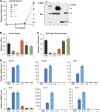
C/EBPβ depletion partially rescues the proliferation defect in Cebpg−/− MEFs. (A) Proliferation assays of Cebpb; Cebpg double-knockout MEFs. MEFs from embryos representing all 9 possible genotypes were seeded at 25,000 cells/well, and proliferation was measured over 6 days. Cebpb−/−; Cebpg−/− cells underwent rapid apoptosis after plating, and nearly all cells died during growth assays. (B) Partial depletion of C/EBPβ restores growth of Cebpg−/− MEFs. WT and mutant cells were infected with two different C/EBPβ shRNA retroviruses. Cells were seeded at 106 cells/plate, and proliferation was assessed by staining after 7 days. Bottom, C/EBPβ Western blot. (C) C/EBPβ knockdown abrogates the increased senescence seen in Cebpg−/− MEFs. SA–β-Gal assays were performed on the cells described for panel B. The data are mean values ± SD (>400 cells were analyzed). (D) Elevated expression of Cxcl1 and Cxcl2 in Cebpg−/− cells requires C/EBPβ. Expression was analyzed by qPCR; data are the mean values ± SEM from 2 replicate measurements assayed in triplicate.
Fig 10
Fig 10
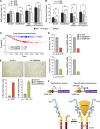
Expression of the γ∼β dimer rescues proliferation and inhibits senescence of Cebpg−/− MEFs. WT and Cebpg−/− cells were infected with retroviruses expressing C/EBPγ or γ∼β and analyzed for proliferation (A), colony formation (B), and senescence (SA–β-Gal staining) (C). Proliferation data are mean values ± SD from a representative experiment assayed in triplicate. (D) Ectopic C/EBPγ and γ∼β expression in Cebpg−/− MEFs. The graph shows Cebpg reverse transcription qPCR assays of RNA from retrovirally infected Cebpg−/− cells. A weak signal is observed in control Cebpg−/− cells because the primers span the Neo insert in the knockout allele (16). (E) Ectopic C/EBP protein expression in WT and Cebpg−/− MEFs. Top, EMSA of nuclear extracts from the cells described for panel D and for Fig. 5C. LIP DNA-binding activity is not observed in Cebpg−/− cells (lane 9), supporting observations from our laboratory that LIP is autoinhibited (25) and can be activated by coexpression of C/EBPγ (unpublished data). Bottom, C/EBPβ Western blot (WB) of the same extracts. The LIP species detected in lanes 3 and 8 is apparently a proteolytic product from the γ∼β dimer. CRM, cross-reacting material.
Fig 11
Fig 11
C/EBPγ is required for proliferation of murine hematopoietic cells and human cancer cells. (A, B) Cell proliferation and CFU-c assays of WT and Cebpg−/− bone marrow cells. Bone marrow was isolated from adult mice on a mixed C57BL/6-129Sv background. The cells were incubated with the indicated combinations of hematopoietic growth factors and evaluated for proliferation (EdU incorporation assays) (A) and colony growth (1.5 × 10⁴ cells/plate) (B). (C) C/EBPγ expression is negatively associated with relapse-free survival of lung adenocarcinoma patients. Graph shows Kaplan-Meier relapse-free survival from a study of lung adenocarcinoma patients (47) stratified according to high (n = 130) and low (n = 74) CEBPG expression in tumors. Dotted lines indicate 95% confidence intervals. Meta-analysis of microarray data are from PrognoScan (26). (D) C/EBPγ depletion impairs proliferation of human A549 lung adenocarcinoma cells. C/EBPγ was ablated by two independent shRNAs (1 and 2), and colony growth was analyzed. Data are the mean values ± SEM from 2 experiments. C/EBPγ mRNA levels are shown in graphs on the right. (E) Increased senescence in C/EBPγ-depleted A549 cells. Four days after plating, cells were stained for SA–β-Gal, and senescent cells were visualized (upper panels) and counted (bottom panels). Data are mean values ± SEM from 2 experiments. (F) Differential regulatory properties of C/EBPβ homo- and heterodimers. In proliferating cells and in the absence of an activating signal such as oncogenic Ras, C/EBPβ occurs predominantly as a heterodimer with C/EBPγ. β:γ heterodimers bind to target C/EBP sites in SASP gene promoters but are transcriptionally inactive. In cells undergoing Ras-induced senescence, p90RSK-mediated phosphorylation on Ser273 in the C/EBPβ leucine zipper stabilizes the LZ interaction and promotes formation of β:β homodimers (15). Activated homodimers are capable of binding p300/CBP, inducing cell cycle arrest, and activating transcription of SASP genes. In contrast, β:γ heterodimers do not recruit p300/CBP and promote cell proliferation and cancer.

References

    1. Campisi J. 2005. Senescent cells, tumor suppression, and organismal aging: good citizens, bad neighbors. Cell 120:513–522 - PubMed
    1. Kuilman T, Michaloglou C, Mooi WJ, Peeper DS. 2010. The essence of senescence. Genes Dev. 24:2463–2479 - PMC - PubMed
    1. Lowe SW, Cepero E, Evan G. 2004. Intrinsic tumour suppression. Nature 432:307–315 - PubMed
    1. Coppe J-P, Desprez P-Y, Krtolica A, Campisi J. 2010. The senescence-associated secretory phenotype: the dark side of tumor suppression. Annu. Rev. Pathol. 5:99–118 - "VSports手机版" PMC - PubMed
    1. Kang TW, Yevsa T, Woller N, Hoenicke L, Wuestefeld T, Dauch D, Hohmeyer A, Gereke M, Rudalska R, Potapova A, Iken M, Vucur M, Weiss S, Heikenwalder M, Khan S, Gil J, Bruder D, Manns M, Schirmacher P, Tacke F, Ott M, Luedde T, Longerich T, Kubicka S, Zender L. 2011. Senescence surveillance of pre-malignant hepatocytes limits liver cancer development. Nature 479:547–551 - PubMed (VSports在线直播)

Publication types

V体育2025版 - MeSH terms

Substances

VSports注册入口 - Grants and funding