Skip to main page content
U.S. flag

An official website of the United States government

Dot gov

The . gov means it’s official VSports app下载. Federal government websites often end in . gov or . mil. Before sharing sensitive information, make sure you’re on a federal government site. .

Https

The site is secure. The https:// ensures that you are connecting to the official website and that any information you provide is encrypted and transmitted securely. V体育官网.

. 2013 Apr 1;2(4):e23564.
doi: 10.4161/onci.23564.

Collaboration of chimeric antigen receptor (CAR)-expressing T cells and host T cells for optimal elimination of established ovarian tumors

Affiliations

Collaboration of chimeric antigen receptor (CAR)-expressing T cells and host T cells for optimal elimination of established ovarian tumors

Paul Spear et al. Oncoimmunology. .

Abstract

Conditioning strategies that deplete host lymphocytes have been shown to enhance clinical responses to some adoptive T-cell therapies. However, host T cells are capable of eliminating tumor cells upon the relief of immunosuppression, indicating that lymphodepletion prior to T-cell transfer may reduce optimal tumor protection elicited by cell treatments that are capable of shaping host immunity. In this study, we show that adoptively transferred T cells bearing a chimeric antigen receptor (CAR) harness endogenous T cells for optimal tumor elimination and the development of a tumor-specific memory T cell response. Mice bearing ID8 ovarian cancer cells were treated with T cells transduced with a NKG2D-based CAR. CAR-expressing T cells increased the number of host CD4+ and CD8+ T cells at the tumor site in a CXCR3-dependent manner and increased the number of antigen-specific host CD4+ T cells in the tumor and draining lymph nodes. In addition, the administration of CAR-expressing T cells increased antigen presentation to CD4+ T cells, and this increase was dependent on interferon γ and granulocyte-macrophage colony-stimulating factor produced by the former. Host CD4+ T cells were sufficient for optimal tumor protection mediated by NKG2D CAR-expressing T cells, but they were not necessary if CD4+ T cells were adoptively co-transferred VSports手机版. However, host CD4+ T cells were essential for the development of an antigen-specific memory T-cell response to tumor cells. Moreover, optimal tumor elimination as orchestrated by NKG2D CAR-expressing T cells was dependent on host CD8+ T cells. These results demonstrate that adoptively transferred T cells recruit and activate endogenous T-cell immunity to enhance the elimination of tumor cells and the development of tumor-specific memory responses. .

Keywords: CD4+ T cells; CD8+ T cells; ID8; NKG2D; adoptive T cell therapy; chNKG2D; immunotherapy; ovarian cancer; tumor immunology V体育安卓版. .


Figures
None
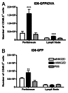
Figure 1. The administration of chNKG2D-expressing T cells increases antigen-specific CD4+ T cells in the peritoneum and tumor-draining lymph nodes. (A and B) Mice bearing ID8-GFP-tOVA- (A) or ID8-GFP-derived (B) tumors were injected with chNKG2D-expressing, wtNKG2D-expressing T cells or PBS i.p. One hour before local T-cell injection, CD45.2+ OT-II cells were injected i.v. Three days after T-cell transfer, the absolute number of CD45.2+ cells in the peritoneum and mediastinal lymph nodes was determined by flow cytometry. The average of each group and SD (n = 8) is shown (**p < 0.01; ***p < 0.001 as compared with animals receiving wtNKG2D-expressing cells).
None
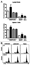
Figure 2. chNKG2D-expressing T cells enhance MHC Class II antigen presentation and the proliferation of CD4+ T cells. (A and B) ID8-GFP or ID8-GFP-tOVA tumor-bearing mice were injected with wtNKG2D-expressing (open), chNKG2D-expressing (filled), interferon γ (IFNγ)-deficient chNKG2D-expressing (hatched), or granulocyte-macrophage colony-stimulating factor (GM-CSF)-deficient chNKG2D-expressing T cells (dotted) i.p. (A) Seven days after T-cell transfer, mediastinal lymph node and spleen cells were isolated and cultured with CFSE-labeled OT-II cells. After 4 d, T-cell proliferation was assessed by flow cytometry. The proliferation of T cells cultured with cells from naïve mice is shown (checked). (B) Representative OT-II CFSE dilution flow cytometry plots as induced by lymph node and spleen cells isolated from naïve mice or from mice bearing ID8-GFP-tOVA tumors treated with wtNKG2D-expressing or chNKG2D-expressing T cells. Data are representative of two individual experiments. The average of each group and SD (n = 8) are shown (**p < 0.01 as compared with ID8-GFP-tOVA tumor-bearing animals receiving wtNKG2D-expressing T cells; p < 0.05 as compared with ID8-GFP tumor-bearing animals receiving wtNKG2D-expressing T cells).
None
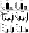
Figure 3. The administration of chNKG2D-expressing T cells increases the number of host T cells at the tumor site in a CXCR3-dependent mechanism. (A and B) Mice bearing ID8-GFP tumors were injected with wtNKG2D-expressing (open), chNKG2D-expressing (filled) or interferon γ (IFNγ)-deficient chNKG2D-expressing T cells (hatched). (A) Peritoneal cells isolated 3 d after T-cell transfer were assessed for CXCL9 and CXCL10 expression by quantitative RT-PCR. (*p < 0.05 as compared with peritoneal cells from animals receiving wtNKG2D-expressing T cells). (B) F4/80+ cells were isolated 3 d after T-cell transfer and cultured for 24 h. Cell-free culture media from the F4/80+ fraction, the F4/80 fraction, and the combined F4/80+ and F4/80 fractions (1:1) were assessed for CXCL9 and CXCL10 production by multiplex protein analysis (*p < 0.05; ***p < 0.001 as compared with cells from animals receiving wtNKG2D-expressing T cells; p < 0.05 as compared with F4/80+ and F4/80 cells from animals receiving chNKG2D-expressing T cells). (C) C57BL/6 or Cxcr3−/− mice were injected with ID8-GFP tumor cells and treated with CD45.1+ chNKG2D-expressing T cells 5 weeks later. Three days after T-cell transfer, a peritoneal wash was performed and the absolute numbers of CD4+ and CD8+ host T cells were determined. The average of each group and SD (n = 8) are shown. Data are representative of two independent experiments (*p < 0.05; **p < 0.01 as compared with C57BL/6 mice receiving wtNKG2D-expressing cells; p < 0.05 as compared with C57BL/6 mice receiving chNKG2D-expressing T cells).
None
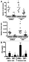
Figure 4. CD4+ T cells are necessary for optimal tumor protection. (A and B) Tumor-bearing mice were injected with anti-CD4 depleting antibodies (GK1.5) on day 33, 39 and 45 and treated with CD45.1+ wtNKG2D-expressing or chNKG2D-expressing T cells on day 35. (A) Eight weeks after tumor-cell injection, the number of solid tumors on the peritoneal wall and number of tumor cells in the peritoneal wash was assessed. (B) Spleen and peritoneal cells were isolated from mice 8 weeks after tumor cell injection and cultured for 24 h. Cell-free supernatants were then assessed for the presence of interferon γ (IFNγ). (C) Tumor bearing mice were injected with anti-CD4 depleting antibodies on day 33 and then treated with T cells on day 35. Peritoneal cells were harvested 7 d after T-cell transfer and CD45.2+CD8b+ cells were assayed for IFNγ expression by flow cytometry. Cumulative data of two independent experiments are shown. The average of each group and SD (n = 8) are shown (*p < 0.01; ***p < 0.001 as compared with mice receiving wtNKG2D-expressing T cells; p < 0.05 as compared with non-depleted mice receiving chNKG2D-expressing T cells).
None
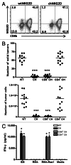
Figure 5. Adoptively transferred CD8+ T cells are sufficient for tumor elimination in the presence of host CD4+ T cells. (A–C) Mice bearing 5-week ID8 tumors were treated with wtNKG2D-expressing, chNKG2D-expressing, purified CD8+ chNKG2D-expressing or purified CD4+ chNKG2D-expressing T cells. (A) chNKG2D-expressing T cells gated on CD3e+ cells were assayed for NKG2D and CD8b expression by flow cytometry. (B) Eight weeks after tumor cell injection, the number of solid tumors on the peritoneal wall and number of tumor cells in the peritoneal wash was assessed. The average of each group and SD (n = 12) are shown. (C) WtNKG2D-expressing, chNKG2D-expressing, CD8+ chNKG2D-expressing and CD4+ chNKG2D-expressing T cells were cultured in standard conditions or together with RMA, RMA-Rae1 or ID8 tumor cells. Cell-free supernatants were assayed for interferon γ (IFNγ) production after 24 h. (*p < 0.01; ***p < 0.001 as compared with wtNKG2D-expressing T cells)
None
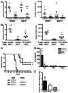
Figure 6. Host CD4+ T cells are required for the development of a tumor-specific memory T-cell response, but not for tumor protection mediated by chNKG2D-expressing T cells. (A) C57BL/6 and MHC Class II-deficient mice bearing 5-week ID8 tumors were treated with wtNKG2D-expressing T cells or chNKG2D-expressing T cells. Eight weeks after tumor cell injection, the number of solid tumors on the peritoneal wall and number of tumor cells in the peritoneal wash was assessed. Cumulative data of two independent experiments are shown. The average of each group and SD (n = 8) are shown (**p < 0.01; ***p < 0.001 as compared with C57BL/6 mice receiving wtNKG2D-expressing T cells; p < 0.05 as compared with C57BL/6 mice receiving chNKG2D-expressing T cells). (B) MHC Class II-deficient mice bearing ID8 tumors were treated with wtNKG2D-expressing, chNKG2D-expressing or purified CD8+ chNKG2D-expressing T cells 5 weeks after tumor inoculation. The number of solid tumors on the peritoneal wall and number of tumor cells in the peritoneal wash was assessed. Cumulative data of two independent experiments are shown. The average of each group and SD (n = 8) are shown. (***p < 0.001 as compared with mice receiving wtNKG2D-expressing T cells; p < 0.05 as compared with mice receiving chNKG2D-expressing T cells). (C) C57BL/6 or MHC Class II-deficient mice bearing ID8 tumors were treated with chNKG2D-expressing T cells and control C57BL/6 mice were treated with Hank’s balanced salt solution (HBSS) 1, 2 and 3 weeks after tumor-cell inoculation. The survival of the mice was measured (n = 8–10 mice) (*p < 0.01 as compared with HBSS-treated mice). (D) Two-hundred days after tumor-cell injection, spleen cells from surviving or naïve mice were cultured with ID8 cells, RMA tumor cells or medium alone. Cell-free supernatants were analyzed for interferon γ (IFNγ) production (***p < 0.001 as compared with spleen cells from naïve mice; p < 0.01 vs. as compared with spleen cells from MHC Class II-deficient mice). (E) Spleen cells from tumor-surviving mice (day 200) or from naïve mice were stimulated with ID8 tumor cells and IFNγ production by CD8b+ T cells was assessed by intracellular flow cytometry (*p < 0.05 as compared with spleen cells from naïve mice).
None
Figure 7. Host CD8+ T cells are necessary for complete tumor elimination mediated by chNKG2D-expressing T cells. (A and B) C57BL/6 and Cd8−/− mice bearing 5-week ID8 tumors were treated with wtNKG2D-expressing or chNKG2D-expressing T cells. (A) Eight weeks after tumor-cell inoculation, the number of solid tumors on the peritoneal wall and number of tumor cells in the peritoneal wash was assessed. (B) Spleen and peritoneal cells were isolated from mice 8 weeks after tumor cell injection and cultured for 24 h. Cell-free supernatants were then assessed for interferon γ (IFNγ) production. The average of each group and SD (n = 8) are shown (***p < 0.001 as compared with C57BL/6 mice receiving wtNKG2D-expressing cells; p < 0.05 as compared with C57BL/6 mice receiving chNKG2D-expressing cells).

References

    1. Gattinoni L, Finkelstein SE, Klebanoff CA, Antony PA, Palmer DC, Spiess PJ, et al. Removal of homeostatic cytokine sinks by lymphodepletion enhances the efficacy of adoptively transferred tumor-specific CD8+ T cells. J Exp Med. 2005;202:907–12. doi: 10.1084/jem.20050732. - "VSports" DOI - PMC - PubMed
    1. Dudley ME, Yang JC, Sherry R, Hughes MS, Royal R, Kammula U, et al. Adoptive cell therapy for patients with metastatic melanoma: evaluation of intensive myeloablative chemoradiation preparative regimens. J Clin Oncol. 2008;26:5233–9. doi: 10.1200/JCO.2008.16.5449. - DOI - PMC - PubMed
    1. Yee C, Thompson JA, Byrd D, Riddell SR, Roche P, Celis E, et al. Adoptive T cell therapy using antigen-specific CD8+ T cell clones for the treatment of patients with metastatic melanoma: in vivo persistence, migration, and antitumor effect of transferred T cells. Proc Natl Acad Sci U S A. 2002;99:16168–73. doi: 10.1073/pnas.242600099. - DOI - PMC - PubMed
    1. Boon T, Van Pel A. Teratocarcinoma cell variants rejected by syngeneic mice: protection of mice immunized with these variants against other variants and against the original malignant cell line. Proc Natl Acad Sci U S A. 1978;75:1519–23. doi: 10.1073/pnas.75.3.1519. - DOI - PMC - PubMed
    1. el-Shami K, Tirosh B, Bar-Haim E, Carmon L, Vadai E, Fridkin M, et al. MHC Class I-restricted epitope spreading in the context of tumor rejection following vaccination with a single immunodominant CTL epitope. Eur J Immunol. 1999;29:3295–301. doi: 10.1002/(SICI)1521-4141(199910)29:10<3295::AID-IMMU3295>3.0.CO;2-N. - DOI - PubMed

"V体育安卓版" Publication types

LinkOut - more resources