Skip to main page content
U.S. flag

An official website of the United States government

Dot gov

The . gov means it’s official. Federal government websites often end in . gov or . mil VSports app下载. Before sharing sensitive information, make sure you’re on a federal government site. .

Https

The site is secure V体育官网. The https:// ensures that you are connecting to the official website and that any information you provide is encrypted and transmitted securely. .

. 2013 May 24:11:128.
doi: 10.1186/1479-5876-11-128.

A short protocol using dexamethasone and monophosphoryl lipid A generates tolerogenic dendritic cells that display a potent migratory capacity to lymphoid chemokines

Affiliations

A short protocol using dexamethasone and monophosphoryl lipid A generates tolerogenic dendritic cells that display a potent migratory capacity to lymphoid chemokines

Paulina García-González et al. J Transl Med. .

Abstract (VSports)

Background: Generation of tolerogenic dendritic cells (TolDCs) for therapy is challenging due to its implications for the design of protocols suitable for clinical applications, which means not only using safe products, but also working at defining specific biomarkers for TolDCs identification, developing shorter DCs differentiation methods and obtaining TolDCs with a stable phenotype. We describe here, a short-term protocol for TolDCs generation, which are characterized in terms of phenotypic markers, cytokines secretion profile, CD4+ T cell-stimulatory ability and migratory capacity. VSports手机版.

Methods: TolDCs from healthy donors were generated by modulation with dexamethasone plus monophosphoryl lipid A (MPLA-tDCs) V体育安卓版. We performed an analysis of MPLA-tDCs in terms of yield, viability, morphology, phenotypic markers, cytokines secretion profile, stability, allogeneic and antigen-specific CD4+ T-cell stimulatory ability and migration capacity. .

Results: After a 5-day culture, MPLA-tDCs displayed reduced expression of costimulatory and maturation molecules together to an anti-inflammatory cytokines secretion profile, being able to maintain these tolerogenic features even after the engagement of CD40 by its cognate ligand. In addition, MPLA-tDCs exhibited reduced capabilities to stimulate allogeneic and antigen-specific CD4+ T cell proliferation, and induced an anti-inflammatory cytokine secretion pattern. Among potential tolerogenic markers studied, only TLR-2 was highly expressed in MPLA-tDCs when compared to mature and immature DCs V体育ios版. Remarkable, like mature DCs, MPLA-tDCs displayed a high CCR7 and CXCR4 expression, both chemokine receptors involved in migration to secondary lymphoid organs, and even more, in an in vitro assay they exhibited a high migration response towards CCL19 and CXCL12. .

Conclusion: We describe a short-term protocol for TolDC generation, which confers them a stable phenotype and migratory capacity to lymphoid chemokines, essential features for TolDCs to be used as therapeutics for autoimmunity and prevention of graft rejection VSports最新版本. .


"V体育平台登录" Figures
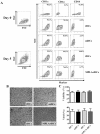
Figure 1
Figure 1
In a 5-day culture human monocytes differentiate into dendritic cells (DCs) according to lineage markers and cellular morphology. After 5 days incubation DCs are CD11c + CD1a + and show lower expression of CD14 compared to monocytes. They also exhibit a typical DC morphology. (A) 5-day monocyte-derived DCs were differentiated under multiple stimulatory conditions: DC maturated with MPLA (mDCs), DCs conditioned with dexamethasone (tDCs), tDCs activated with MPLA (MPLA-tDCs) or immature DCs left untreated (iDCs). Cells were stained with CD14, CD11c and CD1a to assess their development towards fully differentiated DCs. Density plots of monocytes (day 0) and DCs (day 5) after each differentiation scheme are shown. Plots are representative of 5 independent experiments. (B) Cell morphology was evaluated by light microscopy on each DCs group on day 5. Representative microphotographs are shown. (C) Cell viability percentage on day 5 is expressed as the percentage of Annexin V and 7-AAD negative cells (mean ± SEM), while DC yield is expressed as a percentage of DCs obtained on day 5 related to the initial number of monocytes cultured per condition (mean ± SEM) (n = 7).
Figure 2
Figure 2
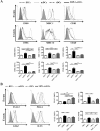
MPLA-tDCs exhibit a semi-mature surface phenotype and differentially express high levels of TLR-2. DCs generated following a 5-day protocol were treated with dexamethasone 48 hours before cell harvest for tolerance induction (tDCs). MPLA was added 24 hours before cell collection to obtain mature DC (mDCs) and also to activate tDCs (MPLA-tDCs). Untreated DCs were used as immature controls (iDCs). (A) After harvesting, DC surface expression levels of costimulatory (CD80 and CD86), antigen presentation (MHC class I and II), maturation (CD83) and functional activator (CD40) molecules were assessed by flow cytometry, which are shown as representative histograms of each marker (top) and graphic analyses of MFI measurements determined for every marker and expressed as mean ± SEM (bottom) (n = 33). (B) TLR-2, PD-L1, GILZ and ILT3 expression on each DC condition was analyzed by flow cytometry to seek for specific tolerogenic DC markers. Representative histograms (left) and graphic analyses for each molecule assessed from 13 independent experiments are shown and represented as mean ± SEM (* P < 0.05; ** P < 0.01; *** P < 0.001).
Figure 3
Figure 3
MPLA-tDCs display an anti-inflammatory cytokine secretion profile with major IL-10 production. MPLA-tDCs show a pro-tolerogenic cytokine secretion profile with high anti-inflammatory cytokines production and reduced pro-inflammatory cytokines secretion. On day 5, harvested DCs were washed and then incubated for another 24 hours in AIM-V medium alone or co-cultured with the NIH-3 T3 murine fibroblast cell line transfected with human CD40L at a 1:1 DCs:fibroblasts ratio. Culture supernatants were collected and further analyzed by ELISA for pro- and anti-inflammatory cytokines detection. Untreated immature DCs (iDCs) as well as MPLA-matured DCs (mDCs) were used as controls. (A) IL-12, IL-23 and TNFα levels determined for all DC groups upon CD40L stimulation. Levels of these cytokines were undetectable in supernatants of DCs without CD40L stimulation. (B) IL-10 and TGFβ1 were detected in supernatants of DCs cultured for 24 hours in AIM-V medium. (C) IL-10 and TGFβ1 were also determined in supernatants of DCs co-incubated with fibroblasts expressing CD40L. Data in A, B and C correspond to mean ± SEM of 13 independent experiments (* P < 0.05; ** P < 0.01; *** P < 0.001).
Figure 4
Figure 4
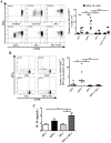
MPLA-tDCs display reduced capability to stimulate allogeneic CD4+ T cell proliferation and IFNγ production. MPLA-tDCs and tDCs show decreased allostimulatory capacity compared to MPLA-matured DCs and similar to iDCs. (A) Alloproliferative response induced by MPLA-tDCs and tDCs was assessed by co-culturing with allogeneic CD4+ T cells stained with CFSE at a 1:2 DC/T cell ratio for 6 days, and proliferation was determined by CFSE dilution through flow cytometry. CD4+ T cells incubated alone or treated with anti-CD3 (OKT3 clone) were used as controls. Representative flow cytometry plots (left) and graphic analysis of DC allostimulatory capacity for all conditions expressed as percentage of CD4+ CFSElow T cells from 7 independent experiments (right) are shown. (B) Production of IFNγ by allogeneic CD4+ T cells was measured on day 6 of culture through intracellular staining after stimulation of cells with PMA/ionomycin for 5 hours and analyzed by flow cytometry. Representative flow cytometry plots (left) and graphic analysis of percentage of IFNγ producing CD4+ CFSElow T cells from 4 independent experiments (right) are shown. Horizontal lines represent median values (*** P < 0.001).
Figure 5
Figure 5
MPLA-tDCs modulate CD4+ T cell responses in an antigen-specific manner. CD4+ T cells co-cultured with autologous PPD-loaded MPLA-tDCs exhibited impaired proliferative response together to a reduced IFNγ production and high IL-10 secretion. (A) Antigen-specific T cell responses were assessed by co-culturing DCs with autologous CD4+ T cells stained with CFSE at a 1:2 DC/T cell ratio for 6 days, and proliferation was determined by CFSE dilution by flow cytometry. CD4+ T cells incubated alone and CD4+ T cells treated with anti-CD3 (OKT) were used as controls. Representative flow cytometry plots (left) and graphic analysis of DC antigen-specific stimulatory ability for all conditions expressed as number of CD4+ CFSElow T cells for 6 independent experiments (right) are shown. (B) Production of IFNγ by autologous T cells was measured on day 6 of culture through intracellular staining after stimulation of cells with PMA/ionomycin for 5 hours and analyzed by flow cytometry. Representative flow cytometry plots (right) and graphic analysis of the number of IFNγ producing CD4+ CFSElow T cells from 6 independent experiments are shown. In A and B horizontal lines represent median values (* P < 0.05; ** P < 0.01; *** P < 0.001). (C) IL-10 secretion levels from DC/T cell culture supernatants were measured using ELISA. Results are represented as the mean ± SEM of 5 independent experiments (* P < 0.05).
Figure 6
Figure 6
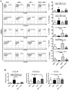
MPLA-activated tDCs acquire secondary lymphoid homing capacity in response to CCR7 and CXCR4 ligands compared to non-activated tDCs. MPLA activation of tDCs induces upregulation of migratory receptors CCR7 and CXCR4 and enhances chemotactic responsiveness to their ligands CCL19 and CXCL12. MPLA was added 24 hours before cell harvest to induce tDCs activation, generating MPLA-tDCs. After cell collection on day 5, DCs chemokine receptors expression profile was evaluated by flow cytometry together with their migratory response to relevant chemokines. tDCs without MPLA activation, mature (mDCs) and immature controls (iDCs) were also studied. (A) Representative density plots for each chemokine among CD11c + DCs (left). Data are also shown as the mean ± SD percentage of CD11c + DCs expressing the respective receptor analyzed from 10 independent experiments (right). (B) DC migratory capacity in response to 250 ng/ml of CCL19, CXCL12 or CCL5, or to medium alone, was assessed through a transwell assay. DC migration for each chemokine is expressed as the mean ± SD of migration index (Migrating DCs towards chemokine/Migrating DCs towards medium) determined in duplicates in 6 independent experiments (* P < 0.05; ** P < 0.01; *** P < 0.001).

References

    1. Ueno H, Klechevsky E, Morita R, Aspord C, Cao T, Matsui T, Di Pucchio T, Connolly J, Fay JW, Pascual V, Palucka AK, Banchereau J. Dendritic cell subsets in health and disease. Immunol Rev. 2007;219:118–142. doi: 10.1111/j.1600-065X.2007.00551.x. - DOI - PubMed
    1. Randolph GJ, Ochando J, Partida-Sanchez S. Migration of dendritic cell subsets and their precursors. Annu Rev Immunol. 2008;26:293–316. doi: 10.1146/annurev.immunol.26.021607.090254. - DOI (V体育安卓版) - PubMed
    1. Delgado-Martín C, Escribano C, Pablos JL, Riol-Blanco L, Rodríguez-Fernández JL. Chemokine CXCL12 Uses CXCR4 and a Signaling Core Formed by Bifunctional Akt, Extracellular Signal-regulated Kinase (ERK)1/2, and Mammalian Target of Rapamycin Complex 1 (mTORC1) Proteins to Control Chemotaxis and Survival Simultaneously in Mature Dendritic Cells. J Biol Chem. 2011;286:37222–37236. doi: 10.1074/jbc.M111.294116. - DOI - PMC - PubMed
    1. Naranjo-Gomez M, Raich-Regue D, Onate C, Grau-Lopez L, Ramo-Tello C, Pujol-Borrell R, Martinez-Caceres E, Borras FE. Comparative study of clinical grade human tolerogenic dendritic cells. J Transl Med. 2011;9:89. doi: 10.1186/1479-5876-9-89. - DOI - PMC - PubMed
    1. Torres-Aguilar H, Aguilar-Ruiz SR, González-Pérez G, Munguía R, Bajaña S, Meraz-Ríos MA, Sánchez-Torres C. Tolerogenic Dendritic Cells Generated with Different Immunosuppressive Cytokines Induce Antigen-Specific Anergy and Regulatory Properties in Memory CD4+ T Cells. J Immunol. 2010;184:1765–1775. doi: 10.4049/jimmunol.0902133. - "VSports注册入口" DOI - PubMed

VSports最新版本 - Publication types

"V体育安卓版" MeSH terms