Skip to main page content
U.S. flag

An official website of the United States government

Dot gov

The . gov means it’s official VSports app下载. Federal government websites often end in . gov or . mil. Before sharing sensitive information, make sure you’re on a federal government site. .

Https

The site is secure V体育官网. The https:// ensures that you are connecting to the official website and that any information you provide is encrypted and transmitted securely. .

. 2013 Aug;145(2):383-95.e1-21.
doi: 10.1053/j.gastro.2013.04.050. Epub 2013 May 2.

"VSports注册入口" Isolation and characterization of intestinal stem cells based on surface marker combinations and colony-formation assay

Affiliations

Isolation and characterization of intestinal stem cells based on surface marker combinations and colony-formation assay

VSports最新版本 - Fengchao Wang et al. Gastroenterology. 2013 Aug.

Abstract

Background & aims: Identification of intestinal stem cells (ISCs) has relied heavily on the use of transgenic reporters in mice, but this approach is limited by mosaic expression patterns and difficult to directly apply to human tissues VSports手机版. We sought to identify reliable surface markers of ISCs and establish a robust functional assay to characterize ISCs from mouse and human tissues. .

Methods: We used immunohistochemistry, real-time reverse-transcription polymerase chain reaction, and fluorescence-activated cell sorting (FACS) to analyze intestinal epithelial cells isolated from mouse and human intestinal tissues. We compared different combinations of surface markers among ISCs isolated based on expression of Lgr5-green fluorescent protein V体育安卓版. We developed a culture protocol to facilitate the identification of functional ISCs from mice and then tested the assay with human intestinal crypts and putative ISCs. .

Results: CD44(+)CD24(lo)CD166(+) cells, isolated by FACS from mouse small intestine and colon, expressed high levels of stem cell-associated genes. Transit-amplifying cells and progenitor cells were then excluded based on expression of GRP78 or c-Kit. CD44(+)CD24(lo)CD166(+) GRP78(lo/-) putative stem cells from mouse small intestine included Lgr5-GFP(hi) and Lgr5-GFP(med/lo) cells V体育ios版. Incubation of these cells with the GSK inhibitor CHIR99021 and the E-cadherin stabilizer Thiazovivin resulted in colony formation by 25% to 30% of single-sorted ISCs. .

Conclusions: We developed a culture protocol to identify putative ISCs from mouse and human tissues based on cell surface markers VSports最新版本. CD44(+)CD24(lo)CD166(+), GRP78(lo/-), and c-Kit(-) facilitated identification of putative stem cells from the mouse small intestine and colon, respectively. CD44(+)CD24(-/lo)CD166(+) also identified putative human ISCs. These findings will facilitate functional studies of mouse and human ISCs. .

Keywords: CBC; CFE; CoSC; Differentiation; FACS; Flow Cytometry Analysis; GRP; IEC; IHC; ISC; PC; Paneth cell; Single-Cell Sorting; Stemness; colonic stem cell; colony-forming efficiency; crypt base columnar; fluorescence-activated cell sorting; green fluorescent protein; immunohistochemistry; intestinal epithelial cell; intestinal stem cell; qRT-PCR; quantitative reverse-transcription polymerase chain reaction. V体育平台登录.


Figures

Figure 1
Figure 1
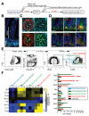
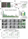
Combination of CD44, CD24, and CD166 preliminarily excludes small intestinal differentiated cells. (A) Experimental scheme of excluding villus and crypt differentiated cells. (B) Immunohistochemistry (IHC) staining shows gradient expression of CD44 in crypts but not in villi. (C) Confocal cross section of crypt bottom, showing strong staining of CD44 (bright blue) in the junctions of Lgr5-GFPhi stem cells (green, arrowheads) and weak staining between PCs (red, arrows). (D) IHC staining of CD24 and CD166 (green) shows higher expression at the crypt base. (E) FACS strategy for sequentially gating single cells, live IECs, CD44+ cells, and cells with different CD24 and CD166 expression. (F) qRT-PCR analysis showing the stem cell and differentiation marker expression in 4 subpopulations gated from CD44+ cells versus total live IECs by heat map (left) and column chart (right). Data are presented as mean ± SEM (n = 3). Scale bars = 50 μm (B and D); 20 μm (C).
Figure 2
Figure 2
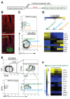
GRP78 excludes progenitors in the CD44+ CD24loCD166+ population. (A) Experimental scheme of excluding progenitor and differentiated cells from CD44, CD24, and CD166 cells using GRP78+. (B) IHC staining shows high expression of GRP78 (red) in the villi and upper crypt but low expression in the stem cell zone at the crypt base. (C) Single live IECs divided into 3 subgates of CD44hiGRP78−/lo, CD44lo GRP78+, and CD44 GRP78+. (D) Heat map of qRT-PCR results shows the gene expression relative to single live IECs in 3 subgates (C) (n = 2). (E) Two subgates of CD44+CD24loCD166+GRP78−/lo and CD44+CD24loCD166+GRP78+. (F) Heat map of qRT-PCR results shows the gene expression profiling in CD44+CD24loCD166+GRP78−/lo cells relative to the CD44+CD24loCD166+GRP78+ population (n = 3). Scale bars = 50 μm (B).
Figure 3
Figure 3
CD44, CD24, CD166, c-Kit combination identifies CoSCs. (A) Experimental scheme of gradually purifying colonic ISCs. (B) IHC staining shows expression pattern of CD44, CD24, CD166, and c-Kit in colon. The cross section shows the expression of CD44 (white) in Muc2+ (red) cells adjacent to Lgr5-GFPhi (green) cells (arrowheads) and in the conjunction of Lgr5-GFPlo cells (arrows). (C) Sequential FACS plots and gates as indicated. (D) qRT-PCR results show gene expression in the CD44med and CD44hi cells versus CD44lo/− cells (n = 2). (E) Gene expression in CD44hiCD24loCD166lo and CD44hiCD24loCD166hi populations relative to the CD44hiCD24hiCD166med population (E)and further separation with c-Kit (F) (n = 3). Scale bars = 50 μm (B); 10 μm (B, magnification).
Figure 4
Figure 4
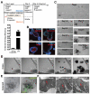
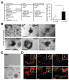
Small molecules enable robust culturing of bona fide single ISCs or CoSCs. (A) Strategies to culture single Lgr5-GFPhi cells and comparison of Wnt3a and CHIR99021 have an impact on CFE. Data are presented as mean ± SEM (P = .0004, t test, n = 3). (B) IHC staining shows the differentiated cell type in the enteroid derived from single ISCs. (C) Representative images recording the process of single Lgr5-GFPhi cell–forming enteroids. (D) A growth process from a single Lgr5-GFPhi CoSC into colonoid. (E) An open colonoid structure derived from a single Lgr5-GFPhi CSC. AF, autofluorescence. (F) Colonoids containing differentiated cell types identified by electron microscopic analysis. Scale bars = 50 μm (B); 100 μm (C, D, E); 4 μm (F).
Figure 5
Figure 5
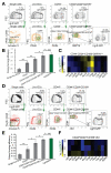
The surface marker combination identifies the ISC population with CFE comparable to that of Lgr5-GFPhi cells. (A) Lgr5-GFPhi cells in parent gates were largely confined in daughter gates, showing the gradual selection by surface marker combination. (B) CFE analysis of different subpopulations. Data are presented as mean ± SEM, (*P < .05, **P < .01, n = 4, t test). (C) The gene profiling of CD44+CD24loCD166+ GRP78−/lo cells, taking Lgr5-GFPhi ISCs as control (n = 3). (D) Colonic Lgr5-GFPhi cells in parent gates were largely confined in daughter gates, showing the gradual purification by surface marker combination. (E) CFE analysis of series of gates. Data are presented as mean ± SEM (*P < .05, **P < .01, n = 4, t test). (F) The gene profiling of CD44hiCD24loCD166+cKit cells, taking Lgr5-GFPhi CoSCs as control (n = 3).
Figure 6
Figure 6
CD44+CD24loCD166+ GRP78−/lo identifies ISCs independent of Lgr5-GFPhi cells. (A) The mosaic distribution of Lgr5-GFP+ crypt in jejunum. Gradient GFP expression in Lgr5-GFP+ crypt. (B) Lgr5-GFPneg, Lgr5-GFPlo/med, and Lgr5-GFPhi gates in the CD44+CD24loCD166+ GRP78−/lo population. (C) qRT-PCR results show the gene expression of Lgr5-GFPneg and Lgr5-GFPhi cells relative to Lgr5-GFPlo/med cells from the CD44+CD24loCD166+ GRP78−/lo population. Data are presented as mean ± SEM (n = 4, *P < .05, **P < .01, ***P < .001). (D) CFE analysis of the Lgr5-GFPneg, Lgr5-GFPlo/med, and Lgr5-GFPhi cells in the gates of B. Data are represented as mean ± SEM (*P < .05, n = 3, t test). (E) A growth process of single cells sorted from Lgr5-GFPneg, Lgr5-GFPlo/med, and Lgr5-GFPhi gates in B, showing that Lgr5-GFPlo/med cells can generate Lgr5-GFPhi enteroid. (F) FACS analysis of enteroids derived from single Lgr5-GFPneg, Lgr5-GFPlo/med, and Lgr5-GFPhi cells, showing that single Lgr5-GFPlo/med cells can generate enteroids containing Lgr5-GFPhi cells as single Lgr5-GFPhi cells did. Data are presented as mean ± SEM (n = 3, **P < .01).
Figure 7
Figure 7
The new culture strategy enables highly efficient human crypt culture and maintenance. (A) Crypt culture strategy. CHIR99021 significantly increased the efficiency of culturing human small intestinal crypts. Data are presented as mean ± SEM (n = 6, *P .011, t test). (B) Representative growth process of human small intestinal crypt culture with addition of a single dose of GSK inhibitor CHIR99021 and other factors. (C) Human enteroid can be passaged up to 10 times over a 3-month period, as shown by their growth after passaging from day 0 to day 7. (D) IHC staining show the cellular proliferation and differentiated cell types in cultured enteroid. Positive staining is indicated by the arrow in the inset. Scale bars = 200 μm (B and C); 50 μm (D).

References

    1. Barker N, van Es JH, Kuipers J, et al. Identification of stem cells in small intestine and colon by marker gene Lgr5. Nature. 2007;449:1003–1007. - PubMed
    1. Sangiorgi E, Capecchi MR. Bmi1 is expressed in vivo in intestinal stem cells. Nat Genet. 2008;40:915–920. - PMC - PubMed
    1. Montgomery RK, Carlone DL, Richmond CA, et al. Mouse telomerase reverse transcriptase (mTert) expression marks slowly cycling intestinal stem cells. Proc Natl Acad Sci U S A. 2011;108:179–184. - PMC - PubMed
    1. Tian H, Biehs B, Warming S, et al. A reserve stem cell population in small intestine renders Lgr5-positive cells dispensable. Nature. 2011;478:255–259. - PMC - PubMed
    1. Powell AE, Wang Y, Li Y, et al. The Pan-ErbB negative regulator Lrig1 is an intestinal stem cell marker that functions as a tumor suppressor. Cell. 2012;149:146–158. - PMC - PubMed

Publication types

MeSH terms