Skip to main page content
U.S. flag

An official website of the United States government

Dot gov

The . gov means it’s official VSports app下载. Federal government websites often end in . gov or . mil. Before sharing sensitive information, make sure you’re on a federal government site. .

Https

The site is secure V体育官网. The https:// ensures that you are connecting to the official website and that any information you provide is encrypted and transmitted securely. .

. 2013 Apr 25;4(4):e611.
doi: 10.1038/cddis.2013.135.

V体育2025版 - Calcium-activated and apoptotic phospholipid scrambling induced by Ano6 can occur independently of Ano6 ion currents

Affiliations

"VSports app下载" Calcium-activated and apoptotic phospholipid scrambling induced by Ano6 can occur independently of Ano6 ion currents

A Kmit (VSports手机版) et al. Cell Death Dis. .

Abstract

Immune cells and platelets maintain plasma membrane phospholipid asymmetry. Upon activation, this asymmetry is disrupted by phospholipid scrambling (PS), which is a major step during activation of immune cells, hemostasis and apoptosis. Anoctamin 6 (Ano6; TMEM16F) causes chloride (Cl(-)) and cation currents and is required for Ca(2+)-dependent PS. It is defective in blood cells from patients with Scott syndrome, a rare bleeding disorder. We examined if Cl(-) currents and PS are related, whether both processes are Ca(2+) dependent, and whether Ca(2+)-independent scrambling during intrinsic and extrinsic apoptosis is controlled by Ano6. Ca(2+) increase by ionomycin activated Ano6 Cl(-) currents and PS in normal lymphocytes, but not in B-lymphocytes from two different patients with Scott syndrome. Fas ligand (FasL) did not increase intracellular Ca(2+), but activated Cl(-) currents in normal but not in Scott lymphocytes. Whole-cell currents were inhibited by Cl(-) channel blockers and by siRNA knockdown of Ano6 VSports手机版. In contrast, intrinsic mitochondrial apoptosis by ABT-737 did not induce Cl(-) currents in lymphocytes. PS was not inhibited by blockers of Ano6 or removal of Cl(-) ions. Remarkably, Ca(2+)-independent scrambling due to extrinsic (FasL) or intrinsic (ABT-737) apoptosis was unchanged in Scott cells. We conclude that: (i) Ano6 Cl(-) currents are activated by increase in cytosolic Ca(2+), or Ca(2+) independent by stimulation of Fas receptors; (ii) Ca(2+)-dependent PS induced by Ano6 does not require Cl(-) currents; (iii) Ca(2+)-independent PS does not require Ano6; (iv) Ano6 is necessary for Ca(2+)-dependent PS, but not by increasing intracellular Ca(2+). .


Figures
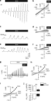
Figure 1
Figure 1
Ca2+-dependent activation of whole-cell currents in normal but not Scott lymphocytes. Original recordings of whole-cell currents measured in normal lymphocytes (a) and lymphocytes from two patients with Scott disease (c and e). Cells were kept under current clamp and were voltage clamped in intervals from −100 to +50 mV in steps of 10 mV. Ionomycin (1 μM) activated a whole-cell current only in normal lymphocytes (a). Current/voltage relationships were obtained in normal (b) and Scott lymphocytes (d and f). Note the activation of a large whole-cell current by ionomycin (filled circles) in normal but not in Scott lymphocytes. (g and h) Activation of a whole-cell Cl current by ionomycin (1 μM) in normal B lymphocytes, using 145 mM NMDG+Cl in pipette and bath. Note the outward rectification of the whole-cell current and the pronounced inhibition by 20 μM AO1. (i) Inhibition of ionomycin-activated outward currents by replacement of extracellular Cl with gluconate (5 Cl). (j) Inhibition of ionomycin-induced whole-cell currents (current densities) and depolarization in normal B lymphocytes after knockdown of Ano6 expression with siRNA. Mean±S.E.M. (number of cells). ‘*' denotes significant activation by ionomycin or effect of siRNA for Ano6 (si-Ano6); P<0.05; paired t-test
Figure 2
Figure 2
Lack of Ano6 in Scott lymphocytes abrogates Cl currents but does not affect Ca2+ increase. (a) Whole-cell overlay currents obtained in wt lymphocytes and lymphocytes from two patients with Scott disease, under control conditions and after stimulation with ionomycin (1 μM). Cells were voltage clamped from −100 to +50 mV in steps of 10 mV. Arrowhead indicates currents at clamp voltage of 0 mV. (b) Summary of whole-cell current densities and membrane voltages under control conditions and after stimulation with ionomycin (filled bars). (c) Continuous recordings of 380/340 ratios (Fura-2) and effects of 1 or 2 μM ionomycin (Iono1, Iono2), and Ca2+ removal in wt and Scott lymphocytes. (d) Summary of [Ca2+]i upon application of 1 or 2 μM ionomycin. (e) Effects of 10 μM ionomycin on [Ca2+]i measured in cells in suspensions (Fluo-4). Mean±S.E.M. (number of cells). ‘*' denotes significant increase when compared with control; P<0.05; paired t-test
Figure 3
Figure 3
Ca2+ activates Ano6 Cl conductance in normal but not Scott lymphocytes. (ac) Ionomycin-activated whole-cell currents in normal and Scott lymphocytes. Inhibition of currents by replacement of extracellular Cl with impermeable gluconate (except of 5 mM; 5 Cl), or by application of tannic acid (TA; 20 μM), NPPB (100 μM), CaCCinh-AO1 (AO1; 10 μM), or 4,4′-diisothiocyano-2,2′-stilbenedisulfonic acid (100 μM). Dashed lines indicate currents before stimulation with ionomycin (control). No currents were activated in ScottUSA lymphocytes (b), whereas currents were largely reduced in ScottUK lymphocytes (c). (d) Activation of Ano6 currents by high (100 μM) pipette [Ca2+] in a control lymphocyte and inhibition by removal of extracellular Cl (5 Cl). The black arrowhead in d indicates the time point that was chosen to read currents for generation of the summary bars shown in a, b, c and e. (e) Summary of current densities, indicating activation of Ano6 by pipette Ca2+ (black bars) and inhibition by low Cl. Mean±S.E.M. (number of cells). ‘*' denotes significant difference compared with control; P<0.05; paired t-test
Figure 4
Figure 4
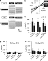
Activation Ano6 Cl currents by activation of Fas receptors. (a) Whole-cell current overlays obtained in normal lymphocytes before and after incubation with FasL (0.5 μg/ml; 2 h), and inhibition by Cl replacement (5 Cl) and tannic acid (TA; 20 μM). Cells were voltage clamped from −100 to +50 mV in steps of 10 mV. Arrowhead indicates currents at clamp voltage of 0 mV. (b) Current/voltage relationship of the FasL-activated whole-cell current and effect of TA. (ce) Calculated whole-cell conductances in normal, ScottUSA, and ScottUK lymphocytes and effects of 5 Cl, TA and NPPB. Dashed line indicates conductance before incubation with FasL (control). Mean±S.E.M. (number of cells). ‘*'denotes significant inhibition; P<0.05; paired t-test. ‘#'denotes significant activation by FasL; P<0.05; unpaired t-test
Figure 5
Figure 5
Phospholipid scrambling by ionomycin but not by FasL is impaired in Scott lymphocytes. (a) Percentage of phosphatidylserine (PS)-exposing lymphocytes after 10 min stimulation with 10 μM ionomycin in presence of 1 mM CaCl2 for normal and Scott lymphocytes as measured after labeling with annexin A5 for 5 min. (b) Effect of NPPB (100 μM), TA (50 μM) and replacement of Cl by gluconate on ionomycin-induced PS exposure in healthy control lymphocytes. (c) Percentage PS-exposing lymphocytes after 4 h incubation with 0.5 μg/ml FasL in presence of 1 mM CaCl2 for control and Scott lymphocytes in presence or absence of 20 μM Q-VD-Oph. (d) No effects of NPPB (100 μM), TA (50 μM) and Cl replacement on FasL-induced PS exposure in healthy control lymphocytes were observed. (e) Caspase-3 activity of control and Scott lymphocytes after 4 h stimulation with FasL as measured by a caspase activity assay. (f) Percentage of PS-exposing lymphocytes after incubation with 50 μM ABT-737. Mean±S.D. (n=3–12). ‘#' denotes significant activation of PS exposure or caspase; P<0.05; unpaired t-test

References

    1. Yang YD, Cho H, Koo JY, Tak MH, Cho Y, Shim WS, et al. TMEM16A confers receptor-activated calcium-dependent chloride conductance. Nature. 2008;455:1210–1215. - PubMed
    1. Schroeder BC, Cheng T, Jan YN, Jan LY. Expression cloning of TMEM16A as a calcium-activated chloride channel subunit. Cell. 2008;134:1019–1029. - PMC - PubMed
    1. Caputo A, Caci E, Ferrera L, Pedemonte N, Barsanti C, Sondo E, et al. TMEM16A, a membrane protein associated with calcium-dependent chloride channel activity. Science. 2008;322:590–594. - PubMed
    1. Tian Y, Schreiber R, Kunzelmann K. Anoctamins are a family of Ca2+ activated Cl- channels. J Cell Sci. 2012;125:4991–4998. - PubMed
    1. Ferrera L, Caputo A, Galietta LJ. TMEM16A protein: a new identity for Ca(2+)-dependent Cl channels. Physiology. 2010;25:357–363. - PubMed

Publication types

MeSH terms

Substances

V体育2025版 - Supplementary concepts