<kbd dir="tfR7K"></kbd> V体育ios版 - Skip to main page content
U.S. flag

An official website of the United States government

Dot gov

The . gov means it’s official. Federal government websites often end in VSports app下载. gov or . mil. Before sharing sensitive information, make sure you’re on a federal government site. .

Https

The site is secure V体育官网. The https:// ensures that you are connecting to the official website and that any information you provide is encrypted and transmitted securely. .

. 2013 Apr 15;23(4):516-26.
doi: 10.1016/j.ccr.2013.03.018.

Relapse or eradication of cancer is predicted by peptide-major histocompatibility complex affinity (VSports)

Affiliations

"V体育官网" Relapse or eradication of cancer is predicted by peptide-major histocompatibility complex affinity

"VSports最新版本" Boris Engels et al. Cancer Cell. .

Abstract (VSports app下载)

Cancers often relapse after adoptive therapy, even though specific T cells kill cells from the same cancer efficiently in vitro VSports手机版. We found that tumor eradication by T cells required high affinities of the targeted peptides for major histocompatibility complex (MHC) class I. Affinities of at least 10 nM were required for relapse-free regression. Only high-affinity peptide-MHC interactions led to efficient cross-presentation of antigen, thereby stimulating cognate T cells to secrete cytokines. These findings highlight the importance of targeting peptides with high affinity for MHC class I when designing T cell-based immunotherapy. .


Conflict of interest statement

The authors have no conflict of interests.

Figures
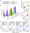
Figure 1
Figure 1. Six transduced cancer cell lines that express antigens at high levels were effectively killed in vitro
(A) Diagram of fusion proteins constructed to express antigen in MC57 cancer cells. Triple repeats of peptide and AAY proteasomal cleavage sites were fused to fluorescent proteins: OVA257, SIY, mouse tyrosinase369–377 (Tyr369), mouse or human gp10025–33 (mgp10025 and hgp10025, respectively) and EGP; a Cerulean fusion gene was generated only for SIY. (B) Flow cytometric analysis of peptide-EGFP fusion proteins expressed by the transduced MC57 fibrosarcoma lines. (C) MC57-mgp100/SIY expressed mgp10025 and SIY antigens as EGFP and Cerulean fusion proteins, respectively. The HLA-A2/Db chimeric protein HHD was co-transduced with the Tyr369-EGFP fusion protein to generate MC57-TyrHHD. Parental MC57 (gray) was analyzed for comparison. (D) Cytolysis of MC57 target cells overexpressing SIY, mgp10025, hgp10025, EGP, or Tyr369 and HHD (Tyr) by 2C, pmel, AFH (Tyr-negative) or FH (Tyr-positive) T cells in a 4.5 h 51Cr-release assay. Cancer cells expressing non-cognate peptide were used as negative controls. These data are compiled of three experiments and are representative for seven independent experiments. See Figure S1 for induction of vitiligo by FH and pmel T cells.
Figure 2
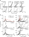
Figure 2. Targeting SIY, OVA257 or Tyr369 eradicated large tumors while targeting mgp10025, hgp10025 or EGP caused initial tumor regression but was followed by relapse
(A) Cancer cell lines formed progressively growing tumors within one week. TCR-transgenic mice with irrelevant specificity were challenged s.c. with 2×10⁶ cancer cells. MC57-SIY, -mgp100, -mgp100/SIY, -hgp100 and -EGP were injected into OT-I mice; MC57-OVA grew in 2C mice and MC57-TyrHHD grew in AOTA (Tyr-deficient, non-self) and OTA (Tyr-positive, self). Graphs represent single mice in 11 experiments, listed as non-treated controls in Tables 1, S1 and S3. (B) At least 2 weeks after cancer cell injection, when tumors reached about 500 mm3, each mouse was treated once with cognate T cells (treatment between days 13 and 26, depending on tumor size, as indicated by the horizontal bars (H)). Average size of tumors at day of treatment: 2C: 720 mm3, ranging from 448 to 995 mm3; OT-I: 608 mm3, ranging from 440 to 715 mm3; FH: 517 mm3, ranging from 250 to 848 mm3; pmel, targeting mgp10025: 601 mm3, ranging from 325 to 980 mm3; targeting hgp10025: 470 mm3, ranging from 264 to 936 mm3; targeting EGP: 337 mm3, ranging from 180 to 600 mm3. MC57-TyrHHD was grown in OTA (self) and treated with FH T cells (self). The number of rejected tumors per number of tumors treated is indicated. Data are derived from 15 independent experiments, compiled in Tables 1 and S1. See also Figure S2. * Tumors were isolated and analyzed for antigen expression (see Figure 3).
Figure 3
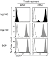
Figure 3. Outgrowth of antigen-loss variants after pmel T cell treatment of cancer cells expressing hgp10025 but not of cancers expressing mgp10025 or EGP
Cancer cells of relapsed tumors expressing mgp10025, EGP (both gray) or hgp10025 (black) were isolated after pmel T cell treatment, adapted to culture, and analyzed for peptide-EGFP fusion gene expression (left panels). MC57 cells (white histogram) cultured in vitro and MC57-mgp100, MC57-EGP (both gray) or MC57-hgp100 (black) cells isolated from non-treated mice (right panels) were analyzed as controls. Isolated lines from mgp100- or hgp100-expressing tumors are representative for four lines each, the isolate from the EGP-expressing tumor is representative for two lines; all lines were isolated after relapse (respective tumors were marked with * in Figure 2B). The repeatability in independent experiments strongly suggests that loss of antigen expression from the hgp10025 cancer cells was not an artifact caused by adaptation or post-isolation culturing. See also Figure S3.
Figure 4
Figure 4. T cells transferred to treat the tumors expressing the different peptides showed the same phenotype of activated T cells
T cells of 2C AFH and pmel TCR-transgenic mice were tested for their activation status in “naïve”, untreated mice (splenocytes), at day 3 after peptide activation in vitro, and on day 4 after adoptive transfer (tumor infiltrating cells). Cells were analyzed by flow cytometry for expression of CD44 and CD62L, and gated on CD8+ T cells expressing the cognate Vβ-chain: Vβ8 for 2C, Vβ11 for AFH and Vβ13 for pmel. Data are representative for two or three independent experiments for the data in vivo and in vitro data, respectively.
Figure 5
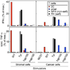
Figure 5. Only SIY and Tyr369 are cross-presented, as detected by cytokine secretion by T cells stimulated by stromal cells isolated from untreated tumors
CD11b+ stromal cells were isolated from established untreated tumors and were co-cultured with peptide-activated T cells. Enriched stromal cells from tumors grown from MC57-SIY, -hgp100 and -mgp100 cells (all grown in OT-I mice) and -TyrHHD (grown in AOTA mice (non-self)) were co-cultured with 2C pmel AFH (non-self) or FH (self) TCR-transgenic T cells. Stromal cells from the various tumors were compared to cultured cancer cells expressing the same antigen. Supernatants were harvested after 24 h of co-culture and amounts of IFN-γ and TNF-α measured by ELISA. Data are shown as percent of maximal cytokine secretion (anti-CD3 and -CD28 antibody stimulation, defined as 100 %). For TNF-α, cytokine secretion by stromal cells without T cells was subtracted to obtain specific TNF-α secretion by T cells (443 to 975 pg/ml by 1×10⁵ cells cultured in 200 µl for 24 h, depending on experiment). Unstimulated T cells served as negative control (below 0.7 % for all responders, not shown). Data shown are combined from two experiments and are representative for four independent experiments. See also Figure S4. * not done
Figure 6
Figure 6. Death of stromal cells in T cell-treated SIY and Tyr369 expressing tumors
MC57-SIY, -TyrHHD, -hgp100 and -mgp100 tumors were treated with 2C, AFH (non-self) or pmel T cells, respectively. Five days after adoptive transfer, single cell suspensions were generated from treated and untreated tumors and analyzed by flow cytometry. Histograms show CD11b+ T cells stained with propidium iodide (PI) to identify cell death. Numbers indicate the percentage of dead cells among CD11b+ cells. Data are representative for four independent experiments, with single mice per group.

Comment in

References

    1. Anders K, Buschow C, Herrmann A, Milojkovic A, Loddenkemper C, Kammertoens T, Daniel P, Yu H, Charo J, Blankenstein T. Oncogene-targeting T cells reject large tumors while oncogene inactivation selects escape variants in mouse models of cancer. Cancer Cell. 2011;20:755–767. - PMC - PubMed
    1. Antony PA, Piccirillo CA, Akpinarli A, Finkelstein SE, Speiss PJ, Surman DR, Palmer DC, Chan CC, Klebanoff CA, Overwijk WW, et al. CD8+ T cell immunity against a tumor/self-antigen is augmented by CD4+ T helper cells and hindered by naturally occurring T regulatory cells. J Immunol. 2005;174:2591–2601. - PMC - PubMed
    1. Assarsson E, Sidney J, Oseroff C, Pasquetto V, Bui HH, Frahm N, Brander C, Peters B, Grey H, Sette A. A quantitative analysis of the variables affecting the repertoire of T cell specificities recognized after vaccinia virus infection. J Immunol. 2007;178:7890–7901. - PubMed
    1. Bowerman NA, Colf LA, Garcia KC, Kranz DM. Different strategies adopted by K(b) and L(d) to generate T cell specificity directed against their respective bound peptides. J Biol Chem. 2009;284:32551–32561. - PMC (V体育2025版) - PubMed
    1. Budhu S, Loike JD, Pandolfi A, Han S, Catalano G, Constantinescu A, Clynes R, Silverstein SC. CD8+ T cell concentration determines their efficiency in killing cognate antigen-expressing syngeneic mammalian cells in vitro and in mouse tissues. J Exp Med. 2010;207:223–235. - V体育安卓版 - PMC - PubMed

VSports最新版本 - Publication types

MeSH terms

Substances