Skip to main page content
U.S. flag

An official website of the United States government

Dot gov

The VSports app下载. gov means it’s official. Federal government websites often end in . gov or . mil. Before sharing sensitive information, make sure you’re on a federal government site. .

Https

The site is secure V体育官网. The https:// ensures that you are connecting to the official website and that any information you provide is encrypted and transmitted securely. .

Review
. 2013 Jun;62(1):95-162.
doi: 10.1111/prd.12010.

V体育ios版 - Lessons learned and unlearned in periodontal microbiology

Review

Lessons learned and unlearned in periodontal microbiology

"V体育官网" Ricardo Teles et al. Periodontol 2000. 2013 Jun.

V体育官网 - Abstract

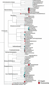
Periodontal diseases are initiated by bacterial species living in polymicrobial biofilms at or below the gingival margin and progress largely as a result of the inflammation elicited by specific subgingival species. In the past few decades, efforts to understand the periodontal microbiota have led to an exponential increase in information about biofilms associated with periodontal health and disease. In fact, the oral microbiota is one of the best-characterized microbiomes that colonize the human body. Despite this increased knowledge, one has to ask if our fundamental concepts of the etiology and pathogenesis of periodontal diseases have really changed VSports手机版. In this article we will review how our comprehension of the structure and function of the subgingival microbiota has evolved over the years in search of lessons learned and unlearned in periodontal microbiology. More specifically, this review focuses on: (i) how the data obtained through molecular techniques have impacted our knowledge of the etiology of periodontal infections; (ii) the potential role of viruses in the etiopathogenesis of periodontal diseases; (iii) how concepts of microbial ecology have expanded our understanding of host-microbe interactions that might lead to periodontal diseases; (iv) the role of inflammation in the pathogenesis of periodontal diseases; and (v) the impact of these evolving concepts on therapeutic and preventive strategies to periodontal infections. We will conclude by reviewing how novel systems-biology approaches promise to unravel new details of the pathogenesis of periodontal diseases and hopefully lead to a better understanding of their mechanisms. .


Figures

Figure 1
Figure 1
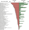
 Cumulative mean counts (×105) of 41 bacterial species in supragingival samples taken from 38 periodontally healthy subjects and from 17 subjects with periodontitis before professional removal of the dental biofilms (Pre), immediately after cleaning (time 0) and after 1, 2, 4 and 7 days of redevelopment (Post). The subjects refrained from oral‐hygiene procedures for the 7‐day test period. Samples were removed, precleaning and immediately postcleaning, from the mesio‐buccal aspect of each tooth (excluding third molars). In addition, supragingival samples were obtained from up to seven teeth in randomly selected quadrants, 1, 2, 4 and 7 days after tooth cleaning. All samples were individually analyzed for their content of 41 taxa using checkerboard DNA–DNA hybridization. Species counts in the samples were averaged within each subject at each time point and were then averaged across subjects in the two clinical groups. The plots present the cumulative mean values at each time point in each clinical group. The species were ordered and color‐coded according to previously described microbial complexes ( 337 ). Printed with permission from Uzel et al. ( 373 ).
Figure 2
Figure 2
 Diagrammatic representation of the relationships of species within microbial complexes, and between the microbial complexes in supragingival biofilm samples. This diagram was based on the results of nine cluster and two community ordination analyses using the baseline data from 187 subjects. Modified with permission from Haffajee et al. ( 136 ).
Figure 3
Figure 3
 Diagrammatic representation of the relationships of species within microbial complexes, and between the microbial complexes in supragingival biofilm samples. This diagram was based on the results of nine cluster and two community ordination analyses using the long‐term plaque‐redevelopment data from 93 subjects with post‐therapy microbiological data from 3 to 24 months. Modified with permission from Haffajee et al. ( 136 ).
Figure 4
Figure 4
 Plot of the total mean proportions of each of the seven supragingival complexes described by Haffajee et al. ( 136 ). The samples were divided, according to total‐DNA probe counts, into 10 groups using the 10, 20, 30, 40, 50, 60, 70, 80 and 90th percentiles of the total counts as cut‐off points. The x‐axis values represent the mean values for total‐DNA probe counts of each group and the y‐axis represents the sum of the proportions comprised by the species in each microbial complex. Printed with permission from Haffajee et al. ( 139 ).
Figure 5
Figure 5
 Mean counts (×105) of 40 test species in supragingival biofilm samples from the mesiobuccal surface of each tooth of 187 subjects. The upper panel represents the maxilla and the lower panel represents the mandible. Species counts were averaged across subjects for each tooth separately. A significant difference among teeth (P < 0.001, Kruskal–Wallis test) was observed for all species after adjusting for 40 comparisons ( 338 ). The species were ordered and color‐coded according to supragingival microbial complexes ( 136 ). In this cumulative plot, the total height at each sample location provides the mean total‐DNA probe count at that site. The cartoons of each tooth are presented to depict the location of each tooth sample. The full genus and species names of the 40 taxa listed in the Figure are as follows: Actinomyces naeslundii, Actinomyces israelii, Actinomyces odontolyticus, Actinomyces gerencseriae, Actinomyces oris, Veillonella parvula, Neisseria mucosa, Capnocytophaga gingivalis, Eikenella corrodens, Capnocytophaga sputigena, Capnocytophaga ochracea, Selenomonas noxia, Campylobacter gracilis, Prevotella melaninogenica, Prevotella nigrescens, Prevotella intermedia, Campylobacter rectus, Campylobacter showae, Fusobacterium nucleatum ssp. vincentii, Fusobacterium nucleatum ssp. nucleatum, Fusobacterium nucleatum ssp. polymorphum, Fusobacterium periodonticum, Tannerella forsythia, Porphyromonas gingivalis, Eubacterium nodatum, Treponema denticola, Parvimonas micra, Eubacterium saburreum, Streptococcus gordonii, Streptococcus sanguinis, Streptococcus oralis, Streptococcus mitis, Streptococcus intermedius, Streptococcus constellatus, Streptococcus anginosus, Aggregatibacter actinomycetemcomitans, Propionibacterium acnes, Leptotrichia buccalis, Treponema socranskii and Gemella morbillorum. Modified with permission from Haffajee et al. ( 140 ) (bacterial species names have been updated).
Figure 6
Figure 6
 Cumulative mean counts (×105) of 41 bacterial species in samples taken from 55 subjects with natural teeth and from 62 subjects with full‐mouth dentures. Samples were taken before professional removal of the dental biofilms (Pre), immediately after cleaning (time 0) and after 1, 2, 4 and 7 days of redevelopment (Post). The subjects refrained from oral‐hygiene procedures for the 7‐day test period. Samples were removed before, and immediately after cleaning from the mesio‐buccal aspect of each natural tooth (excluding third molars) and from each denture tooth. In addition, samples were obtained from up to seven teeth in randomly selected quadrants at 1, 2, 4 and 7 days after tooth cleaning. All samples were individually analyzed for their content of 41 taxa using checkerboard DNA–DNA hybridization. Species counts in the samples were averaged within each subject at each time point and were then averaged across subjects in the two clinical groups. The plots present the cumulative mean values at each time point in each clinical group. The species were ordered and color‐coded according to previously described microbial complexes ( 337 ). Printed with permission from Teles et al. ( 360 ).
Figure 7
Figure 7
 Cumulative mean counts (×105) of 41 bacterial species in subgingival samples taken from 38 periodontally healthy subjects and from 17 subjects with periodontitis before professional removal of the dental biofilms (Pre), immediately after cleaning (time 0) and after 1, 2, 4 and 7 days of redevelopment (Post). Species counts in the samples were averaged within each subject at each time point and were then averaged across subjects in the two clinical groups. The plots present the cumulative mean values at each time point in each clinical group. The species were ordered and color‐coded according to previously described microbial complexes ( 337 ). Printed with permission from Uzel et al. ( 373 ).
Figure 8
Figure 8
 Mean counts (×105) of subgingival taxa detected in 30 periodontally healthy subjects during experimental gingivitis. Subgingival plaque samples were collected from the mesio‐buccal surface of four randomly selected teeth at different time points. The baseline visit occurred 21 days after a preparation phase to achieve gingival health and minimal plaque accumulation (Day 0). Days 3, 7, 10, 14 and 21 correspond to different time points after interruption of oral‐hygiene practices (experimental gingivitis phase), and Day 35 corresponds to the resolution of the experimental gingivitis. Counts of 40 subgingival species were measured for each sample using checkerboard DNA–DNA hybridization. Data were averaged within a subject and were then averaged across subjects at each time point separately. The species were ordered and grouped according to the complexes described by Socransky et al. ( 337 ). The figure was adapted from data from Lee et al. ( 199 ).
Figure 9
Figure 9
 Mean counts (×105) of subgingival taxa detected in 123 periodontally healthy subjects enrolled in a 3‐year preventive program. Subgingival plaque samples were collected from the mesio‐buccal surface of every tooth present at baseline and at 1, 2 and 3 years after therapy. Counts of 40 subgingival species were measured for each sample using checkerboard DNA–DNA hybridization. Sites were grouped into three categories: (i) sites that did not show bleeding on probing in any of the visits (yellow) (n = 1,489); (ii) sites that had bleeding on probing at one or two visits (orange) (n = 1,593); and (iii) sites that had bleeding on probing at three or four visits (red) (n = 309). Data were averaged within subjects, across subjects and then across the different visits within each site category, separately, in order to obtain a summary measure of cumulative exposure to the 40 taxa over time. The species were ordered and grouped according to the complexes described by Socransky et al. ( 337 ). The figure was generated from data from Bogren et al. ( 38 ).
Figure 10
Figure 10
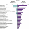
 Bacterial taxa detected in sites with different pocket‐depth categories at baseline. Sites with a pocket depth of <4 mm are shown in blue, those with a pocket depth of 4–6 mm are shown in orange and those with a pocket depth of >6 mm are shown in red. Only taxa that presented a significantly different prevalence across site categories were plotted (Kruskal‐Wallis, P < 0.05).
Figure 11
Figure 11
 Bacterial taxa detected in 42 subjects with chronic periodontitis (subject level analysis), before and after periodontal therapy. Results are shown as mean prevalence ± standard error of the mean. Only taxa that presented a significantly different prevalence between the two time‐point categories were plotted (Wilcoxon test, P < 0.05).
Figure 12
Figure 12
 Bacterial taxa detected in sites from 42 subjects with chronic periodontitis, before and after therapy (site‐level analysis). Results are shown as mean prevalence ± standard error of the mean. Only taxa that presented a significantly different prevalence between the two time‐point categories were plotted (Wilcoxon test, P < 0.05).
Figure 13
Figure 13
 An RNA‐oligonucleotide quantification technique membrane showing hybridization of clinical samples with oligonucleotide probes. Probes for uncultured/unnamed species (‘orange’ group of bacteria) are listed across the top. Each horizontal lane represents the total nucleic acids (TNA) from a sample from the indicated numbered tooth. Standards comprised a mixture of ‘complementary’ sequences from all the test taxa at 0.004 and 0.04 pM. Teeth marked with an asterisk (*) were absent. Printed with permission from Teles et al. ( 361 ).
Figure 14
Figure 14
 Weighted correlation network analysis results based on data obtained using checkerboard DNA–DNA hybridization. The images show the Cytoscape representation of the correlation networks for the four modules identified by weighted correlation network analysis. Checkerboard analysis was performed for 40 species of oral bacteria on a total of 2,565 individual subgingival samples from patients with periodontitis. The scale free topology model fit value (R2) was 0.40, the maximum value in the analysis. The identified modules correlated well with microbial complexes previously described by Socransky et al. ( 337 ). Printed with permission from Duran‐Pinedo et al. ( 85 ).
Figure 15
Figure 15
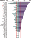
 Prevalence of uncultured/unnamed taxa and ‘reference’ culturable species (Fusobacterium nucleatum, Porphyromonas gingivalis, Veillonella parvula and Actinomyces naeslundii) examined in samples from periodontally healthy individuals (aqua) (n = 8) and from patients with chronic periodontitis (purple) (n = 8). Significant differences are indicated with an asterisk (chi square test, P < 0.002). Red vertical bars highlight taxa that were present in fewer than 10% of sites. Taxa present in fewer than 10% of sites were excluded from subsequent analyses.
Figure 16
Figure 16
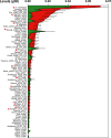
 Levels of selected taxa in sites from periodontally healthy patients (green) (n = 8) and from patients with chronic periodontitis (red) (n = 8). Values are given as pM ± standard error of the mean. Significant differences are indicated with an asterisk (Mann–Whitney U‐test, P < 0.05).
Figure 17
Figure 17
 Levels of selected taxa in shallow sites (green) (pocket depth <4 mm) in healthy sites of periodontally healthy individuals and in deep sites (red) (pocket depth >4 mm) of patients with chronic periodontitis. Values are given as pM ± standard error of the mean. Only taxa with a significantly different prevalence between the site categories were plotted (Mann–Whitney U‐test, P < 0.0001).
Figure 18
Figure 18
 Representative detail images of combinatorial labeling and spectral imaging–fluorescence in‐situ hybridization‐labeled semidispersed human dental plaque. The color in the raw spectral images represents a merge of six different fluorophore channels. The color in the segmented image represents one of 15 taxa. Scale bar = 10 μm. Printed with permission from Valm et al. ( 375 ).
Figure 19
Figure 19
 Mean counts (×105) of subgingival taxa in the clusters and not‐in‐cluster group detected in 31 subjects with generalized aggressive periodontitis. The counts of 40 subgingival species were measured at each of up to 14 sites in each subject and were employed in a cluster analysis using the chord coefficient and an average unweighted linkage sort. Five clusters (CL1–CL5) were formed at >33% similarity. Four subjects presented at least one site with a microbial profile that did not fit any cluster (NIC). The numbers above each panel represent the number of subjects with at least one site with the microbial profile that defined that cluster. The numbers in parentheses represent the total number of sites in each cluster. After the clusters were identified, data were averaged within a subject and then averaged across subjects in each cluster group separately. The numbers above each panel represent the number of subjects with at least one site with the microbial profile that defined that cluster. The numbers in parentheses represent the total number of sites in each cluster. The pie charts indicate the mean proportion of each cytokine in the five cluster groups and in the not‐in‐cluster subjects. Significance of differences for each species among cluster groups was sought using the Kruskal–Wallis test. *P < 0.05, **P < 0.01, ***P < 0.001, and adjusted for 40 comparisons ( 338 ). The species were ordered and grouped according to the complexes described by Socransky et al. ( 337 ). Significance of differences for the proportion of each cytokine among clusters was also examined using the Kruskal–Wallis test. GM‐CSF, granulocyte–macrophage colony‐stimulating factor; IFN‐γ, interferon gamma; IL, interleukin; IL‐1b, interleukin‐1beta; TNF‐a, tumor necrosis factor‐alpha. Printed with permission from Teles et al. ( 363 ).
Figure 20
Figure 20
 Illustration of the purine‐degradation pathway and distribution of the breakdown products in health and periodontal disease. The levels of inosine, hypoxanthine, xanthine, uric acid, guanosine and guanine in gingival crevicular fluid samples from each site category (healthy, gingivitis and periodontitis) are shown in box plots for groups (representing the data distribution of the 22 participants in the study) and in scatter plots for each individual subject. The metabolites that were up‐regulated and down‐regulated by periodontal disease are indicated by up and down block arrows, respectively. For the box plots, the top and the bottom of the box represent the 75th and 25th percentiles, respectively. The top and the bottom bars (‘whiskers’) represent the entire spread of the data points for the participants, excluding ‘extreme’ points, which are indicated with squares. The filled triangle indicates the mean value, and the open triangle indicates the median value. P < 0.05 is marked with an asterisk. AMP, adenosine monophosphate; G, gingivitis sites; GMP, guanosine monophosphate; H, healthy sites; P, periodontitis sites; XMP, xanthine monophosphate. The analytical variations for the compounds measured were below 15%. Adapted with permission from Barnes et al. ( 31 ).
Figure 21
Figure 21
 Metatranscriptomics from pooled samples from 74 sites from a periodontally healthy subject (no bleeding on probing, no gingival redness and pocket depth <2 mm) and pooled samples from 27 diseased sites from a subject with untreated chronic periodontitis (pocket depth >6 mm with bleeding on probing). Phylogenetic assignments of metatranscriptomic reads were performed using software for analyzing metagenomes [MEtaGenome ANalyzer (MEGAN)]. Illumina sequences from metatranscriptomic analysis were assigned to phylogenetic groups using blat results. Numbers of reads were normalized to database size, which are represented by different sizes of the pie charts. Reads from healthy patients are in white and reads from patients with periodontal disease are in red. Green semicircles represent the statistical significance of the differences after Holm–Bonferroni correction. The taxa identified were clustered according to phyla and genus.

V体育2025版 - References

    1. Consensus report. Periodontal diseases: pathogenesis and microbial factors. Ann Periodontol 1996: 1: 926–932. - PubMed
    1. Proceedings of the 1996 World Workshop in Periodontics. Lansdowne, Virginia, July 13–17, 1996. Ann Periodontol 1996: 1: 1–947. - PubMed
    1. Aas JA, Paster BJ, Stokes LN, Olsen I, Dewhirst FE. Defining the normal bacterial flora of the oral cavity. J Clin Microbiol 2005: 43: 5721–5732. - PMC - PubMed
    1. Abiko Y, Sato T, Mayanagi G, Takahashi N. Profiling of subgingival plaque biofilm microflora from periodontally healthy subjects and from subjects with periodontitis using quantitative real‐time PCR. J Periodontal Res 2010: 45: 389–395. - PubMed
    1. Abramson JS, Mills EL. Depression of neutrophil function induced by viruses and its role in secondary microbial infections. Rev Infect Dis 1988: 10: 326–341. - PubMed