Skip to main page content
U.S. flag

An official website of the United States government

Dot gov

The . gov means it’s official. Federal government websites often end in . gov or . mil. Before sharing sensitive information, make sure you’re on a federal government site VSports app下载. .

Https

The site is secure. The https:// ensures that you are connecting to the official website and that any information you provide is encrypted and transmitted securely. V体育官网.

. 2013 Mar 28;153(1):206-15.
doi: 10.1016/j.cell.2013.02.024. Epub 2013 Feb 28.

Cand1 promotes assembly of new SCF complexes through dynamic exchange of F box proteins

Affiliations

Cand1 promotes assembly of new SCF complexes through dynamic exchange of F box proteins

Nathan W Pierce (VSports手机版) et al. Cell. .

Abstract

The modular SCF (Skp1, cullin, and F box) ubiquitin ligases feature a large family of F box protein substrate receptors that enable recognition of diverse targets. However, how the repertoire of SCF complexes is sustained remains unclear. Real-time measurements of formation and disassembly indicate that SCF(Fbxw7) is extraordinarily stable, but, in the Nedd8-deconjugated state, the cullin-binding protein Cand1 augments its dissociation by one-million-fold. Binding and ubiquitylation assays show that Cand1 is a protein exchange factor that accelerates the rate at which Cul1-Rbx1 equilibrates with multiple F box protein-Skp1 modules. Depletion of Cand1 from cells impedes recruitment of new F box proteins to pre-existing Cul1 and profoundly alters the cellular landscape of SCF complexes VSports手机版. We suggest that catalyzed protein exchange may be a general feature of dynamic macromolecular machines and propose a hypothesis for how substrates, Nedd8, and Cand1 collaborate to regulate the cellular repertoire of SCF complexes. .


Figures

Figure 1
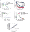
Figure 1. FRET Reveals Properties of SCF Assembly
(A) Fluorescence emission spectra from excitation at 430 nm of 70 nM CFPCul1–Rbx1, 70 nM Fbxw7TAMRA–Skp1, a mixture of the two, or buffer alone revealed FRET with 30% efficiency upon complex formation. Signals were normalized to peak donor emission at 478 nm. (B) The change in donor fluorescence versus time in a stopped flow apparatus with 5 nM CFPCul1–Rbx1 and varying concentrations of Fbxw7TAMRA–Skp1. Signal changes were fit to single exponential curves. (C) The rate of signal change in (B) versus the concentration of Fbxw7TAMRA–Skp1. Fitting the data to (kobs = kon*[Fbxw7] + koff) gave kon of 4 × 10⁶ M−1 s−1 regardless of Cul1’s neddylation status. Error bars: +/− SD, n≥3. (D) 700 nM Skp2–Skp1 (chase) competed FRET away if pre-incubated with 70 nM Fbxw7TAMRA–Skp1 before, but not after addition of 70 nM CFPCul1 for 5 min. (E) Fluorescence emission at 478 nm versus time after addition of chase to pre-incubated CFPCul1–Rbx1 and Fbxw7TAMRA–Skp1 normalized to peak donor emission in (D). Single exponential fit with a fixed end point of 1 gave koff of 8.5 × 10⁻⁷ s−1. KD is thus 2 × 10⁻¹³ M. Error bars: +/− SD, n=3. See also Figure S1.
Figure 2
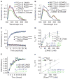
Figure 2. Cand1 Actively Removes Fbxw7–Skp1 From Cul1 by Altering Off Rate
(A) As in Figure 1A and Figure 1D except with the addition of 100 nM Cand1. (B) As in (A), except using neddylated CFPCul1. (C) The change in donor fluorescence versus time in a stopped flow apparatus upon addition of 150 nM Cand1 to 50 nM CFPCul1–Rbx1 pre-incubated with 50 nM Fbxw7TAMRA–Skp1. (D) The single exponential observed rates of SCF disassembly for various Cand1 concentrations mixed with 5 nM CFPCul1–Rbx1 or 5nM neddylated CFPCul1–Rbx1 pre-incubated with 5 nM Fbxw7TAMRA–Skp1. Chase indicates 700 nM Skp2–Skp1. Error bars: +/− SD, n≥3. (E) As in Figure 1E except with 150 nM or 300 nM Cand1 and 700 nM Skp2–Skp1 chase mixed with 70 nM neddylated CFPCul1 pre-incubated with 70 nM Fbxw7TAMRA–Skp1. Error bars: range of values, n=2. (F) As in Figure 1C, except with 150 nM Cand1 pre-incubated with 5 nM CFPCul1–Rbx1. Error bars: +/− SD, n≥3. See also Figure S2.
Figure 3
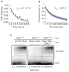
Figure 3. F-box proteins Rapidly Remove Cand1 from Cul1
(A) GST-Rbx1–Cul1–Cand1TAMRA (100 nM) was supplemented with 1 μM Cand1. At indicated times, aliquots were removed and incubated with glutathione resin for 15 min. Resin-associated proteins were fractionated by SDS-PAGE and detected by fluorography. (B) The ratio of released Cand1TAMRA to total Cand1 over time was fit to a single exponential giving koff of 1.2 × 10⁻⁵ s−1. Error bars: +/− SD, n=3. (C) GST-Rbx1–Cul1–Cand1TAMRA (100 nM) pre-incubated with glutathione resin was supplemented with buffer or 1 μM of indicated proteins. Bound and released proteins were collected at indicated times and distribution of Cand1TAMRA was evaluated as in (A). (D) Summary of the rates measured here. Transient complexes are in brackets. See also Figure S3.
Figure 4
Figure 4. Cand1 Functions as an F-box protein Exchange Factor
(A) Fluorescence emission at 478 nm versus time after addition of 210 nM Fbxw7TAMRA–Skp1 to 70 nM CFPCul1 pre-incubated with 70 nM β-TRCP–Skp1. A single exponential fit gave koff of 5 × 10⁻⁵ s−1. Error bars: range of values, n=2. (B) The change in donor fluorescence versus time in a stopped flow apparatus upon addition of 150 nM Cand1 to 70 nM CFPCul1–Rbx1 pre-incubated first with 70 nM β-TRCP–Skp1 and second with 210 nM Fbxw7TAMRA–Skp1. (C) Cul1–Rbx1 (150 nM) was pre-incubated with 500 nM Fbxw7–Skp1 (lanes 1–6) or 660 nM β-TRCP–Skp1 (lanes 7–18) for 5 min, followed by addition of 600 nM radiolabeled cycE peptide substrate and either 660 nM β-TRCP–Skp1 (lanes 1–6) or 500 nM Fbxw7–Skp1 (lanes 7–18). Either buffer (lanes 1–12) or 200 nM Cand1 (lanes 13–18) were then added, and reactions were incubated an additional 5 min prior to initiation of an ubiquitylation assay (all lanes) by supplementation of all lanes with 60 μM Ubiquitin, 1μM Ubiquitin E1, and 10 μM Cdc34b. See also Figure S4.
Figure 5
Figure 5. Cellular Cand1 Shapes Steady State and Dynamic SCF Landscape
(A) Tet-FLAGCul controlkd and Tet-FLAGCul1 Cand1kd cells (kd refers to knockdown) grown in medium with isotopically light or heavy lysine plus arginine, respectively, were induced with 1 μg/mL tetracycline for 1 hour and lysed in 1 μM MLN4924 and 2 mM o-phenanthroline 24 hours later. Two experiments were performed according to this protocol. In the first experiment, we used ‘pseudoMRM’ mass spectrometry to measure the relative amounts of 14 observable F-box proteins in total cell lysate from Cand1-depleted and control cells (white bars). In the second experiment, we retrieved FLAGCul1 and measured the relative amounts of 34 F-box proteins in the immunoprecipitates (black bars). All isotopic ratios were normalized to FLAGCul1’s (0.94), which was set to 1.0. For both experiments, results represent the ratio Cand1kd:controlkd of each protein in anti-FLAG IP measured by mass spectrometry. Each protein had ≥ 2 peptides. Error bars represent standard errors of overall protein group ratios, calculated from bootstrap analysis of two biological replicates (the second replicate was performed as a label-swap). Abundance changes in Cul1 IP for all the proteins listed in Fig5a except for Fbxo8, Fbxw9, and Fbxw4 achieved p-values < 0.05. Fbxo44a and b correspond to IPI00647771 and IPI00414844, respectively. Statistical analysis is provided in Table S1. (B) Immunoblot validation with indicated antibodies of results in (A). (C) The same cells used in (A) were transfected with a plasmid that encodes FLAGCry1. Forty-eight hours later, a chase was initiated by addition of 40 μg/mL cycloheximide. Cells were harvested at the indicated times and their content of FLAGCry1 and GAPDH was evaluated by SDS-PAGE and immunoblotting (left panel), and quantified (right panel). (D) The same cells used in (A) and grown in isotopically light lysine plus arginine were induced with 1 μg/mL tetracycline for 1 hour at t=0 hours, treated with 5 μM epoxomicin at t=48 hours, shifted to isotopically heavy lysine plus arginine at t=49 hours, and lysed at t=61 hours in 1 μM MLN4924 and 2 mM o-phenanthroline. Two experiments were performed according to this protocol. In the first experiment shown here, we used data-dependent mass spectrometry to discover and measure the fraction of F-box proteins in FLAGCul1 IPs that was heavy (i.e. made in the 12 hours prior to lysis). In the second experiment (Figure S5B), we used pseudoMRM to target 9 F-box proteins (italicized) and measure the fraction of heavy-labeled species in total cell lysate from Cand1-depleted and control cells. Each protein had ≥ 2 peptides. Error bars represent standard errors of overall protein group ratios, calculated from bootstrap analysis of two biological replicates. The F-box proteins shown to the left of the dotted line are those for which the different association observed in control and Cand1kd cells achieved a p-value <0.05. See also Figure S5 and Table S2.
Figure 6
Figure 6. Hypothesis for Control of CRL Assembly by Substrate, Cand1, and Nedd8
Rapid exchange of multiple CRL adaptor-bound substrate receptors occurs in the Cand1 exchange regime through the formation and decay of transient ternary complexes shown in brackets. Cand1 and adaptor are drawn as deformed in these complexes, to emphasize the proposal that they clash sterically, yielding an unstable state. In the presence of substrates, CRLs that pass through an intermediate state become neddylated and enter a stable state where ubiquitylation of substrates occurs. Loss of substrates facilitates recruitment of CSN, removal of Nedd8, and a return to the exchange regime effected by Cand1.

Comment in

References

    1. Bandau S, Knebel A, Gage ZO, Wood NT, Alexandru G. UBXN7 docks on neddylated cullin complexes using its UIM motif and causes HIF1α accumulation. BMC Biol. 2012;10:36. - PMC - PubMed
    1. Bennett EJ, Rush J, Gygi SP, Harper JW. Dynamics of Cullin-RING Ubiquitin Ligase Network Revealed by Systematic Quantitative Proteomics. Cell. 2010;143:951–965. - PMC - PubMed
    1. Bergqvist S, Alverdi V, Mengel B, Hoffmann A, Ghosh G, Komives EA. Kinetic enhancement of NF-kappaBxDNA dissociation by IkappaBalpha. Proceedings of the National Academy of Sciences. 2009;106:19328–19333. - PMC - PubMed
    1. Bornstein G, Ganoth D, Hershko A. Regulation of neddylation and deneddylation of cullin1 in SCFSkp2 ubiquitin ligase by F-box protein and substrate. Proc Natl Acad Sci USa. 2006;103:11515–11520. - VSports最新版本 - PMC - PubMed
    1. Bosu DR, Feng H, Min K, Kim Y, Wallenfang MR, Kipreos ET. C. elegans CAND-1 regulates cullin neddylation, cell proliferation and morphogenesis in specific tissues. Developmental Biology. 2010;346:113–126. - "V体育ios版" PMC - PubMed

"VSports在线直播" Publication types

Substances (V体育平台登录)