"VSports手机版" Skip to main page content
U.S. flag

An official website of the United States government

Dot gov

The . gov means it’s official. Federal government websites often end in . gov or . mil. Before sharing sensitive information, make sure you’re on a federal government site VSports app下载. .

Https

The site is secure. The https:// ensures that you are connecting to the official website and that any information you provide is encrypted and transmitted securely V体育官网. .

. 2013;8(1):e55612.
doi: 10.1371/journal.pone.0055612. Epub 2013 Jan 31.

VSports在线直播 - Cigarette smoke-induced collagen destruction; key to chronic neutrophilic airway inflammation?

Affiliations

Cigarette smoke-induced collagen destruction; key to chronic neutrophilic airway inflammation?

Saskia A Overbeek et al. PLoS One. 2013.

Erratum in

  • PLoS One. 2013;8(10). doi:10.1371/annotation/0faf4c12-4fd4-474c-b5a4-16e49370aff3

"VSports app下载" Abstract

Background: Cigarette smoking induces inflammatory responses in all smokers and is the major risk factor for lung disease such as chronic obstructive pulmonary disease (COPD). In this progressive disease, chronic inflammation in the lung contributes to lung tissue destruction leading to the formation of chemotactic collagen fragments such as N-acetylated Proline-Glycine-Proline (N-ac-PGP). The generation of this tripeptide is mediated by a multistep pathway involving matrix metalloproteases (MMPs) 8 and 9 and prolyl endopeptidase (PE). Here we investigated whether cigarette smoke extract (CSE) stimulates human PMNs to breakdown whole matrix collagen leading to the generation of the chemotactic collagen fragment N-ac-PGP. VSports手机版.

Methodology/principal findings: Incubating PMNs with CSE led to the release of chemo-attractant CXCL8 and proteases MMP8 and MMP9. PMNs constitutively expressed PE activity as well as PE protein. Incubating CSE-primed PMNs with collagen resulted in collagen breakdown and in N-ac-PGP generation. Incubation of PMNs with the tripeptide N-ac-PGP resulted in the release of CXCL8, MMP8 and MMP9 V体育安卓版. Moreover, we tested whether PMNs from COPD patients are different from PMNs from healthy donors. Here we show that the intracellular basal PE activity of PMNs from COPD patients increased 25-fold compared to PMNs from healthy donors. Immunohistological staining of human lung tissue for PE showed that besides neutrophils, macrophages and epithelial cells express PE. .

Conclusions: This study indicates that neutrophils activated by cigarette smoke extract can breakdown collagen into N-ac-PGP and that this collagen fragment itself can activate neutrophils, which may lead in vivo to a self-propagating cycle of neutrophil infiltration, chronic inflammation and lung emphysema. MMP-, PE- or PGP-inhibitors can serve as an attractive therapeutic target and may open new avenues towards effective treatment of COPD. V体育ios版.


Conflict of interest statement (VSports最新版本)

Competing Interests: JG is employed by a commercial company, Danone Research. However, this does not alter the authors’ adherence to all the PLOS ONE policies on sharing data and materials VSports最新版本.

Figures

Figure 1
Figure 1. Neutrophil viability after CSE-stimulation for 9 or 16 hours.
Significant cell viability loss was observed after 9 and 16 hours incubating neutrophils with CSE OD 0.24 (* p<0.05, Friedman, Dunns, n = 3).
Figure 2
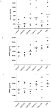
Figure 2. CSE induces the release of CXCL8, MMP8 and MMP9 from human PMNs.
(A) 105 freshly isolated buffy coat PMNs were stimulated for 9 hours with cigarette smoke extract (CSE; OD 0.03–0.24) or LPS (100 ng/ml; positive control). CXCL8, MMP8 and MMP9 ELISA's were performed on the supernatants. CSE induced the release of CXCL8 from fresh cells (** p<0.01 repeated measures ANOVA+Tukey CSE vs. control; ∧ p<0.01 paired t-test LPS vs. control). (B) CSE induced the release of MMP8 from fresh cells (* p<0.05 repeated measures ANOVA+Tukey CSE vs. control; ∧∧ p<0.01 t-test LPS vs. control). (C) CSE induced the release of MMP9 from fresh cells (* p<0.05 paired t-test CSE OD 0.12 vs. control; ∧∧ p<0.01 paired t-test LPS vs. control). Legend: each symbol represents a different donor (n = 5). Individual data are shown, horizontal bars represent mean values. The data presented here all passed the normality test.
Figure 3
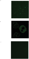
Figure 3. Human PMNs contain prolyl endopeptidase (PE).
Representative photomicrographs (n = 3) of an immunofluorescent staining for PE (green color) in PMNs of healthy volunteers (A and B (magnified from figure A)). Panel C displays unstained cells (negative control). PE is located in the cytoplasm in a granular pattern. Magnification 200×.
Figure 4
Figure 4. Human PMN incubation with CSE does not affect the level and activity of PE in cell lysates.
(A) 106 isolated PMNs were stimulated for 9 hours with CSE (OD 0.06 or 0.12). PE and GAPDH Western blots were performed on the cell lysates. PE in human neutrophils was a monomer and migrated at 75 kDa, which was similar to rhPE (not depicted). Incubation of PMNs with CSE did not change the optical density of the bands when compared to the control. (n = 2) (B) Freshly isolated PMNs (106 cells) were stimulated for 16 hours with indicated reagents. PE activity was measured in lysates using Z-Gly-Pro-AMC as a substrate. Control was standardized to 1. Intracellular PE activity does not change after CSE exposure when compared to the control. (n = 5).
Figure 5
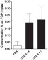
Figure 5. Human PMNs can generate N-ac-PGP de novo from collagen type I.
106 PMNs were incubated for 16 hours at 37°C with collagen type I dialyzed solution (1 mg/ml) and CSE (OD 0.06 or 0.12). Bestatin (50 µg/ml) was added every 3 hours. At time point 16 hours, the N-ac-PGP levels were determined in supernatants from the incubated PMNs. All samples were filtered through a 10-kDa filter, washed with 20 µl of 1 N HCl and analyzed using ESI- LC-MS/MS for levels of N-ac-PGP. Detection limit N-ac-PGP: <0.025 ng/ml. (n = 2).
Figure 6
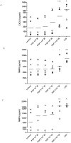
Figure 6. N-ac-PGP induces the release of CXCL8, MMP8 and MMP9 from human PMNs.
105 freshly isolated buffy coat PMNs were stimulated for 9 hours with N-ac-PGP (10−4–3·10−3 M) or LPS (100 ng/ml). CXCL8, MMP8 and MMP9 ELISA's were performed on the supernatants. (A) N-ac-PGP induced the release of CXCL8 from fresh cells (*** p<0.001 repeated measures ANOVA+Tukey N-ac-PGP vs. control; ∧ p<0.05 paired t-test LPS vs. control). (B, C) N-ac-PGP induced the release of MMP8 from fresh cells (* p<0.05 paired t-test CSE OD 0.12 vs. control; ∧∧ p<0.01 paired t-test LPS vs. control). (C) N-ac-PGP induced the release of MMP9 from fresh cells (** p<0.01 repeated measures ANOVA+Tukey N-ac-PGP vs. control; ∧ p<0.01 paired t-test LPS vs. control). Legend: each symbol represents a different donor (n = 4–5). Individual data are shown, horizontal bars represent mean values. The data presented here all passed the normality test.
Figure 7
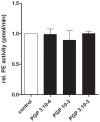
Figure 7. Human PMN incubation with N-ac-PGP does not affect the activity of released or intracellular PE.
Freshly isolated PMNs (106 cells) were stimulated for 16 hours with indicated reagents. PE activity was measured in supernatants and lysates using Z-Gly-Pro-AMC as a substrate. Control was standardized to 1. Intracellular PE activity does not change after N-ac-PGP (3·10−4–3·10−3 M) exposure when compared to the control (n = 3).
Figure 8
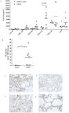
Figure 8. The basal PE activity of PMNs from COPD patients is a 25-fold higher when compared to healthy donors.
(A) 105 freshly isolated PMNs from healthy donors (block dots, n = 5) and COPD patients (white squares, n = 7) were stimulated for 9 hours with cigarette smoke extract (CSE; OD 0.03–0.24). A CXCL8 ELISA was performed on the supernatants. PMNs from COPD patients tend to produce higher amounts of CXCL8 after CSE incubation (p = 0.0560, t-test CSE OD 0.12 donor vs. COPD). Individual data are shown, horizontal bars represent mean values. (B) The PE activity was measured in lysates of unstimulated PMNs (106 cells) using Z-Gly-Pro-AMC as a substrate. The basal PE activation of PMNs from COPD patients (white squares, n = 7) is significantly higher than the PE activity of PMNs from healthy donors (block dots, n = 3) (* p<0.05 Mann-Whitney). (C–F) Localization of PE in the human lung. Representative photomicrographs of an immunohistological staining for PE (brown color, DAB staining) in lung tissue of (C) a current smoker, (D) ex-smoker, (E) COPD patient with GOLD stage II and (F) a COPD patient with GOLD stage IV. Magnification, ×200.

References (VSports在线直播)

    1. Barnes PJ (2004) Mediators of chronic obstructive pulmonary disease. Pharmacol Rev 56: 515–548. - PubMed
    1. Stockley RA, Mannino D, Barnes PJ (2009) Burden and pathogenesis of chronic obstructive pulmonary disease. Proc Am Thorac Soc 6: 524–526. - PubMed
    1. Barnes PJ (2010) New therapies for chronic obstructive pulmonary disease. Med Princ Pract 19: 330–338. - PubMed
    1. Quint JK, Wedzicha JA (2007) The neutrophil in chronic obstructive pulmonary disease. J Allergy Clin Immunol 119: 1065–1071. - PubMed
    1. Mannino DM, Buist AS (2007) Global burden of COPD: risk factors, prevalence, and future trends. Lancet 370: 765–773. - PubMed

Publication types

MeSH terms

LinkOut - more resources