Skip to main page content
U.S. flag

An official website of the United States government

Dot gov

The . gov means it’s official VSports app下载. Federal government websites often end in . gov or . mil. Before sharing sensitive information, make sure you’re on a federal government site. .

Https

The site is secure. The https:// ensures that you are connecting to the official website and that any information you provide is encrypted and transmitted securely V体育官网. .

Clinical Trial
. 2013 Jan 29;6(260):ra7.
doi: 10.1126/scisignal.2003057.

"V体育安卓版" Vemurafenib potently induces endoplasmic reticulum stress-mediated apoptosis in BRAFV600E melanoma cells

Affiliations
Clinical Trial

"V体育安卓版" Vemurafenib potently induces endoplasmic reticulum stress-mediated apoptosis in BRAFV600E melanoma cells

"V体育安卓版" Daniela Beck et al. Sci Signal. .

Abstract

The V600E mutation in the kinase BRAF is frequently detected in melanomas and results in constitutive activation of BRAF, which then promotes cell proliferation by the mitogen-activated protein kinase signaling pathway. Although the BRAFV600E kinase inhibitor vemurafenib has remarkable antitumor activity in patients with BRAFV600E-mutated melanoma, its effects are limited by the onset of drug resistance. We found that exposure of melanoma cell lines with the BRAFV600E mutation to vemurafenib decreased the abundance of antiapoptotic proteins and induced intrinsic mitochondrial apoptosis. Vemurafenib-treated melanoma cells showed increased cytosolic concentration of calcium, a potential trigger for endoplasmic reticulum (ER) stress, which can lead to apoptosis. Consistent with an ER stress-induced response, vemurafenib decreased the abundance of the ER chaperone protein glucose-regulated protein 78, increased the abundance of the spliced isoform of the transcription factor X-box binding protein 1 (XBP1) (which transcriptionally activates genes involved in ER stress responses), increased the phosphorylation of the translation initiation factor eIF2α (which would be expected to inhibit protein synthesis), and induced the expression of ER stress-related genes. Knockdown of the ER stress response protein activating transcription factor 4 (ATF4) significantly reduced vemurafenib-induced apoptosis. Moreover, the ER stress inducer thapsigargin prevented invasive growth of tumors formed from vemurafenib-sensitive melanoma cells in vivo VSports手机版. In melanoma cells with low sensitivity or resistance to vemurafenib, combination treatment with thapsigargin augmented or induced apoptosis. Thus, thapsigargin or other inducers of ER stress may be useful in combination therapies to overcome vemurafenib resistance. .


Figures (V体育2025版)
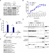
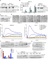
Figure 1
Figure 1. Vemurafenib induces intrinsic mitochondrial apoptosis in BRAF-mutant melanoma cells
(A) Western blot analysis for phosphorylated (p)-ERK, ERK, p-AKT, and AKT was performed on melanoma cells with BRAF (SKMEL19, 451LU) or NRAS mutation (MV3) that were treated with vemurafenib (Vem) or DMSO (control) for 24 hours (h). (B) Growth assessment using 4-methylumbelliferyl heptanoate of melanoma cells treated with vemurafenib for 72 h. One representative experiment of four is shown (mean ± SD). (C) Apoptosis (as assessed by the subG1 fraction) was quantified in melanoma cells treated with vemurafenib or DMSO for 72 h. Bar graphs show the quantification of results from 4 independent experiments (mean ± SD, Mann-Whitney test, 95% CI; * P < 0.05; ns=not significant). (D) Western blot analysis for caspase-9, caspase-3, cleaved caspase-3, and PARP was performed on melanoma cells treated with vemurafenib or DMSO for the indicated time periods or with staurosporine (Stauro, 1 μM) for 15 h. Black arrow: uncleaved protein. White arrow: cleaved protein. (E) Western blot analysis for Bcl-2 and Mcl-1 was performed on melanoma cells treated with vemurafenib or DMSO for 72 h. Western blots in (A, D, E) show a representative experiment (n=3); β-actin or α-tubulin served as the loading control.
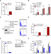
Figure 2
Figure 2. Vemurafenib triggers ER stress
(A) Western blot analysis to detect BIM isoforms was performed on BRAF-mutated (SKMEL19, 451LU) melanoma cells treated with vemurafenib (+) or DMSO (-). (B) Western blot analysis for BIM isoforms was performed on BRAF-mutated melanoma cells (SKMEL19) transfected with control siRNA or siRNA directed against BIM prior to treatment with vemurafenib or DMSO. Apoptosis (subG1 fraction) was quantified by propidium iodide staining. Bar graphs show the quantification of results from 3 independent experiments (mean ± SD, Mann-Whitney test, 95% CI; * P < 0.05). (C) Western blot analysis for PUMA and Noxa was performed on BRAF-mutated (SKMEL19, 451LU) melanoma cells treated with vemurafenib or DMSO for 24 h. (D) EIectron microscopy of melanoma cells treated with vemurafenib, DMSO, or thapsigargin (TG) for 6 h. DMSO-treated cells show a normal-sized endoplasmic reticulum (arrows). Vemurafeniband thapsigargin-treated cells show dilation of the endoplasmic reticulum (arrows). Scale bar = 0.5 μm, n=2 experiments. (E) Melanoma cells loaded with Fluo3-AM were exposed to the indicated treatments and monitored by flow cytometry. Changes in fluorescence intensity over time reflect changes in cytosolic Ca2+ concentrations. As a positive control, melanoma cells were treated with ionomycin, a selective Ca2+ ionophore. One representative experiment is shown (n=3). (F, G) Western blot analysis for p-eIF2α, eIF2α, and spliced XBP1 (XBP1s) was performed at the indicated time points on BRAF-mutated (SKMEL19, 451LU) melanoma cells treated with vemurafenib or DMSO (D). (H) Real-time PCR to detect the expression of p8, ATF4, ATF3, CHOP, and TRB3 relative to the DMSO control was performed in melanoma cells treated with vemurafenib or DMSO for 12 h. Mean ± SD; n=3 experiments. (I) Western blot analysis for GRP78 was performed on melanoma cells treated with vemurafenib or DMSO for the indicated times. (A, B, C, F, G, I) Western blots show one representative experiment (n=3); β-actin served as the loading control.
Figure 3
Figure 3. ER stress plays a role in vemurafenib-induced apoptosis
(A, B) BRAF-mutated melanoma cells (SKMEL19) transfected with control or p8-targeted siRNA were treated with vemurafenib (6 μM) or DMSO. After 15 h, real-time PCR analysis (A) was performed to measure p8 mRNA expression in vemurafenib-treated cells relative to DMSO-treated (control) cells. Abundance of p8 protein were detected by Western blot analysis 18 h after treatment (one representative experiment of three is shown). 24, 48, and 72 h after treatment, apoptosis (subG1 fraction) was quantified (B). Bar graphs show the quantification of results from 4 independent experiments (mean ± SD). (C, D) Melanoma cells (SKMEL19) were transfected with Ctrl-GFP-plasmid (pEGFP-N1, 27kDa) or p8-GFP-plasmid (pEGFP-C2-p8, 37kDa). After 24 h, Western blot analysis for GFP (C) was performed (one representative experiment of three is shown). Apoptosis (subG1 fraction) was quantified in GFP-positive cells treated with vemurafenib (6 μM) or DMSO for 48 h (D). Bar graphs show the quantification of results from 4 independent experiments (mean ± SD). (E, F) Real-time PCR to detect ATF4 mRNA expression was performed in melanoma cells (SKMEL19) transfected with control or ATF4 siRNA and treated with vemurafenib (3 μM) or DMSO (E). Western blot analysis for ATF4 was performed 48 h after treatment (one representative experiment of three is shown). 24, 48, and 72 h after treatment, apoptosis (as assessed by the subG1 fraction) was quantified (F). Bar graphs show the quantification of results from 4 independent experiments (mean ± SD). P values for (B, D, F) were determined by Mann-Whitney test, 95% CI; * P < 0.05; ns=not significant.
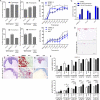
Figure 4
Figure 4. Inducers of ER stress trigger apoptosis and inhibit invasive tumor growth of melanoma cells
(A) Real-time PCR to detect p8 and CHOP mRNA expression was performed in BRAF-mutated (SKMEL19, 451LU) and NRAS-mutated (MV3) melanoma cells treated with DMSO (control), 60 nM thapsigargin, or 595 nM tunicamycin for 12 h. One representative experiment of three is shown (means ± SD). (B) Growth assessment using 4-methylumbelliferyl heptanoate of melanoma cells treated with the indicated concentrations of thapsigargin or tunicamycin for 72 h. One representative experiment of three is shown (mean ± SD). (C) Apoptosis (subG1 fraction) was quantified in melanoma cells treated with thapsigargin or tunicamycin for 72 h. Bar graphs show the quantification of results from 3 independent experiments (mean ± SD). (D) NRAS-mutated melanoma cells (MV3) seeded onto dermal reconstructs were treated with DMSO (control) or thapsigargin. Red, Ki67 (n=3 experiments). Scale bar = 100 μm. (E to J) Melanoma cells were injected into the rhombencephalic brain vesicle of stage 13/14 HH chick embryos. After 48 and 72 h, embryos were treated with DMSO (control, top row) or thapsigargin (TG, bottom row). HE: hematoxylin and eosin stain at 5× magnification (E and H). HMB45: immunohistochemistry with the melanoma-specific marker HMB45, at 40× magnification(F and I). MIB1: immunohistochemistry for the proliferation marker Ki67 with the MIB1 antibody at 5× magnification (G and J). n=2 experiments, 8 embryos per treatment. (K) Growth assessment of melanoma cells treated with the indicated concentrations of vemurafenib, thapsigargin, or both for 72 h. Bar graphs show the quantification of results from 3 independent experiments (mean ± SD, Mann-Whitney test, 95% CI; * P < 0.05, ** P < 0.001, *** P < 0.0001).
Figure 5
Figure 5. ER stress inducers overcome acquired resistance to vemurafenib
(A) Growth assessment of SKMEL19-Vem-S and SKMEL19-Vem-R melanoma cells treated with the indicated concentrations of vemurafenib for 72 h. One representative experiment of 4 is shown (mean ± SD). (B) Apoptosis (subG1 fraction) was quantified in SKMEL19-Vem-S and SKMEL19-Vem-R cells treated with vemurafenib or DMSO for 72 h. Bar graphs show the quantification of results from 3 independent experiments (mean ± SD). (C, D, E, F) Western blot analysis for p-ERK, ERK, p-AKT, AKT, GRP78, p-eIF2α, eIF2α, and BIM isoforms was performed at the indicated time points in SKMEL19-Vem-S or SKMEL19-Vem-R cells treated with vemurafenib (6 μM) or DMSO, β-tubulin or β-actin served as loading control. One representative experiment of three is shown. (C) The ratio of the amount of p-AKT to that of AKT for each lane from three experiments is indicated in the accompanying bar graph (mean ± SD, Mann-Whitney test, 95% CI; * P < 0.05). (G) Growth assessment of SKMEL19-Vem-S and SKMEL19-Vem-R cells treated with the indicated concentrations of thapsigargin for 72 h. One representative experiment of 3 is shown (mean ± SD). (H) Apoptosis (subG1 fraction) was quantified in SKMEL19-Vem-R cells treated with vemurafenib, DMSO, or thapsigargin for 72 h. Bar graphs show the quantification of results from 3 independent experiments (mean ± SD). (I) Western blot analysis for p-ERK, ERK, p-AKT, AKT, and β-actin was performed in SKMEL19-Vem-S and SKMEL19-Vem-R cells treated with thapsigargin (TG) or DMSO for 24 h. One representative experiment of 3 is shown.
Figure 6
Figure 6. Proposed mechanism of vemurafenib action
Vemurafenib induces ER stress by increasing the concentration of cytosolic Ca2+. eIF2α is phosphorylated, thereby globally inhibiting translation. The expression of ER stress-related genes and the abundance of the pro-apoptotic protein Bim are increased. Furthermore, vemurafenib inhibits phosphorylation of ERK, which enhances downstream pro-apoptotic signaling. Dashed lines represent indirect effects.

References

    1. Balch CM, Gershenwald JE, Soong SJ, Thompson JF, Atkins MB, Byrd DR, Buzaid AC, Cochran AJ, Coit DG, Ding S, Eggermont AM, Flaherty KT, Gimotty PA, Kirkwood JM, McMasters KM, Mihm MC, Jr., Morton DL, Ross MI, Sober AJ, Sondak VK. Final version of 2009 AJCC melanoma staging and classification. J Clin Oncol. 2009;27:6199. published online EpubDec 20. - PMC - PubMed
    1. Davies H, Bignell GR, Cox C, Stephens P, Edkins S, Clegg S, Teague J, Woffendin H, Garnett MJ, Bottomley W, Davis N, Dicks E, Ewing R, Floyd Y, Gray K, Hall S, Hawes R, Hughes J, Kosmidou V, Menzies A, Mould C, Parker A, Stevens C, Watt S, Hooper S, Wilson R, Jayatilake H, Gusterson BA, Cooper C, Shipley J, Hargrave D, Pritchard-Jones K, Maitland N, Chenevix-Trench G, Riggins GJ, Bigner DD, Palmieri G, Cossu A, Flanagan A, Nicholson A, Ho JW, Leung SY, Yuen ST, Weber BL, Seigler HF, Darrow TL, Paterson H, Marais R, Marshall CJ, Wooster R, Stratton MR, Futreal PA. Mutations of the BRAF gene in human cancer. Nature. 2002;417:949. published online EpubJun 27. - PubMed
    1. Satyamoorthy K, Li G, Gerrero MR, Brose MS, Volpe P, Weber BL, Van Belle P, Elder DE, Herlyn M. Constitutive mitogen-activated protein kinase activation in melanoma is mediated by both BRAF mutations and autocrine growth factor stimulation. Cancer research. 2003;63:756. published online EpubFeb 15. - PubMed
    1. Dai DL, Martinka M, Li G. Prognostic significance of activated Akt expression in melanoma: a clinicopathologic study of 292 cases. J Clin Oncol. 2005;23:1473. published online EpubMar 1. - PubMed
    1. Meier F, Schittek B, Busch S, Garbe C, Smalley K, Satyamoorthy K, Li G, Herlyn M. The RAS/RAF/MEK/ERK and PI3K/AKT signaling pathways present molecular targets for the effective treatment of advanced melanoma. Front Biosci. 2005;10:2986. - PubMed

Publication types

MeSH terms (V体育安卓版)