"VSports" Skip to main page content
U.S. flag

An official website of the United States government

Dot gov

The . gov means it’s official VSports app下载. Federal government websites often end in . gov or . mil. Before sharing sensitive information, make sure you’re on a federal government site. .

Https

The site is secure. The https:// ensures that you are connecting to the official website and that any information you provide is encrypted and transmitted securely. V体育官网.

. 2013 Jan;15(1):1-10.
doi: 10.1593/neo.121550.

Concurrent AURKA and MYCN gene amplifications are harbingers of lethal treatment-related neuroendocrine prostate cancer (V体育ios版)

Affiliations

Concurrent AURKA and MYCN gene amplifications are harbingers of lethal treatment-related neuroendocrine prostate cancer

Juan Miguel Mosquera et al. Neoplasia. 2013 Jan.

Abstract

Neuroendocrine prostate cancer (NEPC), also referred to as anaplastic prostate cancer, is a lethal tumor that most commonly arises in late stages of prostate adenocarcinoma (PCA) with predilection to metastasize to visceral organs. In the current study, we explore for evidence that Aurora kinase A (AURKA) and N-myc (MYCN) gene abnormalities are harbingers of treatment-related NEPC (t-NEPC). We studied primary prostate tissue from 15 hormone naïve PCAs, 51 castration-resistant prostate cancers, and 15 metastatic tumors from 72 patients at different stages of disease progression to t-NEPC, some with multiple specimens. Histologic evaluation, immunohistochemistry, and fluorescence in situ hybridization were performed and correlated with clinical variables. AURKA amplification was identified in overall 65% of PCAs (hormone naïve and treated) from patients that developed t-NEPC and in 86% of metastases. Concurrent amplification of MYCN was present in 70% of primary PCAs, 69% of treated PCAs, and 83% of metastases. In contrast, in an unselected PCA cohort, AURKA and MYCN amplifications were identified in only 5% of 169 cases. When metastatic t-NEPC was compared to primary PCA from the same patients, there was 100% concordance of ERG rearrangement, 100% concordance of AURKA amplification, and 60% concordance of MYCN amplification. In tumors with mixed features, there was also 100% concordance of ERG rearrangement and 94% concordance of AURKA and MYCN co-amplification between areas of NEPC and adenocarcinoma. AURKA and MYCN amplifications may be prognostic and predictive biomarkers, as they are harbingers of tumors at risk of progressing to t-NEPC after hormonal therapy. VSports手机版.


Figures
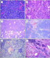
Figure 1
Figure 1
Morphologic spectrum of t-NEPC. (A) Small cell carcinoma of the prostate. The tumor is composed of sheets of uniform cells with scant cytoplasm, hyperchromatic nuclei, coarse chromatin, and unapparent nucleoli. (B) Large cell neuroendocrine carcinoma of the prostate. Tumor is composed of sheets and ribbons of cells with abundant cytoplasm, large nuclei with coarse chromatin, brisk mitotic activity, and foci of necrosis; pseudorosettes are also apparent. (C) Metastatic poorly differentiated adenocarcinoma of the prostate without neuroendocrine differentiation, treated (metastatic CRPC). Sheets of tumor cells with pale eosinophilic cytoplasm and abundant mitotic figures are seen within fibroadipose tissue. (D) Poorly differentiated adenocarcinoma of the prostate with neuroendocrine differentiation, treated (CRPC). Note the vaguely organoid pattern of tumor cells, which have amphophilic cytoplasm and prominent nucleoli. (E) Poorly differentiated adenocarcinoma of the prostate with focal areas of neuroendocrine differentiation, treated (CRPC). Areas of tumor cells with neuroendocrine differentiation are interspersed and demonstrate basophilic appearance. (F) Mixed t-NEPC and adenocarcinoma of prostate, treated (CRPC). Areas of small cell carcinoma and poorly differentiated adenocarcinoma are seen (H&E stain, original magnification, x200).
Figure 2
Figure 2
Prostate cancer with Paneth cell-like neuroendocrine differentiation harbors AURKA amplification. (A–F) Six cases of localized prostate cancer with Paneth cell-like change were identified and used as separate controls of low-grade neuroendocrine differentiation. On H&E stain, tumor cells with Paneth cell-like neuroendocrine differentiation are easily identified and contain distinct large eosinophilic granules in the cytoplasm. One case (A) demonstrated AURKA and MYCN amplifications. The other five cases (B–F) harbored AURKA amplification only (insets). ERG rearrangement, one through insertion (D) and one through deletion (A), is identified in two of these cases (insets). Clusters of tumor cells with Paneth cell-like neuroendocrine differentiation are located around asterisks, and more focal areas are marked with arrowheads (H&E stain, original magnification, x400; FISH images, original magnification, x600).
Figure 3
Figure 3
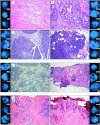
AURKA and MYCN amplifications in primary prostatic adenocarcinoma predict the development of t-NEPC. (A–D) Top panel illustrates several specimens from a patient at different stages of disease progression to t-NEPC. (A and B) Images of hormone naïve prostate cancer with areas of Gleason score 3 + 3 = 6 (A) and 4 + 5 = 9 (B) at initial diagnosis. Concurrent AURKA (upper inset) and MYCN (middle inset) amplifications are present in both areas. (C) Subsequent metastasis/local recurrence in the bladder demonstrates poorly differentiated adenocarcinoma without neuroendocrine differentiation, exhibiting both AURKA and MYCN amplifications (upper and middle insets, respectively). (D) Five years after treatment, the patient presents with metastatic large cell neuroendocrine carcinoma in pelvic soft tissue. The tumor has organoid appearance focally forming pseudorosettes, and cells have abundant cytoplasm and prominent nucleoli. The tumor has both AURKA and MYCN amplifications (upper and middle insets, respectively). Clonal origin is confirmed by ERG rearrangement through translocation in all tumors (lower inset). (E and F) Center panel illustrates prostatectomy specimen from a patient with initial diagnosis of PCA Gleason score 4 + 5 = 9 (E), which has concurrent AURKA and MYCN amplifications (upper and middle insets, respectively). A liver biopsy 7 years after (F) shows metastatic small cell carcinoma, which harbors AURKA and MYCN coamplification as well. Clonal origin is confirmed by ERG rearrangement through deletion in both tumors (lower inset). (G and H) Lower panel illustrates needle biopsies from a patient with initial diagnosis of (G) PCA Gleason score 3 + 4 = 7 with intraductal spread (IDC-P) with amplification of AURKA (upper inset) but not MYCN (middle inset). Eight years after initial diagnosis and intermittent treatment, the patient developed pancytopenia and bone lytic lesions, which biopsy demonstrates (H) metastatic small cell carcinoma (frozen tissue artifact present), consistent with spread from known prostatic primary. In addition to AURKA amplification (upper inset), clonal origin is confirmed by ERG rearrangement through translocation in both tumors (lower inset). The metastatic tumor demonstrates MYCN amplification (middle inset) (H&E stain, original magnification, x200; FISH images, original magnification, x600).
Figure 4
Figure 4
Concordance of AURKA and MYCN amplifications in tumors with mixed areas of neuroendocrine carcinoma and poorly differentiated adenocarcinoma. Representative image of local recurrence of castration-resistant prostatic carcinoma with areas of mixed small cell carcinoma (right) and adenocarcinoma (left). Both areas demonstrate concordance of AURKA and MYCN amplifications (upper and middle insets, respectively). Clonal origin is supported by ERG rearrangement through translocation in both areas (lower inset) (H&E stain, original magnification, x200; FISH images, original magnification, x600).
Figure 5
Figure 5
Concurrent AURKA and MYCN gene amplifications are harbingers of lethal t-NEPC. AURKA and MYCN gene amplifications evaluated by FISH are not present in benign prostate tissue and identified only in 5% of unselected primary prostate cancers. In contrast, 67% of primary tumors from patients who clinically develop t-NEPC harbor AURKA amplification, 70% of which also demonstrate concurrent MYCN amplification. Similar frequency of AURKA/MYCN amplification is present in t-NEPC. Metastatic t-NEPC harbors AURKA amplification in 86% of cases, with 83% MYCN co-amplification.

References (VSports在线直播)

    1. Jemal A, Bray F, Center MM, Ferlay J, Ward E, Forman D. Global cancer statistics. CA Cancer J Clin. 2011;61(2):69–90. - PubMed
    1. Brawn PN, Speights VO. The dedifferentiation of metastatic prostate carcinoma. Br J Cancer. 1989;59(1):85–88. - PMC - PubMed
    1. Ismail AH, Landry F, Aprikian AG, Chevalier S. Androgen ablation promotes neuroendocrine cell differentiation in dog and human prostate. Prostate. 2002;51(2):117–125. - PubMed
    1. Ito T, Yamamoto S, Ohno Y, Namiki K, Aizawa T, Akiyama A, Tachibana M. Up-regulation of neuroendocrine differentiation in prostate cancer after androgen deprivation therapy, degree and androgen independence. Oncol Rep. 2001;8(6):1221–1224. - PubMed
    1. Shen R, Dorai T, Szaboles M, Katz AE, Olsson CA, Buttyan R. Transdifferentiation of cultured human prostate cancer cells to a neuroendocrine cell phenotype in a hormone-depleted medium. Urol Oncol. 1997;3(2):67–75. - PubMed

Publication types

VSports在线直播 - MeSH terms

LinkOut - more resources