<font draggable="NI8py"></font><var lang="1ByMKoby"><style lang="fTE85f"></style></var> Skip to main page content
U.S. flag

An official website of the United States government

Dot gov

The . gov means it’s official. Federal government websites often end in . gov or VSports app下载. mil. Before sharing sensitive information, make sure you’re on a federal government site. .

Https

The site is secure. The https:// ensures that you are connecting to the official website and that any information you provide is encrypted and transmitted securely. V体育官网.

. 2013 Mar 1;190(5):2121-8.
doi: 10.4049/jimmunol.1202145. Epub 2013 Jan 25.

VSports最新版本 - A role for Ly108 in the induction of promyelocytic zinc finger transcription factor in developing thymocytes

Affiliations

"VSports在线直播" A role for Ly108 in the induction of promyelocytic zinc finger transcription factor in developing thymocytes

Mala Dutta et al. J Immunol. .

Abstract

The promyelocytic zinc finger transcription factor (PLZF) is required for the development of activated phenotypes in NKT and other innate T lymphocytes. Although strong TCR stimulation has been implicated in the induction of PLZF, the factors regulating PLZF expression are incompletely understood. We show in this study that costimulation of preselection double-positive thymocytes through the signaling lymphocyte activation molecule family receptor Ly108 markedly enhanced PLZF expression compared with that induced by TCR stimulation alone. Costimulation with Ly108 increased expression of early growth response protein (Egr)-2 and binding of Egr-2 to the promoter of Zbtb16, which encodes PLZF, and resulted in PLZF levels similar to those seen in NKT cells. In contrast, costimulation with anti-CD28 failed to enhance Egr-2 binding and Zbtb16 expression. Moreover, mice lacking Ly108 showed decreased numbers of PLZF-expressing CD4(+) T cells. Together, these results support a potential role for Ly108 in the induction of PLZF. VSports手机版.


Figures

Figure 1
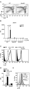
Figure 1. Ly108 costimulation enhances PLZF expression in developing PS-DP thymocytes
(A) Expression of Zbtb16 (encoding PLZF) evaluated by qRT-PCR of WT PS-DP thymocytes stimulated with media alone (NS; non-stimulated) or plate-bound αCD3 for 24 or 48h. (B) Zbtb16 expression of WT PS-DP thymocytes stimulated with media alone (NS), αCD3±αLy108 or αLy108 alone. RQ is relative quantitation. Values in A–B were normalized to NS WT samples. Similar results were obtained using freshly isolated PS-DP cells as normalization controls (Supplemental Fig. S2A). Data are the means ± SEM of 5 independent experiments, using cells from ≥3 mice each. (C) Zbtb16 expression in WT and β2m-deficient PS-DP thymocytes stimulated with plate-bound αCD3±αLy108. Data are from two independent experiments. Cells in these experiments were also negatively selected to remove TCRγδ+ cells. (D) Zbtb16 expression in WT and SAP-deficient (Sh2d1a−/−) PS-DP thymocytes in the presence or absence of Ly108Fc fusion protein were stimulated with LPS-activated B cells (which express high surface levels of Ly108) plus αCD3. Data are the means ± SEM of 3 independent experiments, using cells from ≥3 mice each.
Figure 2
Figure 2. Ly108 is required for full induction of PLZF in vitro
(A) Characterization of Ly108-deficient (Slamf6−/−) mice. Representative flow plots of thymocyte populations from WT and Slamf6−/− mice stained for CD4 and CD8. Frequencies and absolute numbers of thymocyte populations (N=6) stained for DP, DN CD4+ and CD8+ cells are depicted in Table 1. (B) Zbtb16 expression in WT, Slamf6−/− and SAP-deficient (Sh2d1a−/−) thymocytes stimulated as in Fig.1. Values were normalized to NS WT samples. Similar results were obtained using freshly isolated PS-DP cells as controls (Supplemental Fig. S2A). Data are the means ± SEM of 3 independent experiments, using cells from ≥3 mice each. (C) Representative histograms of intracellular PLZF staining in PS-DP thymocytes from WT and Slamf6−/− mice, NS (solid histogram) or stimulated with plate-bound αCD3±αLy108 for 48 h. Staining controls are shown in Supplemental Fig. S2E. (D) MFI ± SD of PLZF from 3 mice per genotype. (E) Intracellular PLZF expression in PS-DP thymocytes stimulated as in (C) compared to PLZF+CD1d-PBS57 tetramer+ iNKT cells. Histograms are representative of triplicates for each condition. Data in (E) were generated with a different antibody lot than in (C).
Figure 3
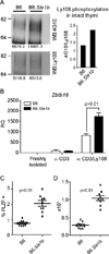
Figure 3. Ly108-deficient mice have fewer PLZF+CD4+ thymocytes
Percentages (A) and absolute numbers (B) of CD1d-PBS57 Tetramer+ and PLZF+, CD4+ thymocytes in WT and Slamf6−/− mice. (C–D) PLZF levels in CD1d-PBS57 Tetramer+ iNKT cells from WT and Slamf6−/− mice. Data are the mean ± SD of 3 mice per group.
Figure 4
Figure 4. Increased PLZF expressing cells observed in a mouse model with enhanced Ly108 signaling
(A) Enhanced phosphorylation of Ly108 in intact thymi of B6.Sle1b mice. Ly108 was immunoprecipitated from lysates of intact thymi of C57Bl/6J (B6) and B6.Sle1b mice and probed for phosphotyrosine using 4G10 antibody (top) and for total Ly108 protein (bottom). Quantitation of relative tyrosine phosphorylation is shown on the right. (B) Zbtb16 expression in C57Bl/6J (B6) and B6.Sle1b PS-DP thymocytes stimulated for 48 hrs with plate-bound αCD3±αLy108 as in Figure 1. Data are representative of three independent experiments. Percentages (C) and absolute numbers (D) of PLZF+ CD4+ thymocytes in C57Bl/6J (B6) and B6.Sle1b mice.
Figure 5
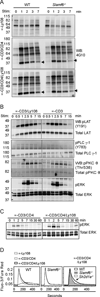
Figure 5. Ly108 costimulation prolongs TCR signaling
(A) Rested WT and Slamf6−/− thymocytes, pre-incubated with biotinylated αLy108 (top), αCD3+αCD4 (middle), and αCD3+αCD4+αLy108 (bottom), were stimulated with streptavidin for indicated times and lysates immunoblotted for α-phosphotyrosine (4G10). Arrows indicate predicted migration of LAT (left). Quantitation of 37 kDa band corresponding to LAT in (A) is shown in supplemental Fig. S3A (B) WT thymocytes were stimulated with αCD3±αLy108 as in (A) and lysates probed for pLAT (Y191), LAT; pPLCγ-1 (Y783), PLCγ-1; pPKCθ (Thr 538), PKCθ; pERK and ERK. Quantitation of band intensities are shown in supplemental Fig. S3B. (C) WT thymocytes were stimulated with αCD3+αCD4±αLy108 for extended times and probed for phospho and total Erk proteins. (D) Ca2+ flux of WT and Slamf6−/− PS-DP thymocytes stimulated with biotin-conjugated αCD3/CD4±αLy108 followed by streptavidin, as indicated by the ratio of Fluo-3/Fura-Red. Arrow indicates enhanced intracellular Ca2+ levels seen in WT, but not Ly108- or SAP-deficient thymocytes, upon αCD3/CD4/Ly108 stimulation. A–D are representative of 3 independent experiments.
Figure 6
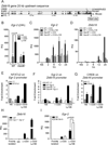
Figure 6. Ly108 induces Egr-2 expression and binding to the Zbtb16 promoter
(A) Mouse and human Zbtb16 sequences, 20 kb upstream of first exon, were aligned and analyzed for conserved binding sites for Egr-2 and CREB. (B) Egr-2 expression in WT or Slamf6−/− PS-DP thymocytes stimulated as in 1B for 24 h, normalized to NS controls. Data are the mean ± SEM from 6 independent experiments. Egr-2 (C) or Zbtb16 (D) expression by qRT-PCR from WT and Slamf6−/− PS-DP thymocytes stimulated for 2, 6, 12 or 24 h. Time courses are representative of 4 independent experiments. (E–G) Chromatin immunoprecipitations (ChIPs) from thymocytes stimulated as in 1B. (E) NFATc2 binding on the Egr-2 promoter. (F) Egr-2 binding on the Zbtb16 promoter. (G) CREB binding on the Zbtb16 promoter. ChIPs show average of 2 replicates ± SD from one of 2 or more independent experiments. (H) Zbtb16 expression in Egr-1−/−Egr-2fl/fl Lck Cre or littermate control PS-DP thymocytes stimulated as in Fig. 1B for 48 hours and normalized to NS controls. (I) Egr-2 expression in C57Bl/6J (B6) and B6.Sle1b PS-DP thymocytes stimulated with plate-bound αCD3±αLy108 for 48 hrs. Data are representative of three independent experiments.
Figure 7
Figure 7. CD28 costimulation does not enhance PLZF expression
(A–C) PS-DP thymocytes were stimulated with αCD3±αCD28 or ±αLy108 and evaluated for Egr-2 expression by qRT-PCR at 18h (A), Egr-2 binding at the Zbtb16 promoter by ChIP at 18h (B), or Zbtb16 expression at 24h (C). Data show the mean of 2 replicates ± SD from one of two or more independent experiments.

"VSports app下载" References

    1. Subleski JJ, Jiang Q, Weiss JM, Wiltrout RH. The split personality of NKT cells in malignancy, autoimmune and allergic disorders. Immunotherapy. 2011;3:1167–1184. - PMC - PubMed
    1. Kovalovsky D, Uche OU, Eladad S, Hobbs RM, Yi W, Alonzo E, Chua K, Eidson M, Kim HJ, Im JS, Pandolfi PP, Sant'Angelo DB. The BTB-zinc finger transcriptional regulator PLZF controls the development of invariant natural killer T cell effector functions. Nat Immunol. 2008;9:1055–1064. - PMC - PubMed
    1. Savage AK, Constantinides MG, Han J, Picard D, Martin E, Li BF, Lantz O, Bendelac A. The transcription factor PLZF directs the effector program of the NKT cell lineage. Immunity. 2008;29:391–403. - PMC - PubMed
    1. Kreslavsky T, Savage AK, Hobbs R, Gounari F, Bronson R, Pereira P, Pandolfi PP, Bendelac A, von Boehmer H. TCR-inducible PLZF transcription factor required for innate phenotype of a subset of gammadelta T cells with restricted TCR diversity. Proc Natl Acad Sci U S A. 2009;106:12453–12458. - PMC - PubMed
    1. Kovalovsky D, Alonzo ES, Uche OU, Eidson M, Nichols KE, Sant'Angelo DB. PLZF induces the spontaneous acquisition of memory/effector functions in T cells independently of NKT cell-related signals. Journal of immunology. 2010;184:6746–6755. - PubMed

Publication types

MeSH terms