Skip to main page content
U.S. flag

An official website of the United States government

Dot gov

The . gov means it’s official VSports app下载. Federal government websites often end in . gov or . mil. Before sharing sensitive information, make sure you’re on a federal government site. .

Https

The site is secure V体育官网. The https:// ensures that you are connecting to the official website and that any information you provide is encrypted and transmitted securely. .

. 2013 Mar;24(6):683-91.
doi: 10.1091/mbc.E12-09-0705. Epub 2013 Jan 23.

COX19 mediates the transduction of a mitochondrial redox signal from SCO1 that regulates ATP7A-mediated cellular copper efflux (VSports注册入口)

Affiliations

COX19 mediates the transduction of a mitochondrial redox signal from SCO1 that regulates ATP7A-mediated cellular copper efflux

Scot C Leary (VSports在线直播) et al. Mol Biol Cell. 2013 Mar.

Abstract

SCO1 and SCO2 are metallochaperones whose principal function is to add two copper ions to the catalytic core of cytochrome c oxidase (COX). However, affected tissues of SCO1 and SCO2 patients exhibit a combined deficiency in COX activity and total copper content, suggesting additional roles for these proteins in the regulation of cellular copper homeostasis. Here we show that both the redox state of the copper-binding cysteines of SCO1 and the abundance of SCO2 correlate with cellular copper content and that these relationships are perturbed by mutations in SCO1 or SCO2, producing a state of apparent copper overload. The copper deficiency in SCO patient fibroblasts is rescued by knockdown of ATP7A, a trans-Golgi, copper-transporting ATPase that traffics to the plasma membrane during copper overload to promote efflux. To investigate how a signal from SCO1 could be relayed to ATP7A, we examined the abundance and subcellular distribution of several soluble COX assembly factors VSports手机版. We found that COX19 partitions between mitochondria and the cytosol in a copper-dependent manner and that its knockdown partially rescues the copper deficiency in patient cells. These results demonstrate that COX19 is necessary for the transduction of a SCO1-dependent mitochondrial redox signal that regulates ATP7A-mediated cellular copper efflux. .


Figures
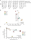
FIGURE 1:
FIGURE 1:
The redox state of the SCO1 CxxxC motif is significantly correlated with cellular copper levels and SCO2 abundance in cultured fibroblasts. (A) Isolated mitochondria (2 mg/ml) from control (n = 3), SCO1, SCO2 (SCO2-3, SCO2-5, SCO2-9, SCO2-11), COX10, and COX15 fibroblasts were incubated in iodoacetamide (I) for 1 h at room temperature, denatured in sample loading buffer in the absence of reductant, and electrophoresed (25 μg/lane) under nonreducing conditions (Leary et al., 2009). The redox state of the SCO1 CxxxC motifs was then detected by immunoblotting with a specific polyclonal antibody (Leary et al., 2004). ox, oxidized disulfides; red, iodoacetamide-modified, reduced thiols. The relative abundance of both redox species was quantified by densitometric analysis of multiple exposures within the linear range of the film (n = 3–4), using ImageQuant software (Leary et al., 2009). The redox state of the SCO1 CxxxC motif was then expressed as the mean ratio of reduced thiols to oxidized disulfides (i.e., Cys red:ox). For SCO2, the electrophoretic mobility of each redox species is identical (Leary et al., 2009), and the immunoreactive band therefore represents total protein abundance. The copper content of fibroblast lines was also measured by ICP-OES and normalized to total cellular zinc. Mutations specific to each patient background are provided in Materials and Methods. (B) The mean ratio of reduced thiols to oxidized disulfides of SCO1 ± SE plotted as a function of total cellular copper content after each parameter had been log transformed. Statistical analysis yielded a high Pearson's r (−0.78) and revealed a significant correlation between these two parameters (p = 0.0062). (C) The mean abundance of SCO2 ± SE quantified by densitometry as described in A and plotted in arbitrary units (A.U.) against the log transformation of the mean ratio of reduced thiols to oxidized disulfides of SCO1 ± SE. Statistical analysis calculated a high Pearson's r (–0.79) and detected a significant correlation between these two parameters (p = 0.0059). For both B and C the data point for SCO1 fibroblasts was omitted from the regression analyses.
FIGURE 2:
FIGURE 2:
The impaired redox regulation of the SCO1 CxxxC motif in SCO fibroblasts signals a state of apparent copper overload that affects ATP7A-dependent cellular copper efflux. (A) Isolated mitochondria (2 mg/ml) from control and ATP7A-1 fibroblasts were incubated in the presence or absence of DTT, treated with IAM or AMS, and then denatured and electrophoresed (25 μg/lane) under nonreducing conditions (Leary et al., 2009). The redox state of the SCO CxxxC motifs was detected by immunoblotting with specific polyclonal antisera raised against each protein. ox, oxidized disulfides; red, iodoacetamide-modified, reduced thiols; red*, the AMS-induced shift in the electrophoretic mobility of reduced thiols, which is caused by the addition of a 0.5-kDa moiety per thiol group. (B) Control (C1, C2), SCO1, SCO2-5, and ATP7A-1 fibroblasts were cultured for 36 h in the absence or presence of 100 μM BCS. Mitochondria were then isolated, treated with IAM or AMS, electrophoresed, and immunoblotted as described in A. For A and B, the relative abundance of both redox species of SCO1 was quantified by densitometric analysis of multiple exposures within the linear range of the film (n = 3–4), using ImageQuant software (Leary et al., 2009). The redox state of each CxxxC motif in IAM- and AMS-treated samples was then expressed as the mean ratio of reduced thiols to oxidized disulfides (i.e., Cys red:ox). For statistical analyses, the Cys red:ox of each IAM- and AMS-treated sample at a given exposure was averaged, and a two-tailed, Student's t test was used to detect significant differences in the redox state of the SCO1 CxxxC motif between (A) control and ATP7A-1 fibroblasts and (B) untreated and BCS-treated fibroblasts, with the associated p values provided in Results. (C, D) Two ATP7A fibroblast lines were stably transduced with an shRNA (SCO1 shRNA 1) that targets the 3′ untranslated region of SCO1 mRNA (Leary et al., 2007) and plated at clonal density, and SCO1 abundance in individual clones (–) was compared with parental (P) and control (C1, C2) lines. Select clones (ATP7A-1, n = 5; ATP7A-3, n = 3) were then retrovirally transduced to overexpress SCO1 P174L, and the phenotypic effects on cellular copper content were quantified by ICP-OES. A one-way analysis of variance (ANOVA) failed to detect a significant effect (i.e., p < 0.05) of manipulating the expression of SCO1 variants on total cellular copper content in either ATP7A patient background. (E, F) Control (C1, C2), SCO1, SCO2-5, and ATP7A-1 fibroblasts were cultured in the absence (–) or presence of one of two different Stealth RNAi duplexes (a, b; see Supplemental Table S1) to knock down ATP7A abundance. After 6 d, cells were harvested and their copper content quantified by ICP-MS. Whole-cell extracts (10 μg/lane) were also prepared and fractionated on a 5–20% gradient gel under denaturing conditions (Leary et al., 2007), and the membrane was blotted with a specific polyclonal antiserum to detect ATP7A. COMMD1 served as an internal loading control.
FIGURE 3:
FIGURE 3:
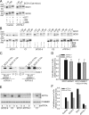
The abundance of several soluble twin Cx9C motif–containing COX assembly factors is sensitive to cellular copper status. (A, B) Whole-cell extracts (15 μg/lane) from control (C1–C4), SCO1, and SCO2-5 fibroblasts (FBs) or myoblasts (MBs) and (C) mitochondrial extracts (10 μg/lane) from control (C1, C2) and SCO2 (n = 4) fibroblasts were electrophoresed by SDS–PAGE on 5–20% gradient gels under denaturing conditions. Membranes were decorated with polyclonal antisera to detect COX17, COX19, COX23, and PET191. Porin and SOD1 served as internal loading controls. (D) Whole-cell extracts (15 μg/lane) from a control fibroblast line cultured for 24 or 48 h in basal media or media supplemented with 100 μM of the Cu(I) chelator BCS or Cu-His were fractionated by SDS–PAGE on 5–20% gradient gels under denaturing conditions. Membranes were then decorated with the polyclonal antisera to detect COX19, COX23, and PET191. Actin served as an internal loading control. (E) Whole-cell extracts (15 μg/lane) from two control (C1, C2) and three ATP7A fibroblast lines (ATP7A-1, -2, and -3) were fractionated by SDS–PAGE on 5–20% gradient gels under denaturing conditions, and membranes were blotted with polyclonal antisera to detect COX19, COX23, and PET191. Actin served as an internal loading control. An equivalent amount of whole-cell extract isolated from SCO1 fibroblasts was included in these analyses as a positive control.
FIGURE 4:
FIGURE 4:
COX19 partitions between mitochondria and the cytosol in a copper-dependent manner. (A) Whole-cell extracts were prepared from SCO1 and ATP7A-1 fibroblasts, as well as a single control fibroblast line (C1) cultured for 24 h in basal media (–), or media supplemented with 100 μM BCS or Cu-His. Denatured extracts (10 μg/lane) were electrophoresed by SDS–PAGE on 5–20% gradient gels under denaturing conditions and membranes decorated with polyclonal antisera to detect COX19, COX23, and PET191. The asterisk denotes residual PET191 immunoreactivity detected upon blotting for COX23. (B) A crude mitochondrial fraction (Mt) was isolated by differential centrifugation from all of the cells described and analyzed in A. The resultant supernatant was subsequently spun for 1 h at 100,000 × g at 4°C to isolate the soluble cytosolic fraction (S) from the insoluble fraction (P), which comprises a minor amount of mitochondria, as well as endoplasmic reticulum, Golgi, and microsomes. The abundance of COX19, COX23, and PET191 in all three fractions (10 μg/lane) was then detected by Western blotting. Exclusive localization of porin and the IMS protein cytochrome c to the Mt fraction argue that the isolated organelles were intact. SOD1 served as a cytosolic marker.
FIGURE 5:
FIGURE 5:
COX19 is critical to the transduction of a SCO1-dependent mitochondrial redox signal that regulates cellular copper homeostasis. (A, B) The abundance of COX19 or PET191 was stably knocked down in control fibroblasts using either a specific shRNA or miRNA (see Supplemental Table S1 for further details) and compared with the parental line (–). Whole-cell extracts were prepared and electrophoresed (10 μg/lane) under denaturing conditions, and membranes were blotted with polyclonal antisera to detect each protein. Actin served as an internal loading control. D3 and D4 are shRNAs targeted to COX19 mRNA, whereas D9 is an shRNA specific to PET191 mRNA. For the miRNA experiments, Q denotes a scrambled, control sequence (see Supplemental Table S1 for further details). (C, D) Quantification of the phenotypic effects of stably knocking down COX19 or PET191 on total cellular copper levels in control, SCO1, SCO2 (SCO2-3, SCO2-5, SCO2-9), COX15, and ATP7A-1 fibroblasts. Data are presented as mean total cellular copper content ± SE. Asterisk denotes a statistically significant difference (p < 0.0001) in total copper content between parental and knockdown cells for control (COX19-1 miRNA, n = 3; D3 COX19 shRNA, n = 5) and SCO2 (D3 COX19 shRNA, n = 3) fibroblasts, detected by a one-way ANOVA and Tukey's HSD post hoc test. Equivalent statistical analyses were not performed for remaining patient fibroblast lines because the data represent single point measurements. (E) Whole-cell extracts were prepared from control (C1, C2) and SCO1 fibroblasts alone and from SCO1 fibroblasts stably expressing an shRNA (SCO1 shRNA 2) that targets the 3′ untranslated region of SCO1 mRNA to knock down the abundance of SCO1 P174L (Leary et al., 2007). Denatured extracts (10 μg/lane) were fractionated by SDS–PAGE on 15% SDS–PAGE gels under denaturing conditions and membranes decorated with polyclonal antisera to detect COX19, COX23, and PET191. Actin served as an internal loading control.

References

    1. Andruzzi L, Nakano M, Nilges MJ, Blackburn NJ. Spectroscopic studies of metal binding and metal selectivity in Bacillus subtilis BSco, a homologue of the yeast mitochondrial protein Sco1p. J Am Chem Soc. 2005;127:16548–16558. - "VSports app下载" PubMed
    1. Antonicka H, Leary SC, Guercin GH, Agar JN, Horvath R, Kennaway NG, Harding CO, Jaksch M, Shoubridge EA. Mutations in COX10 result in a defect in mitochondrial heme A biosynthesis and account for multiple, early-onset clinical phenotypes associated with isolated COX deficiency. Hum Mol Genet. 2003a;12:2693–2702. - PubMed
    1. Antonicka H, Mattman A, Carlson CG, Glerum DM, Hoffbuhr KC, Leary SC, Kennaway NG, Shoubridge EA. Mutations in COX15 produce a defect in the mitochondrial heme biosynthetic pathway, causing early-onset fatal hypertrophic cardiomyopathy. Am J Hum Genet. 2003b;72:101–114. - V体育2025版 - PMC - PubMed
    1. Balatri E, Banci L, Bertini I, Cantini F, Ciofi-Baffoni S. Solution structure of Sco1: a thioredoxin-like protein Involved in cytochrome c oxidase assembly. Structure. 2003;11:1431–1443. - V体育平台登录 - PubMed
    1. Banci L, Bertini I, Calderone V, Ciofi-Baffoni S, Mangani S, Martinelli M, Palumaa P, Wang S. A hint for the function of human Sco1 from different structures. Proc Natl Acad Sci USA. 2006a;103:8595–8600. - PMC (VSports) - PubMed

Publication types

MeSH terms

VSports在线直播 - LinkOut - more resources