Skip to main page content
U.S. flag

An official website of the United States government

Dot gov

The . gov means it’s official. Federal government websites often end in . gov or VSports app下载. mil. Before sharing sensitive information, make sure you’re on a federal government site. .

Https

The site is secure. The https:// ensures that you are connecting to the official website and that any information you provide is encrypted and transmitted securely. V体育官网.

. 2013 Apr;81(4):1129-39.
doi: 10.1128/IAI.01124-12. Epub 2013 Jan 22.

Role of host xanthine oxidase in infection due to enteropathogenic and Shiga-toxigenic Escherichia coli (VSports手机版)

Affiliations

Role of host xanthine oxidase in infection due to enteropathogenic and Shiga-toxigenic Escherichia coli

"VSports app下载" John K Crane et al. Infect Immun. 2013 Apr.

Abstract (V体育官网入口)

Xanthine oxidase (XO), also known as xanthine oxidoreductase, has long been considered an important host defense molecule in the intestine and in breastfed infants. Here, we present evidence that XO is released from and active in intestinal tissues and fluids in response to infection with enteropathogenic Escherichia coli (EPEC) and Shiga-toxigenic E. coli (STEC), also known as enterohemorrhagic E VSports手机版. coli (EHEC). XO is released into intestinal fluids in EPEC and STEC infection in a rabbit animal model. XO activity results in the generation of surprisingly high concentrations of uric acid in both cultured cell and animal models of infection. Hydrogen peroxide (H(2)O(2)) generated by XO activity triggered a chloride secretory response in intestinal cell monolayers within minutes but decreased transepithelial electrical resistance at 6 to 22 h. H(2)O(2) generated by XO activity was effective at killing laboratory strains of E. coli, commensal microbiotas, and anaerobes, but wild-type EPEC and STEC strains were 100 to 1,000 times more resistant to killing or growth inhibition by this pathway. Instead of killing pathogenic bacteria, physiologic concentrations of XO increased virulence by inducing the production of Shiga toxins from STEC strains. In vivo, exogenous XO plus the substrate hypoxanthine did not protect and instead worsened the outcome of STEC infection in the rabbit ligated intestinal loop model of infection. XO released during EPEC and STEC infection may serve as a virulence-inducing signal to the pathogen and not solely as a protective host defense. .


Figures

Fig 1
Fig 1
Biochemical reactions in the pathway for catabolism of nucleosides and purines. Uricase is absent in humans, great apes, and Dalmatian dogs but present in other mammals and birds, as well as many microbes.
Fig 2
Fig 2
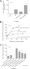
Release of uric acid into supernatant medium of cultured T84 cells in response to EPEC infection. (A) Comparison of uric acid release between commensal E. coli strain HS, wild-type EPEC strain E2348/69, and the plasmid-cured derivative of E2348/69, JPN15. *, significantly greater than JPN15. (B) Effect of increasing multiplicity of infection (MOI) on uric acid released from cultured T84 monolayers by infection with Salmonella enterica serotype Enteritidis, EPEC JCP88, and E. coli HS. (C) Effect of the xanthine oxidase inhibitors allopurinol and oxypurinol on EPEC-induced uric acid release from T84 cells. *, significantly increased compared to the uninfected control; **, significantly decreased compared to JCP88 without allopurinol. uninf, uninfected.
Fig 3
Fig 3
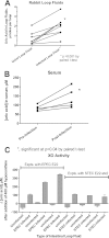
Release of uric acid and xanthine oxidase activity into intestinal loop fluids and serum after infection of rabbits with EPEC E22 or rabbit STEC E22-stx2. (A) Comparison of the uric acid contents of uninfected and EPEC E22-infected ligated rabbit intestinal loops after a 20-h infection. Each line segment represents the uric acid from an uninfected and an infected intestinal loop fluid from the same animal. (B) Increase in serum uric acid in non-surgically altered rabbits infected orally with strain E22 for 7 days compared with levels in the same animal preinfection. (C) Detection of xanthine oxidase (XO) activity in intestinal loop fluids from infected, but not uninfected, ligated ileal loops. XO activity of 6 pairs of intestinal loop fluid samples from 6 rabbits. Uninfected and infected loops shown in adjacent bars as pairs of loop fluids are from the same animal. The first 3 pairs on the left show data from experiments in which some loops were infected with EPEC E22, and the pairs on the right side are from 3 animals for which some loops were infected with STEC E22-stx2. Although asterisks are omitted, in each case, the XO activity was significantly higher in the infected loop than in the uninfected loop fluid. In 5 of 6 cases, the uninfected loop fluid XO activity was a negative number, i.e., uric acid was not generated but instead disappeared in the uninfected loop fluids during the assay, presumably due to uricase activity.
Fig 4
Fig 4
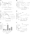
Effects of xanthine oxidase and hypoxanthine on bacterial growth and on Stx production in STEC. (A to C) Graphs of bacterial growth, measured as OD600 values, in response to XO and various concentrations of hypoxanthine or other nucleosides. The x axis in panels A to C is the logarithm of the nucleoside or purine concentration, in moles/liter (M). (A) Growth inhibition in the presence of XO plus hypoxanthine, but not XO plus other nucleosides, on EPEC E2348/69. (B) Comparison of the susceptibilities of 3 strains of E. coli to growth inhibition by various concentrations of hypoxanthine in the presence of a fixed concentration of XO, 1 U/ml. (C) Inhibition of growth under anaerobic conditions in thioglycolate medium for three bacterial strains. (D to F) Effect of XO with or without hypoxanthine on Stx production from human STEC strain Popeye-1 (O157:H7, Stx2 only). (D) Although asterisks are omitted, Stx in the supernatant medium was significantly higher in the presence of XO than in its absence for all 3 concentrations of hypoxanthine tested. (E) Reversal of Stx induction by H2O2-neutralizing agents. A total of 1 U/ml XO and 400 μM hypoxanthine were used. Catalase (added to a final concentration of 600 U/ml) and glutathione (final concentration of 5 mM) reversed the inducing effect of hypoxanthine plus XO. *, significantly less Stx than with hypoxanthine plus XO. (F) Effect of varying the amount of XO in the presence of a fixed concentration of hypoxanthine. *, significant compared to the no-hypoxanthine control for each amount of XO. hypo, hypoxanthine.
Fig 5
Fig 5
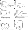
Effects on hypoxanthine plus XO on short-circuit current, electrical resistance, and Stx translocation in polarized T84 cell monolayers studied in the Ussing chamber. Short-circuit current (Isc) represents chloride secretion toward the apical (mucosal or lumenal) side of the tissue in this configuration. (A) Comparison of the short-circuit current triggered by 1 mM hypoxanthine plus XO (black tracing) with that triggered by 1 mM H2O2 (gray tracing), showing similar peak currents and similar offsets of secretion. (B) Hypoxanthine alone, without added XO, triggered a very small short-circuit current of ∼2 μA/cm2. (C) Dose-response relationship between the amount of hypoxanthine added and the short-circuit current; mean ± standard deviation (SD) of 3 tracings per concentration. (D) Effect of catalase on short-circuit current triggered by hypoxanthine plus XO. When 1,200 U/ml catalase was added simultaneously with hypoxanthine and XO (arrow 1, gray tracing), the secretory response was prevented. When hypoxanthine and XO were added first (black arrow) and a current was allowed to develop, subsequent addition of catalase (2nd black arrow) promptly reversed the secretion. (E) Effect of hypoxanthine (hypo) and XO on transepithelial electrical resistance 6 h after addition of hypoxanthine, XO, or both. *, significantly decreased compared to the control and to XO alone. (F) Effect of hypoxanthine (hypoxanth) plus XO on Stx translocation across T84 cell monolayers. T84 cells were grown to confluence in Transwell inserts, reaching a mean initial TER value of 1,648 Ω. Hypoxanthine and XO were added to the apical side of the monolayers, followed by Stx2 3 h later. The amount of Stx2 detectable in the lower basolateral chamber was measured at various times after the start of the experiment. *, significant compared to the control, DMSO vehicle, and XO alone.
Fig 6
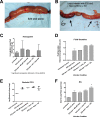
Fig 6
Effects of exogenous XO and hypoxanthine on the outcome of STEC infection in a ligated rabbit ileal loop model of infection. Ten-centimeter segments of ileum were ligated as described in Materials and Methods and infected with 4 × 10⁸ CFU of rabbit STEC E22-stx2 plus XO and hypoxanthine. Twenty hours after infection, loops were collected and photographed and the contents were analyzed. (A) Gross appearance of an ileal loop infected with E22-stx2 but without any other additives, showing distention with fluid but absence of necrosis. (B) One of 2 intestinal loops receiving E22-stx2 plus XO and hypoxanthine (hypoxnth) showed overt necrotic mottling (loop 6, left arrow), while the other loop showed only one small spot of necrosis at the site of the injection (right arrow). (C) Hemoglobin concentrations in loop fluids were assayed after centrifugation of the samples at 16,000 × g for 10 min to remove intact cells and debris. In the presence of STEC bacteria, addition of 1 U/ml XO seemed to reduce the bloody character of the loop fluids, but this did not reach statistical significance. *, in the presence of STEC, XO, and 400 μM hypoxanthine, hemoglobin in the loop fluids was significantly higher than that with the pathogen and XO. (D) Fluid secretion into the loops, as measured by the volume-to-length ratio. hypo, hypoxanthine. (E) Comparison of the numbers of bacteria recovered from each loop (expressed as the logarithm of the number of bacteria recovered per loop), showing the lack of any decrease in CFU in loops receiving hypoxanthine (hypo) and XO. (F) Shiga toxin protein (Stx) content of the loops by enzyme immunoassay, expressed in ng per loop. *, significantly increased compared to E22-stx2 alone. hypo, hypoxanthine.
Fig 7
Fig 7
Hypothetical graph questioning the possibility of an uncanny valley of XO activity in which an intermediate amount of XO activity might be worse for the host than no XO activity (left side of curve) or high XO activity (right side of curve). The concept of the uncanny valley was proposed by Masahiro Mori in an essay in 1970 and is adapted here to microbial pathogenesis.

Comment in

References (V体育ios版)

    1. Hancock J, Salisbury V, Ovejero-Boglione M, Cherry R, Hoare C, Eisenthal R, Harrison R. 2002. Antimicrobial properties of milk: dependence on presence of xanthine oxidase and nitrite. Antimicrob. Agents Chemother. 46:3308–3310 - PMC - PubMed
    1. Martin H, Hancock J, Salisbury V, Harrison R. 2004. Role of xanthine oxidoreductase as an antimicrobial agent. Infect. Immun. 72:4933–4939 - PMC - PubMed
    1. Stevens C, Millar T, Clinch J, Kanczler J, Bodamyali T, Blake D. 2000. Antibacterial properties of xanthine oxidase in human milk. Lancet 356:829–830 - PubMed
    1. Wagner PL, Acheson DWK, Waldor MK. 2001. Human neutrophils and their products induce Shiga toxin production by enterohemorrhagic Escherichia coli. Infect. Immun. 69:1934–1937 - PMC - PubMed
    1. Los JM, Los M, Wêgrzyn A, Wêgrzyn G. 2010. Hydrogen peroxide-mediated induction of the Shiga toxin-converting lambdoid prophage ST2-8624 in Escherichia coli O157:H7. FEMS Immunol. Med. Microbiol. 58(3):322–329 - PubMed

Publication types

MeSH terms

"V体育官网" LinkOut - more resources