V体育官网 - Skip to main page content
U.S. flag

An official website of the United States government

Dot gov

The . gov means it’s official. Federal government websites often end in . gov or . mil. Before sharing sensitive information, make sure you’re on a federal government site VSports app下载. .

Https

The site is secure. The https:// ensures that you are connecting to the official website and that any information you provide is encrypted and transmitted securely V体育官网. .

. 2012 Mar 1;7(3):279-293.
doi: 10.2217/fvl.12.8.

The immune response to human CMV (V体育2025版)

Affiliations

The immune response to human CMV

Corinna La Rosa et al. Future Virol. .

Abstract

This review will summarize and interpret recent literature regarding the human CMV immune response, which is among the strongest measured and is the focus of attention for numerous research groups. CMV is a highly prevalent, globally occurring infection that rarely elicits disease in healthy immunocompetent hosts. The human immune system is unable to clear CMV infection and latency, but mounts a spirited immune-defense targeting multiple immune-evasion genes encoded by this dsDNA β-herpes virus. Additionally, the magnitude of cellular immune response devoted to CMV may cause premature immune senescence, and the high frequencies of cytolytic T cells may aggravate vascular pathologies. However, uncontrolled CMV viremia and life-threatening symptoms, which occur readily after immunosuppression and in the immature host, clearly indicate the essential role of immunity in maintaining asymptomatic co-existence with CMV VSports手机版. Approaches for harnessing the host immune response to CMV are needed to reduce the burden of CMV complications in immunocompromised individuals. .


"VSports手机版" Figures

Figure 1
Figure 1. CMV-seropositive healthy adults
Levels (expressed in percentages as shown inside each plot) of CD8+ T cells specific for MHC class I pp65495–503 tetramer (black dots) in four HLA A*0201 CMV-seropositive healthy adults (each plot represents a different subject). Peripheral blood mononuclear cells were stained with tetramers and analyzed by cytofluorometry as detailed [77]. Note the substantial portion of CD8+ T cells specific for a single HLA A*0201 CMV epitope from the pp65 protein among healthy people with no acute CMV infection [La Rosa et al., Unpublished Data].
Figure 2
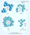
Figure 2. CMV-specific CD8+ T-cell phenotypes
The peculiar differentiation stages of CMV-specific CD8+ T cells during primary infection, memory and latency [44]. Effector cells are markedly cytolytic and have a dominant Th1 cytokine production, which is the permanent CMV signature for both early- and late-stage CMV-specific T cells. In contrast to other viral infections, the memory pool does not contract after viral clearance, but keeps inflating and will eventually dominate the T-cell memory compartment. Although the phenotype of the CMV-specific CD8+ T cells during latency is typical of terminal differentiation, these cells can further expand and produce Th1 cytokines as if they were resting effectors [40]. This unique feature may be crucial in rapidly antagonizing virus replication upon reactivation of latent virus. On the downside, the high expression of IFN-γ may subtly increase the overall proinflammatory status, which may be causally related to pathogenic effects of CMV on the vasculature [55].
Figure 3
Figure 3. Primary CMV infection during immune suppression
Data shown in FACS analysis panels are representative of HLA B*0702 R receiving a SOT (orthotopic liver transplant) from a D+. These patients are prophylactically treated with antivirals, but are at high risk of developing life-threatening late CMV disease upon antiviral discontinuation. The upper panels report data from a CMV viremic D+ R patient at time of CMV disease, and the lower panels are relative to a CMV viremic and asymptomatic D+ R patient. A very high percentage of CD8+ T cells specific for MHC class I pp65265–275 tetramer (black dots, see Figure 1) was detected in both viremic recipients. However, in the case of the D+ R patient with CMV disease, there are significantly higher levels of the negative immune-modulator PD-1 receptor on CMV-specific CD8+ T cells (gray filled curve, obtained as detailed in [70]). Importantly, PD-1 levels measured on CMV-specific T cells in healthy individuals were significantly lower than levels detected in the D+ R cohort, exclusively when these patients were symptomatic for CMV disease [70]. Upregulation of PD-1 receptor impairs T-cell ability to contain viremia and can lead to CMV disease [68,70]. Blocking negative immune modulators may restore protective CMV immunity in SOT recipients [68,70] [La Rosa et al., Unpublished Data]. D+: CMV-seropositive donor; Max: Maximum; R: CMV-naive recipient; SOT: Solid organ transplant.
Figure 4
Figure 4. Immune dysregulation in solid organ transplant patients
The plots represent data from the same cohort of patients described in Figure 3. All four patients shown progressed to CMV disease at the post-TX time point indicated on the x-axis. The left y-axes indicates the levels of IL-10 (pg/ml, solid line) measured in plasma, the right y-axes, the plasma DNAemia (copies/ml, dashed line) [68]. Very high levels of the immunosuppressive cytokine IL-10 were consistently detected in the plasma of liver recipients diagnosed with CMV disease [68,71]. IL-10 upregulation inhibits CMV-specific T-cell proliferation, which can lead to uncontrolled viremia and symptomatic complications [La Rosa et al., Unpublished Data]. Patients died at 10 (UPN 9) and 4.5 (UPN 18) months post-TX. D: CMV disease; GCV: Antiviral ganciclovir; TX: Transplant.
Figure 5
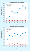
Figure 5. Immunogenicity of a CMV peptide vaccine
The upper and lower plot show vaccine-induced expansion of pp65-specific T cells in two representative HLA A*0201 healthy adults (UPN 212 and 214) immunized with 0.5 mg Tet-CMV, a peptide vaccine developed by DJ Diamond, at the Division of Translational Vaccine Research (City of Hope, CA, USA). The CMV peptide vaccine consists of the HLA A*0201-specific CTL pp65495–503 epitope fused with the tetanus toxin P2 epitope (tt830–843), in the presence of PF03512676 adjuvant [71]. In this Phase Ib clinical trial, which was performed at City of Hope, healthy volunteers were vaccinated by subcutaneous injection, every 3 weeks for a total of four injections (days 0, 21, 42 and 63). Levels of CD8+ T cells in peripheral blood mononuclear cells of UPN 212 and 214 binding either to MHC class I pp65495–503 tetramer (in each plot, square symbol) or to a control HIVgag77–85 tetramer (in each plot, triangle symbol) are indicated on the y-axes (as percentages) [La Rosa et al., Unpublished Data].

References

    1. Cannon MJ, Schmid DS, Hyde TB. Review of cytomegalovirus seroprevalence and demographic characteristics associated with infection. Rev Med Virol. 2010;20(4):202–213. - PubMed
    1. Dolan A, Cunningham C, Hector RD, et al. Genetic content of wild-type human cytomegalovirus. J Gen Virol. 2004;85(Pt 5):1301–1312. - PubMed
    1. Sylwester AW, Mitchell BL, Edgar JB, et al. Broadly targeted human cytomegalovirus-specific CD4+ and CD8+ T cells dominate the memory compartments of exposed subjects. J Exp Med. 2005;202(5):673–685. The most comprehensive study in healthy individuals, which analyzed the broad and heterogeneous CMV-specific cellular immune response. These data provide the first insight into the rules governing immunodominance and cross-reactivity in complex human CMV infection. - PMC - PubMed
    1. Miller-Kittrell M, Sparer TE. Feeling manipulated: cytomegalovirus immune manipulation. Virol J. 2009;6:4. - PMC - PubMed
    1. Poole E, McGregor D, Sr, Colston J, Joseph RS, Sinclair J. Virally induced changes in cellular microRNAs maintain latency of human cytomegalovirus in CD34 progenitors. J Gen Virol. 2011;92(Pt 7):1539–1549. - PubMed

LinkOut - more resources