Skip to main page content
U.S. flag

An official website of the United States government

Dot gov

The . gov means it’s official. Federal government websites often end in VSports app下载. gov or . mil. Before sharing sensitive information, make sure you’re on a federal government site. .

Https

The site is secure V体育官网. The https:// ensures that you are connecting to the official website and that any information you provide is encrypted and transmitted securely. .

. 2013 Feb 15;288(7):4935-46.
doi: 10.1074/jbc.M112.422204. Epub 2013 Jan 2.

Direct interaction of Bax and Bak proteins with Bcl-2 homology domain 3 (BH3)-only proteins in living cells revealed by fluorescence complementation (VSports app下载)

Affiliations

Direct interaction of Bax and Bak proteins with Bcl-2 homology domain 3 (BH3)-only proteins in living cells revealed by fluorescence complementation (V体育2025版)

Laura Vela et al. J Biol Chem. .

Abstract (VSports注册入口)

The key event in the mitochondrial pathway of apoptosis is the activation of Bax and Bak by BH3-only proteins through a molecular mechanism that is still a matter of debate. Here we studied interactions among anti- and proapoptotic proteins of the Bcl-2 family in living cells by using bimolecular fluorescence complementation analysis. Our results indicate that the antiapoptotic proteins Mcl-1 and Bcl-x(L) bind preferably to the BH3-only proteins Bim, PUMA, and Noxa but can also bind to Bak and Bax. We also found a direct interaction between Bim, PUMA, or Noxa with either Bax or Bak during apoptosis induction VSports手机版. In HeLa cells, interaction of Bim with Bax occurs in cytosol, and then Bim-Bax complexes translocate to mitochondria. Complexes of either PUMA or Noxa with Bax or Bak were always detected at mitochondria. Overexpression of Bcl-x(L) or Mcl-1 delayed Bim/Bax translocation to mitochondria. These results reveal the ability of main BH3-only proteins to directly activate Bax and Bak in living cells and suggest that a complex network of interactions regulate the function of Bcl-2 family members during apoptosis. .


Figures

FIGURE 1.
FIGURE 1.
Schematic representation of gene constructs generated for BiFC experiments. A, unique restriction sites in the Multicloning site of pBiFC vectors were used for cloning the proteins of interest (EcoRI, XbaI, SalI, and BglII) fused to corresponding fragment of Venus fluorescent protein. B, secondary structure of proteins and mutations generated for the study of interactions by BiFC technique.
FIGURE 2.
FIGURE 2.
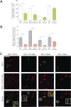
Interactions of Mcl-1 with proapoptotic proteins of the Bcl-2 family. A and B, HeLa cells were cotransfected with vectors expressing the fusion protein VN-Mcl-1 and Bim, PUMA, Bax, or Bak fused to the other half of Venus (VC). Deletion mutants lacking the BH3 domain (ΔBH3) were transfected as indicated. A vector encoding mRFP was also included in the transfection. After transfection, cells were cultured for 24 h, trypsinized, and Venus and mRFP fluorescence was analyzed by flow cytometry. Results are mean ± S.D. of seven independent experiments. *, p < 0.05; **, p < 0.01; ***, p < 0.001. C, HeLa cells were seeded on coverslips and transfected as indicated in A and B. 24 h after transfection, cells were stained with the mitochondrial probe MitoTracker Red (50 nm), fixed, and analyzed by confocal microscopy as indicated under “Experimental Procedures.” Scale bar = 50 μm. The insets show the enlarged x2.5 boxed regions.
FIGURE 3.
FIGURE 3.
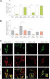
Interactions of Bcl-xLwith proapoptotic proteins of the Bcl-2 family. A and B, HeLa cells were cotransfected with vectors expressing the fusion protein VC- Bcl-xL and Bim, PUMA, Bax, or Bak fused to the other half of Venus (VN). Deletion mutants lacking the BH3 domain (ΔBH3) were transfected as indicated. A vector encoding mRFP was also included in the transfection. After transfection, cells were cultured for 24 h, trypsinized, and then Venus and mRFP fluorescence was analyzed by flow cytometry. Results are mean ± S.D. of seven independent experiments. * p < 0.05; **, p < 0.01; ***, p < 0.001. C, HeLa cells were seeded on coverslips and transfected as indicated in A and B. 24 h after transfection cells were stained with the mitochondrial probe MitoTracker Red (50 nm), fixed, and analyzed by confocal microscopy as indicated under “Experimental Procedures.” Scale bar = 50 μm. The insets show the enlarged (x2.5) boxed regions.
FIGURE 4.
FIGURE 4.
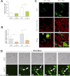
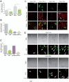
Visualizing direct interactions of Bim with Bak and Bax. A and B, HeLa cells were cotransfected with vectors encoding the fusion protein Bim-VN and Bax or Bak fused to the other half of Venus (VC). Deletion mutants lacking the BH3 domain (ΔBH3) were transfected as indicated. A vector encoding mRFP was also included in the transfection. After transfection, cells were cultured for 24 h, trypsinized, and Venus and mRFP fluorescence was analyzed by flow cytometry. Results are mean ± S.D. of six to ten independent experiments. *, p < 0.05; **, p < 0.01; ***, p < 0.001. C, HeLa cells were seeded on coverslips and transfected as indicated in A and B. 24 h after transfection cells were stained with 50 nm MitoTracker Red, fixed, and analyzed by confocal microscopy as indicated under “Experimental Procedures.” Scale bar = 50 μm. The insets show the enlarged (x2.5) boxed regions. D, time lapse microscopy reveals translocation of Bim-Bax complexes from the cytosol to mitochondria. Cells were plated on μ-Slide 8-well tissue culture plates and transfected with the vectors containing the Bim-VN and Bax-VC constructs. Cells were monitored for 72 h in a Leica AF6000 LX system as indicated under “Experimental Procedures.” Images were acquired with a CCD camera (C9100-02, Hamamatsu) at 7-min intervals, and LAS AF software (Leica) was used to process the images. Also shown is a representative time lapse sequence from supplemental movies Mov1BimBax and Mov2BimBax showing phase-contrast images (upper panel) and Venus fluorescence (lower panel). DIC, differential interference contrast.
FIGURE 5.
FIGURE 5.
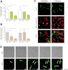
Visualizing direct interactions of PUMA with Bak and Bax. A and B, HeLa cells were cotransfected with vectors expressing the fusion protein PUMA-VN and Bax or Bak fused to the other half of Venus (VC). Deletion mutants lacking the BH3 domain (ΔBH3) or amino acids 1–94 (ΔN) were transfected as indicated. A vector encoding mRFP was also included in the transfection. After transfection, cells were cultured for 24 h, trypsinized, and Venus and mRFP fluorescence was analyzed by flow cytometry. Results are mean ± S.D. of five to ten independent experiments. *, p < 0.05; **, p < 0.01; ***, p < 0.001. C, HeLa cells were seeded on coverslips and transfected as indicated in A and B. 24 h after transfection cells were stained with 50 nm MitoTracker Red, fixed, and analyzed by confocal microscopy as indicated under “Experimental Procedures.” Scale bar = 50 μm. The insets show the enlarged (x2.5) boxed regions. D, time lapse microscopy reveals the interaction of PUMA and Bax in mitochondria prior to cell death induction. Cells were plated on μ-Slide 8-well tissue culture plates and transfected with the vectors containing the PUMA-VN and Bax-VC constructs. Cells were monitored for 72 h in a Leica AF6000 LX system, as indicated under “Experimental Procedures.” Images were acquired with a CCD camera (C9100-02, Hamamatsu) at 7-min intervals, and LAS AF software (Leica) was used to process the images. Also shown is a representative time lapse sequence from supplemental movies Mov3PUMABax and Mov4PUMABax showing phase-contrast images (upper panel) and Venus fluorescence (lower panel). DIC, differential interference contrast.
FIGURE 6.
FIGURE 6.
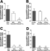
Implication of BH3 and H1α domains of Bak and Bax in their interaction with Bim and PUMA. HeLa cells were transfected with vectors expressing the fusion proteins Bax-VC/Bim-VN (A), Bax-VC/PUMA-VN (B), Bak-VC/Bim-VN (C), or Bak-VC/PUMA-VN (D). Deletion mutants of Bax and Bak (lacking either the BH3 domain, ΔBH3, or the first α helix, ΔH1α) were transfected as indicated (white bars). A vector encoding mRFP was also included in the transfection. After transfection, cells were cultured for 24 h, trypsinized, and then Venus and mRFP fluorescence was analyzed by flow cytometry. Results are mean ± S.D. of four to seven independent experiments. *, p < 0.05; **, p < 0.01; ***, p < 0.001.
FIGURE 7.
FIGURE 7.
Interactions of Noxa with antiapoptotic proteins Mcl-1 and direct interaction with proapoptotic proteins Bak and Bax. A, B, and C, HeLa cells were cotransfected with vectors expressing the fusion protein Noxa-VN and Mcl-1, Bcl-xL, Bax, or Bak fused to the other half of Venus (VC). Deletion mutants lacking the BH3 domain (ΔBH3) or the single-substitution mutant (NoxaL29E) were transfected as indicated. A vector encoding mRFP was also included in the transfection. After transfection, cells were cultured for 24 h, trypsinized, and then Venus and mRFP fluorescence were analyzed by flow cytometry. Results are mean ± S.D. of four independent experiments. *, p < 0.05; **, p < 0.01; ***, p < 0.001. D, HeLa cells were seeded on coverslips and transfected as indicated in A, B, and C. 24 h after transfection, cells were stained with 50 nm MitoTracker Red, fixed, and analyzed by confocal microscopy as indicated under “Experimental Procedures.” Scale bar = 50 μm. The insets show the enlarged boxed regions. E and F, time lapse microscopy reveals the formation of the Noxa-Bax (E) and Noxa-Bak complexes (F) before cell death induction. Cells were plated on μ-Slide 8-well tissue culture plates and transfected with the vectors containing the Noxa-VN and Bax-VC/Bak-VC constructs. Cells were monitored for 72 h in a Leica AF6000 LX system, as indicated under “Experimental Procedures.” Images were acquired with a CCD camera (C9100-02, Hamamatsu) at 7-min intervals, and LAS AF software (Leica) was used to process the images. Also shown is a representative time lapse sequence from supplemental movies Mov5NoxaBax, Mov6NoxaBax, Mov7NoxaBak, and Mov8NoxaBak showing Venus fluorescence (lower panel) and phase-contrast images (upper panel). DIC, differential interference contrast.
FIGURE 8.
FIGURE 8.
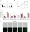
Effect of Bcl-xL or Mcl-1 overexpression on the interactions of BH3-only proteins Bim and PUMA with Bak/Bax. A, HeLa-Vector, HeLa-Bcl-xL, or HeLa-Mcl-1 stable cells lines were transfected with vectors encoding different proapoptotic proteins or corresponding deletion mutants. A phosphatidylserine exposure analysis was performed by annexin V-PE staining and flow cytometry 24 h after transfection. Results are mean ± S.D. of three to four independent experiments. B, time course analysis of the percentage of cells with low mitochondrial membrane potential (ΔΨmlow) after treatment with 100 μm etoposide confirms a delay in cell death when cells overexpress antiapoptotic proteins. C, HeLa cells overexpressing antiapoptotic proteins Bcl-xL or Mcl-1 or containing the empty vector (HeLa-Vector) were cotransfected with the vector pairs indicated together with a vector encoding mRFP. D, time lapse microscopy reveals that overexpression of Bcl-xL delays translocation to the mitochondria of Bim-Bax pairs. HeLa- Bcl-xL cells were plated on μ-Slide 8-well tissue culture plates and transfected with the vectors containing the Bim-VN and Bax-VC constructs. Cells were monitored for 72 h in a Leica AF6000 LX system, as indicated under “Experimental Procedures.” Images were acquired with a CCD camera (C9100-02, Hamamatsu) at 7-min intervals. LAS AF software (Leica) was used to process the images. Also shown is a representative time lapse sequence from supplemental movies Mov9BclXlBimBax and Mov10BclXlBimBax showing phase-contrast images (upper panel) and Venus fluorescence (lower panel).

V体育安卓版 - References

    1. Youle R. J., Strasser A. (2008) The BCL-2 protein family. Opposing activities that mediate cell death. Nat. Rev. Mol. Cell Biol. 9, 47–59 - PubMed
    1. Wei M. C., Zong W.-X., Cheng E. H., Lindsten T., Panoutsakopoulou V., Ross A. J., Roth K. A., MacGregor G. R., Thompson C. B., Korsmeyer S. J. (2001) Proapoptotic BAX and BAK. A requisite gateway to mitochondrial dysfunction and death. Science 292, 727–730 - PMC - PubMed
    1. López-Royuela N., Pérez-Galán P., Galán-Malo P., Yuste V. J., Anel A., Susín S. A., Naval J., Marzo I. (2010) Different contribution of BH3-only proteins and caspases to doxorubicin-induced apoptosis in p53-deficient leukemia cells. Biochem. Pharmacol. 79, 1746–1758 - PubMed
    1. Buckbinder L., Talbott R., Velasco-Miguel S., Takenaka I., Faha B., Seizinger B. R., Kley N. (1995) Induction of the growth inhibitor IGF-binding protein 3 by p53. Nature 377, 646–649 - PubMed
    1. Villunger A., Labi V., Bouillet P., Adams J., Strasser A. (2011) Can the analysis of BH3-only protein knockout mice clarify the issue of “direct versus indirect” activation of Bax and Bak[quest]. Cell Death Differ. 18, 1545–1546 - PMC - PubMed

Publication types (VSports手机版)

"V体育官网入口" MeSH terms