<b dropzone="sv5B8k"></b>
Skip to main page content
U.S. flag

An official website of the United States government

Dot gov

The . gov means it’s official. Federal government websites often end in . gov or . mil VSports app下载. Before sharing sensitive information, make sure you’re on a federal government site. .

Https

The site is secure V体育官网. The https:// ensures that you are connecting to the official website and that any information you provide is encrypted and transmitted securely. .

. 2013 Jan 8;110(2):624-9.
doi: 10.1073/pnas.1213819110. Epub 2012 Dec 24.

Neddylation pathway regulates T-cell function by targeting an adaptor protein Shc and a protein kinase Erk signaling

Affiliations

Neddylation pathway regulates T-cell function by targeting an adaptor protein Shc and a protein kinase Erk signaling

Hyung-seung Jin et al. Proc Natl Acad Sci U S A. .

Abstract

NEDD8 (neural precursor cell expressed, developmentally down-regulated 8) is a ubiquitin-like molecule whose action on modifying protein substrates is critical in various cellular functions but whose importance in the immune system is not well understood. Here we investigated the role of protein neddylation in regulating T-cell function using an in vivo knockdown technique VSports手机版. We found that reduced expression of Ubc12 in CD4(+) T cells led to impaired T-cell receptor/CD28-induced proliferation and cytokine production both in vitro and in vivo, accompanied by reduced Erk activation. These findings were recapitulated by treatment with MLN4924, an inhibitor of NEDD8-activating enzyme. Furthermore, Shc, an adaptor molecule between antigen receptors and the Ras/Erk pathway, was identified as a target for neddylation. Importantly, mice adoptively transferred with Ubc12 knockdown CD4(+) T cells showed markedly ameliorated allergic responses. This study thus identifies an important role for protein neddylation in T-cell function, which may serve as a therapeutic target for inflammatory diseases. .


V体育2025版 - Conflict of interest statement

The authors declare no conflict of interest.

Figures
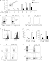
Fig. 1.
Fig. 1.
Ubc12 is required for TCR-induced proliferation and activation. (A) Immunoblot analysis of Ubc12 was performed in sorted GFP+ control (ctl) or GFP+ shUbc12 CD4+ T cells (Inset). Peripheral naïve CD4+ T cells sorted from shRNA expressing chimeric mice were stimulated with various concentration of anti-CD3 and anti-CD28 mAb for 3 d and then pulsed for an additional 8 h with 3H-thymidine. Cell proliferation was measured by 3H-thymidine uptake. (B) Cell cycle analysis of control (Left) and shUbc12 CD4+ (Right) T cells. The cells were activated for 23 h with anti-CD3/CD28 mAbs, then pulsed for 1 h with EdU and analyzed by flow cytometry after staining for EdU and 7-AAD. (C) Control (Left) and shUbc12 CD4+ (Right) violet-labeled CD4+ T cells were stimulated with anti-CD3/CD28 mAbs for 2 d, after which cell division was analyzed by flow cytometry. (D) Flow cytometry for cell surface expression of CD69 (Left) and CD25 (Right) in sorted control and shUbc12 CD4+ T cells stimulated for 16 h with anti-CD3/CD28 mAbs. The results are representative of three repeated experiments. (E) IL-2 (Left) and IFN-γ (Right) production in control and shUbc12 CD4+ T cells stimulated for 48 h with anti-CD3 and anti-CD28, measured by ELISA. (F) Control (Left) and Ubc12 shRNA-expressing (Right) OT-II TCR transgenic mice were generated by reconstitution of retrovirally transduced bone marrow cells from OT-II mice into lethally irradiated recipient mice (n = 3). After 8 w, isolated CD4+ T cells from OT-II control or OT-II Ubc12 knockdown chimeric mice were adoptively transferred into C57BL/6J recipient mice, followed by immunization with OVA plus CFA. At 5 d after immunization, splenocytes and lymph node cells were stimulated with OVA323–339 peptide for 24 h and analyzed by flow cytometry using anti–IL-2 and anti–IFN-γ antibodies. (G) Sorted naïve CD4+ T cells were stimulated with anti-CD3/CD28 mAbs and differentiated in vitro under Th1-inducing (Upper) or Th2-inducing (Lower) conditions. Results are from three repeated experiments.
Fig. 2.
Fig. 2.
MLN4924 treatment blocks TCR-induced proliferation and activation. (A) Flow cytometry of the cell cycle status of CD4+ T cells stimulated with anti-CD3 and anti-CD28 for 16 h in the presence of the indicated concentrations of MLN4924 diluted in DMSO or equivalent volumes of DMSO alone. (B) CD4+ T cells were analyzed for surface expression of CD69 (Upper) and CD25 (Lower) after stimulation with anti-CD3 and anti-CD28 in the presence of the indicated concentrations of MLN4924. (C) IL-2 production by CD4+ T cells stimulated with anti-CD3/CD28 mAbs in the absence or presence of MLN4924, measured by ELISA. (D) Quantitative RT-PCR analysis of IL-2 mRNA. (E) Cell death of stimulated CD4+ T cells examined by 7-AAD staining and FACS analysis.
Fig. 3.
Fig. 3.
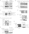
The neddylation pathway is involved in Erk activation. (A) Immunoblot analysis of control (Left) and shUbc12 (Right) CD4+ T cells. The CD4+ T cells sorted from the chimeric mice were stimulated with anti-CD3 and anti-CD28 for the indicated time periods. Cell lysates were separated by SDS/PAGE and immunoblotted with the indicated antibodies. (B) CD4+ T cells were preactivated with anti-CD3 in the presence of the indicated concentrations of MLN4924 for 16 h and then restimulated with a combination of anti-CD3 and anti-CD28. (C) Jurkat E6.1 cells stably expressing FLAG-NEDD8 were treated with MLN4924 for 12 h or left untreated before being collected. Cell lysates were boiled in 1% SDS-containing lysis buffer, then diluted to 0.1% SDS and immunoprecipitated with anti-FLAG–conjugated beads, followed by elution with triple FLAG peptide. The eluates were subjected to Western blot analysis using the indicated antibodies. (D) The three isoforms of Shc. All isoforms contain an N-terminal PTB domain, a CH domain, and a C-terminal SH2 domain. (E) Jurkat cells were transfected with FLAG-tagged p66, p52, or p46 with HA-tagged NEDD8. The samples were processed for immunoprecipitation as described in C. (F) The peptide sequence at the top is derived from MS analysis of p66Shc; an asterisk indicates the modified lysine residue. Jurkat cells were transfected with expression vector encoding WT (wt) p52Shc or p52Shc lysine mutant (K3R). Where indicated, combinations of plasmids expressing HA-NEDD8 and/or Ubc12 were cotransfected. (G) CD4+ T cells were stimulated with anti-CD3/CD28 antibodies and retrovirally transduced with Myc-tagged p52Shc WT-IRES-GFP (WT) or Myc-tagged p52Shc K3R-IRES-GFP (K3R). Whole-cell lysates were immunoprecipitated with anti-ZAP70 antibody and blotted with the indicated antibodies. (H) CD4+ T cells were treated with MLN4924 for 16 h or left untreated. After TCR stimulation, the cell lysates were subjected to coimmunoprecipitation with anti-ZAP70 antibody.
Fig. 4.
Fig. 4.
Role pf the NEDD8 pathway in the development of allergic inflammation. (A) GFP+CD4+ T cells from OT-II control or OT-II Ubc12 knockdown chimeric mice were adoptively transferred into C57BL/6J recipient mice, followed by immunization with OVA plus alum (n = 3). Starting at 10 d after immunization, the mice were challenged intranasally with OVA protein for the next 4 d. The mice were examined at 24 h after the last challenge. (B) Lungs from control mice (Left) and shUbc12 knockdown chimeric mice (Right) were sectioned and stained with H&E to visualize inflammatory infiltrates. (C) Total numbers of eosinophils (Eos), neutrophils (Neu), monocytes (Mon), and lymphocytes (Lym) were calculated by FACS analysis. (D) IL-4 (Left) and IL-5 (Right) concentrations in BAL fluid were measured by ELISA at 24 h after the final OVA challenge. The results are representative of two repeated experiments.

V体育官网入口 - References

    1. Smith-Garvin JE, Koretzky GA, Jordan MS. T cell activation. Annu Rev Immunol. 2009;27:591–619. - PMC - PubMed
    1. Liu YC, Penninger J, Karin M. Immunity by ubiquitylation: A reversible process of modification. Nat Rev Immunol. 2005;5(12):941–952. - "V体育官网入口" PMC - PubMed
    1. Sun SC. Deubiquitylation and regulation of the immune response. Nat Rev Immunol. 2008;8(7):501–511. - PMC - PubMed
    1. Kerscher O, Felberbaum R, Hochstrasser M. Modification of proteins by ubiquitin and ubiquitin-like proteins. Annu Rev Cell Dev Biol. 2006;22:159–180. - PubMed
    1. Harper JW, Schulman BA. Structural complexity in ubiquitin recognition. Cell. 2006;124(6):1133–1136. - V体育2025版 - PubMed

Publication types

MeSH terms (VSports手机版)