Skip to main page content
U.S. flag

An official website of the United States government

Dot gov

The . gov means it’s official. Federal government websites often end in VSports app下载. gov or . mil. Before sharing sensitive information, make sure you’re on a federal government site. .

Https

The site is secure V体育官网. The https:// ensures that you are connecting to the official website and that any information you provide is encrypted and transmitted securely. .

. 2013 Feb;83(2):470-80.
doi: 10.1124/mol.112.081075. Epub 2012 Nov 27.

Mechanism of the antiproliferative activity of some naphthalene diimide G-quadruplex ligands

Affiliations

Mechanism of the antiproliferative activity of some naphthalene diimide G-quadruplex ligands

Sonja M Hampel et al. Mol Pharmacol. 2013 Feb.

Abstract (VSports最新版本)

G-quadruplexes are higher-order nucleic acid structures that can form in G-rich telomeres and promoter regions of oncogenes. Telomeric quadruplex stabilization by small molecules can lead to telomere uncapping, followed by DNA damage response and senescence, as well as chromosomal fusions leading to deregulation of mitosis, followed by apoptosis and downregulation of oncogene expression. We report here on investigations into the mechanism of action of tetra-substituted naphthalene diimide ligands on the basis of cell biologic data together with a National Cancer Institute COMPARE study VSports手机版. We conclude that four principal mechanisms of action are implicated for these compounds: 1) telomere uncapping with subsequent DNA damage response and senescence; 2) inhibition of transcription/translation of oncogenes; 3) genomic instability through telomeric DNA end fusions, resulting in mitotic catastrophe and apoptosis; and 4) induction of chromosomal instability by telomere aggregate formation. .


Figures
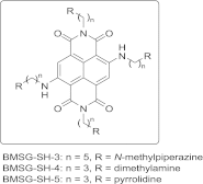
Fig. 1.
Fig. 1.
Structures of BMSG-SH-3–5.
Fig. 2.
Fig. 2.
Cell uptake study of BMSG-SH-3–5 in the breast cancer cell line MCF7. (A1) BMSG-SH-3, 50 μM, 30 min, fluorescence image. (A2) Transmitted light image. (A3) Merge of images A1 and A2. (B1) BMSG-SH-4, 500 nM, 30 min, fluorescence image. (B2) Transmitted light image. (B3) Merge of images B1 and B2. (C1) BMSG-SH-5, 500 nM, 30 min, fluorescence image. (C2) Transmitted light image. (C3) Merge of images C1 and C2. (D) Untreated sample, merge of fluorescence and transmitted light image. Scale: 50 μm.
Fig. 3.
Fig. 3.
Long-term growth inhibition of cancer cell lines in the presence of BMSG-SH-3. (A) A549 cells. (B) MCF7 cells. (C) MIA-Pa-Ca-2 cells. (D) HPAC cells.
Fig. 4.
Fig. 4.
Senescence staining of A549 cells incubated with BMSG-SH-3 (200 nM) for 14 days.
Fig. 5.
Fig. 5.
Cell-cycle analysis of cancer cell lines treated with BMSG-SH-3 and etoposide, which is known to cause DNA damage and mitotic catastrophe. (A1) A549 cells, untreated. (A2) A549 cells treated with etoposide (30 μM) for 14 h. (A3) A549 cells treated with BMSG-SH-3 (1 μM) for 14 h. (B1) MIA-Pa-Ca-2 cells, untreated. (B2) MIA-Pa-Ca-2 cells treated with etoposide (30 μM) for 14 h. (B3) MIA-Pa-Ca-2 cells treated with BMSG-SH-3 (1 μM) for 14 h.
Fig. 6.
Fig. 6.
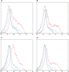
DNA damage in the form of H2AX phosphorylation caused by BMSG-SH-3 in cancer cells. Color code: Green—untreated, unstained. Blue—untreated, anti γ-H2AX stained. Purple— treated, unstained. Red—treated, anti γ-H2AX stained. (A) A549 cells treated with etoposide, 30 μM, 14 h. (B) A549 cells treated with BMSG-SH-3, 1 μM, 14 h. (C) MIA-Pa-Ca-2 cells treated with etoposide, 30 μM, 14 h. (D) MIA-Pa-Ca-2 cells treated with BMSG-SH-3, 1 μM, 14 h.
Fig. 7.
Fig. 7.
(A) Pictures of single cells: 3D fluorescence in situ hybridization of HaCaT-myc cells applying a CY3 (red) labeled telomere-specific PNA probe (left: cell showing double and triple telomeres; right: cell showing a telomeric aggregate). (B) Telomere association (2er, 3er, > 3er) and telomere aggregates (TAs) in HaCaT-myc cells incubated with BMSG-SH-5 (40 nM, 48 h). (C) Representation of the telomeric signal intensity as a measure for telomere length. Each set of blue dots represents the median telomere length within a single cell; the black bar indicates the mean telomere length within each data set. Here, the ligand concentration was 40 nM, and the cells were irradiated with 5 J/m2 UV-C.
Fig. 8.
Fig. 8.
Telomere aggregate (TA) analysis. The figure shows the percentage of telomerically aberrant cells for the differently treated cell samples. Only cells with >3er and TAs are considered “aberrant” since double and triple telomeres can occur also in genomically stable cells. The first number above the bars represents the number of cells with the respective aberration per slide, and the second number represents the total number of observed cells per slide.

References

    1. Balasubramanian S, Hurley LH, Neidle S. (2011) Targeting G-quadruplexes in gene promoters: a novel anticancer strategy? Nat Rev Drug Discov 10:261–275 - PMC - PubMed
    1. Bejugam M, Gunaratnam M, Müller S, Sanders DA, Sewitz S, Fletcher JA, Neidle S, Balasubramanian S. (2010) Targeting the c-Kit promoter G-quadruplexes with 6-substituted indenoisoquinolines. ACS Med Chem Lett 1:306–310 - VSports在线直播 - PMC - PubMed
    1. Bochman ML, Paeschke K, Zakian VA. (2012) DNA secondary structures: stability and function of G-quadruplex structures. Nat Rev Genet 13:770–780 - PMC - PubMed
    1. Boukamp P, Petrussevska RT, Breitkreutz D, Hornung J, Markham A, Fusenig NE. (1988) Normal keratinization in a spontaneously immortalized aneuploid human keratinocyte cell line. J Cell Biol 106:761–771 - VSports app下载 - PMC - PubMed
    1. Boukamp P, Popp S, Krunic D. (2005) Telomere-dependent chromosomal instability. J Investig Dermatol Symp Proc 10:89–94 - PubMed (VSports手机版)

Publication types

"VSports" MeSH terms