Skip to main page content
U.S. flag

An official website of the United States government

Dot gov

The . gov means it’s official. Federal government websites often end in . gov or VSports app下载. mil. Before sharing sensitive information, make sure you’re on a federal government site. .

Https

The site is secure. The https:// ensures that you are connecting to the official website and that any information you provide is encrypted and transmitted securely V体育官网. .

. 2013 Mar;1833(3):468-78.
doi: 10.1016/j.bbamcr.2012.10.019. Epub 2012 Oct 23.

"VSports最新版本" A divalent interaction between HPS1 and HPS4 is required for the formation of the biogenesis of lysosome-related organelle complex-3 (BLOC-3)

Affiliations

A divalent interaction between HPS1 and HPS4 is required for the formation of the biogenesis of lysosome-related organelle complex-3 (BLOC-3) (VSports手机版)

Carmelo Carmona-Rivera et al. Biochim Biophys Acta. 2013 Mar.

"V体育平台登录" Abstract

Hermansky-Pudlak syndrome (HPS) is a group of rare autosomal recessive disorders characterized by oculocutaneous albinism, a bleeding tendency, and sporadic pulmonary fibrosis, granulomatous colitis or infections. Nine HPS-causing genes have been identified in humans. HPS-1 is the most severe subtype with a prevalence of ~1/1800 in northwest Puerto Rico due to a founder mutation in the HPS1 gene. Mutations in HPS genes affect the biogenesis of lysosome-related organelles such as melanosomes in melanocytes and platelet dense granules VSports手机版. Two of these genes (HPS1 and HPS4) encode the HPS1 and HPS4 proteins, which assemble to form a complex known as Biogenesis of Lysosome-related Organelle Complex 3 (BLOC-3). We report the identification of the interacting regions in HPS1 and HPS4 required for the formation of this complex. Two regions in HPS1, spanning amino acids 1-249 and 506-700 are required for binding to HPS4; the middle portion of HPS1 (residues 250-505) is not required for this interaction. Further interaction studies showed that the N-termini of HPS1 and HPS4 interact with each other and that a discrete region of HPS4 (residues 340-528) interacts with both the N- and C-termini of the HPS1 protein. Several missense mutations found in HPS-1 patients did not affect interaction with HPS4, but some mutations involving regions interacting with HPS4 caused instability of HPS1. These observations extend our understanding of BLOC-3 assembly and represent an important first step in the identification of domains responsible for the biogenesis of lysosome-related organelles. .


Figures

Figure 1
Figure 1. Expression and Co-immunoprecipitation of HPS1Δex9
(A) Top. Schematic representation of the HPS1 gene and location of the primers used for PCR analysis to detect HPS1 splice variants. Bottom. A panel of first-strand cDNAs derived from several human tissues (Clontech) was PCR amplified using a primer pair spanning exons 5–9. The two major transcripts were amplified as 766- bp (HPS1) and 667-bp (HPS1Δex9) fragments. β-Actin was utilized as loading control. Additional lower molecular weight bands reflect amplification of minor splice variants. (B) Stably transfected M1 cells expressing Myc3-HPS4 (clone number 26) were transiently transfected with constructs containing either HPS1 full-length or HPS1Δex9. An aliquot of these crude extracts corresponding to 1% of the material available for IP, was analyzed by immunoblotting (IB) using a mouse antibody against the V5 epitope and a mouse monoclonal antibody against the Myc tag. The crude extracts were used in immunoprecipitation (IP) reactions using a mouse monoclonal antibody against the Myc epitope. The immunoprecipitates were analyzed by 4–12% gradient SDS-PAGE followed by immunoblotting using a monoclonal antibody against the V5 epitope. The positions of molecular weight markers are indicated on the left.
Figure 2
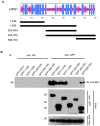
Figure 2. Divalent interaction of HPS1 with HPS4
(A) Top. The consensus secondary structure prediction for HPS1 as determined by the Network Protein Sequence Analysis of the Pôle Bio-informatique Lyonnais. Blue areas represent α-helices, while red areas indicate predicted β-extended strand, and solid purple lines denote random coils. Bottom. GFP-tagged HPS1 constructs used to map the HPS4 binding sites on HPS1 are represented by solid bars with residue numbers indicated on the left. (B) GFP-tagged HPS1 constructs were transiently transfected into M1 clone 26 cells expressing Myc3-tagged HPS4. Coimmunoprecipitation analysis revealed an interaction of HPS4 with the first 249 amino acids of HPS1, suggesting that the N-terminus of HPS1 contains a region required for interaction with full-length HPS4. Interestingly, a construct containing the HPS1 C-terminus (HPS1506-700) also displayed an interaction with HPS4. These results are consistent with HPS4 recognizing two different regions, located at the N-terminus (first 249 amino acids) and the C-terminus (506– 700 amino acids) of HPS1. Co-immunoprecipitation with an irrelevant anti-HA antibody was used as control.
Figure 3
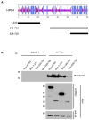
Figure 3. HPS4 shows a dual interaction with HPS1
(A) Top. Consensus secondary structure prediction for HPS4 as determined using the Network Protein Sequence Analysis of the Pôle Bio-informatique Lyonnais. Bottom. Three Myc3-tagged truncations of HPS4 are represented by solid bars with residue numbers indicated on the left. (B) Stably transfected M1 clone expressing HA3-PS1 (clone 5) were transiently transfected with constructs coding for either full length Myc3-HPS4 or truncated proteins. Aliquots of the crude extracts corresponding to 1% of the material available for immunoprecipitation (IP) were analyzed by immunoblotting (IB) using an anti-Myc monoclonal antibody. The extracts were then subjected to an IP with mouse monoclonal anti-Myc. The immunoprecipitates were analyzed by 4–12% gradient SDS-PAGE gel followed by immunoblotting using an antibody against HA-coupled to horseradish peroxidase. IP with irrelevant mouse monoclonal antibodies against GFP was performed as control. The positions of molecular weight markers are indicated on the left.
Figure 4
Figure 4. Mutual interaction of the N-terminal fragments of HPS1 and HPS4, while the central region of HPS4 shows divalent interaction with the N- and C- termini of HPS1
To delineate the structural determinants of the HPS1 and HPS4 interactions, HeLa cells were co-transfected with vectors encoding Myc3- tagged HPS41–230(A), HPS4340–708 (B) or HPS4528–708 (C) truncations in combination with GFP-tagged HPS1 truncations representing N-terminal, middle and C-terminal portions of the protein. The Myc3-HPS4-N terminal region interacts with the N-terminus of HPS1, but not with the C-terminus or the middle region of this protein. Myc3-tagged HPS4340–708 interacts with both the N- and the C- termini of HPS1. The C- terminus of HPS4506–708 did not show any interaction with portions of the HPS1 protein.
Figure 5
Figure 5. HPS1 and HPS4 binding regions exist in soluble and membrane associated forms
Transfected M1 cells were mechanically disrupted by passing them through a 25-gauge needle in buffer A. The crude lysates were centrifuged at 800 x g and the resulting post-nuclear supernatants were centrifuged further to yield cytosolic (cyt) and membrane (memb) fractions as described (Materials and Methods). (A) The presence of GFP-HPS11–249, HPS1506–700 (upper panel) and (B) Myc3-HPS41–230, HPS4340–708 (upper panel) or Lamp-2 (lower panel, integral membrane control) was assessed by immunoblotting. The positions of molecular weight markers are indicated on the left. Transfected M1 cells were fixed, and GFP tagged proteins were visualized directly (C and D) to detect GFP-HPS11–249, HPS1506–700. Transfected M1 cells were stained with mouse anti-Myc antibody (E and F) to detect Myc3-HPS41–230 and HPS4340–708, followed by Alexa 488- conjugated anti-mouse IgG. Bar, 10μm.
Figure 6
Figure 6. Co-immunoprecipitation of HA-tagged HPS1 and HPS1–16bp
(A) The consensus secondary structure prediction for the wild type and the predicted mutant protein HPS116bp–ins was determined using the Network Protein Sequence Analysis of the Pôle Bio-informatique Lyonnais. Note that the predicted secondary structures of the N- terminus of wild type and mutant HPS1 proteins are identical up to amino acid 500. The frameshift mutation in the HPS1 gene causes the loss of the last 200 amino acids compared to wild type HPS1 and the addition of 89 new residues due to the resulting frameshift. Blue areas denote α-helices, red areas indicate predicted β-extended strand, and solid purple lines represent random coils. (B) Stably transfected M1 cells expressing Myc3-HPS4 were transiently transfected with either HA3-HPS1 or HA3-HPS116bp-ins. Aliquots of the extracts corresponding to 1% of the material available for IP were analyzed by immunoblotting (IB) using an anti-HA mAb. The extracts were then subjected to an immunoprecipitation (IP) with mouse monoclonal anti-HA as indicated. Irrelevant mouse monoclonal antibodies (GFP) were included as controls. The immunoprecipitates were analyzed by 4–12% SDS-PAGE gradient gels followed by immunoblotting using an antibody against the Myc-tag. The positions of molecular weight markers are indicated on the left. Serum-starved HPS-1 skin fibroblasts were treated with or without 20 μM wortmannin or LY294002 for 8 h in reduced serum medium. (C) Western blot analysis of HPS1 from cell extracts after wortmannin treatment. Proteins (100 μg per lane) extracted with cell lysis buffer were separated by SDS-PAGE (4–12%) in the presence of β-mercaptoethanol and transferred to nitrocellulose membranes. Treatment with different concentrations of wortmannin suppressed phosphorylation of Akt (control of treatment). A faint band of the expected molecular weight of 64 KDa was observed after treatment with 20 μM wortmannin, corresponding to the truncated form of HPS1 (D) Fibroblasts were fixed with 4% PFA, non specific sites were blocked and a dual incubation with mouse monoclonal anti-HPS1 and rabbit polyclonal anti-HPS4 was performed for 30 min in a humid chamber. Staining with secondary Alexa 488-conjugated goat anti- mouse and Alexa 555- conjugated goat anti-rabbit for 30 min. Treatment with LY294002 for 8 hours showed an intracellular expression of truncated form of HPS1 and a restoration of HPS4 protein as compared with untreated defective cells. Bar, 10μm.
Figure 7
Figure 7. Proposed model for the interaction of HPS1 and HPS4
The results of the co-immunoprecipitation analyses (Figures 1–4 and 6) are consistent with a model where the N- and C- termini of HPS1and the N-terminal and middle region of HPS4 are involved in the interactions, leading to BLOC-3 formation. Specifically, N-termini of HPS4 and HPS1 interact with each other, and the central portion of HPS4 interacts with both the N- and the C- termini of HPS1.
Figure 8
Figure 8. Missense mutations in HPS1 within putative interacting regions cause protein instability
(A) M1 cells expressing Myc3-HPS4 were transiently transfected with GFP-HPS1 or GFP-HPS1 constructs containing mutations identified in HPS patients. Aliquots of the extracts representing 1% of the material available for IP were analyzed by immunoblotting (IB) using anti-GFP. The extracts were then subjected to an immunoprecipitation (IP) with mouse monoclonal anti-Myc as indicated. Irrelevant IgG were included as controls. The immunoprecipitates were analyzed by 4–12% SDS-PAGE gradients followed by immunoblotting using an antibody against GFP. The positions of molecular weight markers are indicated on the left. (B) Transiently transfected M1 cells were treated with chloroamphenicol and cycloheximide. Proteins were chased for different periods (0, 1, 3, and 6 h) at 37°C. Equivalent amounts of homogenate were resolved by SDS-PAGE followed by western blotting. (C) Bands were quantified using ImageJ 1.44 software and the percentage of each mutant at each chase time was calculated relative to time zero (T0).

References

    1. Hermansky F, Pudlak P. Albinism associated with hemorrhagic diathesis and unusual pigmented reticular cells in the bone marrow: report of two cases with histochemical studies. Blood. 1959;14:162–169. - PubMed
    1. Schinella RA, Greco MA, Cobert BL, Denmark LW, Cox RP. Hermansky-Pudlak Syndrome with granulomatous colitis. Ann Intern Med. 1980;9:220–23. - PubMed
    1. Gahl WA, Brantly M, Kaiser-Kupfer MI, Iwata F, Hazelwood S, Shotelersuk V, Duffy LF, Kuehl EM, Troendle J, Bernardini I. Genetic defect and clinical characteristics of patients with a form of oculocutaneous albinism (Hermansky- Pudlak syndrome) N Engl J Med. 1998;338:1258–1264. - "VSports手机版" PubMed
    1. Brantly M, Avila NA, Shotelersuk V, Lucero C, Huizing M, Gahl WA. Pulmonary function and high-resolution CT finding in patients with an inherited form of pulmonary fibrosis, Hermansky-Pudlak syndrome, due to mutations in HPS-1. Chest. 2000;117:129–136. - PubMed (VSports)
    1. Oh J, Bailin T, Fukai K, Feng GH, Ho L, Mao JI, Frenk E, Tamura N, Spritz RA. Positional cloning of a gene for Hermansky-Pudlak syndrome, a disorder of cytoplasmic organelles. Nat Genet. 1996;14:300–306. - PubMed

Publication types

MeSH terms

Substances