Skip to main page content (V体育平台登录)
U.S. flag

An official website of the United States government

Dot gov

The . gov means it’s official VSports app下载. Federal government websites often end in . gov or . mil. Before sharing sensitive information, make sure you’re on a federal government site. .

Https

The site is secure. The https:// ensures that you are connecting to the official website and that any information you provide is encrypted and transmitted securely. V体育官网.

. 2012 Nov;122(11):4012-24.
doi: 10.1172/JCI62746. Epub 2012 Oct 8.

A novel murine infection model for Shiga toxin-producing Escherichia coli

Affiliations

V体育官网入口 - A novel murine infection model for Shiga toxin-producing Escherichia coli

V体育ios版 - Emily M Mallick et al. J Clin Invest. 2012 Nov.

Abstract

Enterohemorrhagic E. coli (EHEC) is an important subset of Shiga toxin-producing (Stx-producing) E. coli (STEC), pathogens that have been implicated in outbreaks of food-borne illness and can cause intestinal and systemic disease, including severe renal damage. Upon attachment to intestinal epithelium, EHEC generates "attaching and effacing" (AE) lesions characterized by intimate attachment and actin rearrangement upon host cell binding. Stx produced in the gut transverses the intestinal epithelium, causing vascular damage that leads to systemic disease. Models of EHEC infection in conventional mice do not manifest key features of disease, such as AE lesions, intestinal damage, and systemic illness. In order to develop an infection model that better reflects the pathogenesis of this subset of STEC, we constructed an Stx-producing strain of Citrobacter rodentium, a murine AE pathogen that otherwise lacks Stx. Mice infected with Stx-producing C VSports手机版. rodentium developed AE lesions on the intestinal epithelium and Stx-dependent intestinal inflammatory damage. Further, the mice experienced lethal infection characterized by histopathological and functional kidney damage. The development of a murine model that encompasses AE lesion formation and Stx-mediated tissue damage will provide a new platform upon which to identify EHEC alterations of host epithelium that contribute to systemic disease. .


Figures (V体育官网入口)

Figure 1
Figure 1. C. rodentiumstx2dact) produces high levels of Shiga toxin upon prophage induction at levels comparable to EHEC isolates.
Stx2 in supernatants (S) or pellets (P) of untreated or mitomycin C–treated cultures of the indicated strains was measured by capture ELISA, and results are expressed relative to bacterial number (see Methods). ND, not detected. Cr, C. rodentium. Data shown are the averages ± SEM of quadruplicate samples. Data show results of 1 experiment representative of 3 independent experiments.
Figure 2
Figure 2. C. rodentiumstx2dact) causes lethal infection in mice.
(A) Colonization of 5-week-old C57BL/6 mice by C. rodentiumstx2dact), C. rodentiumstx2dact::kanR), and C. rodentium (DBS100) was determined by viable stool counts. Shown are the average CFU (±SEM) of 5 mice. (B) Fecal water content of mock-infected 5-week-old mice or mice infected with C. rodentiumstx2dact), C. rodentiumstx2dact::kanR), or C. rodentium (DBS100). Shown are the averages (±SEM) of groups of 5 mice. Range of average fecal water content of uninfected mice is represented by the vertical bar at 0 days after infection. *P < 0.05 compared with all other groups as determined by 2-way ANOVA followed by Bonferroni post tests. (C) Body weight during infection of 8-week-old mice, expressed as percent change from day 0. Shown are the averages (±SEM) of 5 mice per group. **P < 0.01, ***P < 0.001 compared with all other groups as determined by 2-way ANOVA followed by Bonferroni post tests. (D) Percent survival of groups of five 8-week-old mice that were mock infected or infected with C. rodentiumstx2dact), C. rodentiumstx2dact::kanR), or C. rodentium (DBS100). For all panels, results are representative of at least 5 independent experiments.
Figure 3
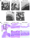
Figure 3. Stx-mediated intestinal damage during murine infection with C. rodentiumstx2dact).
(A) TEM of the proximal large intestine of C57BL/6 mice 6 days after mock infection or infection by the indicated strain. White arrowheads indicate the presence of actin-rich pedestals. Scale bars: 0.5 μm. (B) H&E-stained large intestinal section of mock-infected mice or mice infected with the designated strain 7 to 10 days after infection; original magnification, ×100 (top row), ×200 (bottom row, left and right panels), ×400 (bottom row, middle panel). Scale bars: 50 μm. Black box indicates area of complete crypt loss and abscess formation. Asterisks indicate goblet cells that are prominent in normal intestinal tissue and diminished in mice infected with C. rodentiumstx2dact). Arrowhead indicates infiltration of inflammatory cells into the mucscularis mucosa and muscularis propria layers.
Figure 4
Figure 4. Stx-mediated systemic induction of proinflammatory cytokines during murine infection with C. rodentiumstx2dact).
The concentrations of TNF-α, KC, MIP-1β, IL-17, RANTES, and MCP-1 in serum at 7–10 days after infection in mock-infected mice or mice infected with the indicated strain were determined by Luminex (see Methods). Data are pooled from 2 independent experiments, each using 3- to 8-week-old female mice and 5–10 mice per group. **P < 0.01, ***P < 0.001, 1-way ANOVA and Tukey’s multiple comparison tests.
Figure 5
Figure 5. Stx-mediated renal damage during murine infection with C. rodentiumstx2dact).
(A) Renal cytokines in mock-infected mice or mice infected with the indicated strain. Data are pooled from 3 independent experiments. *P < 0.05, **P < 0.01. (B) Micrographs (original magnification, ×600) of H&E-stained kidney sections from mock-infected mice or the indicated infected mice infected at 7–10 days after infection. Arrowheads indicate attenuation and sloughing of epithelial cells within the proximal tubules. Scale bars: 50 μm. (C) BUN of 8-week-old mice, each represented as a single data point, infected with indicated strain or mock infected at day of necropsy (7–10 days after infection). Shown is the pool of 4 independent experiments using 5 mice per group. **P < 0.01. (D) Proteinuria in mock-infected mice or the indicated infected mice. Data shown are average protein indices ± SEM of 5 mice per group. The range of protein measured was 0–500 mg/dl, with 0 indicating undetectable protein, 0.5 indicating trace, 1.0 indicating ~30 mg/dl, 2.0 indicating ~100 mg/dl, and 3.0 indicating ~500 mg/dl. Shown is a representative of 5 independent experiments. *P < 0.05, **P < 0.01 compared with C. rodentiumstx2dact::kanR). Hematuria in mock-infected mice or the indicated infected mice infected. Shown are the averages (±SEM) of groups of 10 mice. ***P < 0.001 compared with the other groups. Urine Kim-1 concentration in mock-infected mice or the indicated infected mice. Shown are the averages (±SEM) of groups of 3–6 mice. **P < 0.01 compared with the other groups.
Figure 6
Figure 6. Tir is required for colonization and disease upon C. rodentiumstx2dact) infection of mice.
(A) Colonization of 8-week-old C57BL/6 mice that were mock infected mice or infected with the indicated strain was determined by plating stool for viable counts. Shown are the averages CFU (±SEM) of 5 mice. Data are representative of 5 independent experiments. Statistical significance was determined using 2-way ANOVA and Bonferroni post tests and indicates that C. rodentiumstx2dacttir colonized mice significantly (*P < 0.05) less efficiently than C. rodentiumstx2dact) and C. rodentiumstx2dacttir/pTir). (B) Body weight during infection of 8-week-old mice is expressed as percent change from day 0. Shown are the averages (±SEM) of 5 mice per group. Data are representative of 5 independent experiments. (C) Percent survival of mock-infected 8-week-old mice or mice infected with C. rodentiumstx2dact), C. rodentiumstx2dacttir, or C. rodentiumstx2dacttir/pTir. Groups of 5 mice were analyzed. Data are representative of 5 independent experiments. (D) H&E-stained large intestinal sections of mock-infected mice or mice infected with the indicated strain. Scale bars: 50 μm. Asterisks indicate goblet cells that are prominent in normal intestinal tissue. Arrowhead indicates infiltration of inflammatory cells into the mucscularis mucosa and muscularis propria layers. (E) H&E-stained micrograph (original magnification, ×600) of kidney sections from mock-infected mice or mice infected with the indicated strain. Arrowhead indicates attenuation and sloughing of epithelial cells within the proximal tubules. Scale bars: 50 μm.

V体育2025版 - References

    1. Bielaszewska M, et al. Characterisation of the Escherichia coli strain associated with an outbreak of haemolytic uraemic syndrome in Germany, 2011: a microbiological study. Lancet Infect Dis. 2011;11(9):671–676. - PubMed
    1. Kaper JB, Nataro JP, Mobley HL. Pathogenic Escherichia coli. Nat Rev Microbiol. 2004;2(2):123–140. doi: 10.1038/nrmicro818. - DOI - PubMed
    1. Pennington H. Escherichia coli O157. Lancet. 2010;376(9750):1428–1435. doi: 10.1016/S0140-6736(10)60963-4. - "V体育官网入口" DOI - PubMed
    1. Amirlak I, Amirlak B. Haemolytic uraemic syndrome: an overview. Nephrology. 2006;11(3):213–218. doi: 10.1111/j.1440-1797.2006.00556.x. - DOI - PubMed
    1. Karmali MA, Gannon V, Sargeant JM. Verocytotoxin-producing Escherichia coli (VTEC). . Vet Microbiol. 2009;140(3-4):360–370. doi: 10.1016/j.vetmic.2009.04.011. - DOI (VSports手机版) - PubMed

Publication types

MeSH terms

Associated data