Skip to main page content
U.S. flag

An official website of the United States government

Dot gov

The VSports app下载. gov means it’s official. Federal government websites often end in . gov or . mil. Before sharing sensitive information, make sure you’re on a federal government site. .

Https

The site is secure. The https:// ensures that you are connecting to the official website and that any information you provide is encrypted and transmitted securely. V体育官网.

. 2012 Sep 28;151(1):111-22.
doi: 10.1016/j.cell.2012.07.036.

TMEM16F forms a Ca2+-activated cation channel required for lipid scrambling in platelets during blood coagulation

Affiliations

V体育安卓版 - TMEM16F forms a Ca2+-activated cation channel required for lipid scrambling in platelets during blood coagulation

V体育官网 - Huanghe Yang et al. Cell. .

Abstract

Collapse of membrane lipid asymmetry is a hallmark of blood coagulation VSports手机版. TMEM16F of the TMEM16 family that includes TMEM16A/B Ca(2+)-activated Cl(-) channels (CaCCs) is linked to Scott syndrome with deficient Ca(2+)-dependent lipid scrambling. We generated TMEM16F knockout mice that exhibit bleeding defects and protection in an arterial thrombosis model associated with platelet deficiency in Ca(2+)-dependent phosphatidylserine exposure and procoagulant activity and lack a Ca(2+)-activated cation current in the platelet precursor megakaryocytes. Heterologous expression of TMEM16F generates a small-conductance Ca(2+)-activated nonselective cation (SCAN) current with subpicosiemens single-channel conductance rather than a CaCC. TMEM16F-SCAN channels permeate both monovalent and divalent cations, including Ca(2+), and exhibit synergistic gating by Ca(2+) and voltage. We further pinpointed a residue in the putative pore region important for the cation versus anion selectivity of TMEM16F-SCAN and TMEM16A-CaCC channels. This study thus identifies a Ca(2+)-activated channel permeable to Ca(2+) and critical for Ca(2+)-dependent scramblase activity during blood coagulation. PAPERFLICK: .


Figures (V体育平台登录)

Figure 1
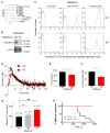
Figure 1. TMEM16F Knockout Mice Exhibit Deficiencies in Ca2+-Dependent Scramblase Activity in Platelets, Defects in Blood Coagulation, and Protection against FeCl3-Induced Carotid Thrombosis
(A) Phylogenetic tree of the human TMEM16 family generated by ClustalW2 with default settings. Percentage of amino acid identity with TMEM16A is indicated in parentheses. Scale bar, 0.1 nucleotide substitutions per site. (B) Immunoblot analysis of TMEM16F expression in the platelets of wild-type (WT) and TMEM16F KO mice. (C) Flow cytometry analysis of platelets from WT and TMEM16F KO mice treated with 0, 1, and 10 μM A23187 and FITC-Annexin-V for 30 min. (D) Tissue-factor-induced thrombin generation curves for WT and TMEM16F KO mice. (E) Endogenous thrombin potential (ETP, the area under the thrombin generation curve) of platelets from WT and TMEM16F KO mice induced by tissue factor. *p < 0.05. Error bar represents SEM. (F) Thrombin generation peak height from the thrombin generation curves induced by tissue factor. *p < 0.05. Error bar represents SEM. (G) Bleeding time measured by a hemostatic challenge assay. **p < 0.005. n.s., not significant. Error bar represents SEM. (H) Carotid artery thrombosis by the 4% FeCl3 injury model. Flow versus time after FeCl3 application is shown. p = 0.0002 log rank test. See also Figure S1.
Figure 2
Figure 2. A Ca2+-Activated, Outwardly Rectifying Current Present in Megakaryocytes of WT Mice Was Eliminated from TMEM16F KO Mice
(A) Megakaryocytes are CD41+ and morphologically distinct from the other cell types in the mouse bone marrow. (B) Representative macroscopic current traces of inside-out patches excised from megakaryocytes of WT mice (top) and TMEM16F KO mice (bottom). The patches were exposed to 100 μM [Ca2+]i with symmetrical 140 mM NaCl. The currents were elicited by voltage ramps from −80 to +80 mV with a rate (dV/dt) = 0.36 V/s. (C) A Ca2+-sensitive, outwardly rectifying current was observed in megakaryocytes of WT mice, but not TMEM16F KO mice (p < 0.0001). I+80mV and |I−80mV| are the absolute values of current amplitudes at +80 and −80 mV, respectively. n equals 15 for WT and 14 for TMEM16F KO. Error bar represent SEM. (D) Representative macroscopic current traces of inside-out patches excised from megakaryocytes of WT (left) and TMEM16F KO (right) mice in the absence or presence of 100 μM [Ca2+]i. Ca2+-induced currents that were derived by subtracting the currents recorded with 0 [Ca2+]i from the currents recorded with 100 μM [Ca2+]i are shown at the bottom. Testing potentials were from −80 mV to +100 mV with 20 mV increments. Both holding and repolarizing potentials were −80 mV. Dotted lines indicate the zero current level. Both sides of the membrane patch were exposed to 140 mM NaCl solutions. (E) I-V relationship of the subtracted peak current as shown in (D).
Figure 3
Figure 3. Functional Expression of TMEM16F in Axolotl Oocytes Elicits a Ca2+- and Voltage-Dependent Current that Resembles the Endogenous TMEM16F Current in Megakaryocytes of WT Mice
(A) Surface expression of murine TMEM16F (mTMEM16F)-eGFP in an Axolotl oocyte. The eGFP signal (green) in the representative confocal image is in close proximity to the signal for Alexa-633-conjugated Concanavalin A (blue), which selectively binds to glycoproteins in the vitelline membrane of the Axolotl oocyte. (B) Representative macroscopic current traces of inside-out patches excised from uninjected and mTMEM16F-expressing Axolotl oocytes in the absence or presence of 100 μM [Ca2+]i. Ca2+-induced currents that were derived by subtracting the currents recorded with 0 [Ca2+]i from the currents recorded with 100 μM [Ca2+]i are shown at the bottom. Testing potentials were from −80 mV to +100 mV with 10 mV increments. Both holding and repolarizing potentials were −80 mV. Dotted lines indicate the zero current level. Both sides of the membrane patch were exposed to 140 mM NaCl solutions. (C) I-V relationship of the subtracted peak current shown in (B). See also Figure S2.
Figure 4
Figure 4. TMEM16F Encodes a Ca2+-Activated, Nonselective Cation Channel Rather Than a CaCC
(A) Reversal potential (ERev) of mTMEM16A (m16A) and mTMEM16F (m16F) channels as a function of [NaCl]i. ERev was measured as a function of [NaCl]i for channels recorded from inside-out patches, as shown in Figures S3A and S3B. The extracellular concentration of NaCl was kept constant at 140 mM. The solid lines were calculated according to the Goldman-Hodgkin-Katz (GHK) equation ΔErev = 59×log[(PNa×[Na]o + PCl×[Cl]i)/(PNa×[Na]i + PCl×[Cl]o)] with PNa/PCl = 6.7 for mTMEM16F and PNa/PCl = 0.14 for mTMEM16A, wherin [Na]o, [Cl]o, [Na]I, and [Cl]i are extracellular and intracellular Na+ and Cl concentrations, respectively. n = 6–14. Error bar represents SEM. (B and C) Representative inside-out patch-clamp recordings from Axolotl oocytes expressing mTMEM16F (B) and megakaryocytes of WT mice (C) with symmetrical 140 mM NaMES. The membrane patches were exposed to 100 μM [Ca2+]I, and testing potentials were from −80 mV to +100 mV with 20 mV increments. Both holding and repolarizing potentials were −80 mV. (D) Both heterologously expressed mTMEM16F channels and endogenous TMEM16F channels in megakaryocytes are nonselective for cations. The relative cation permeability (PX/PNa) was calculated according to the GHK equation: ΔErev = 59×log[(PNa×[Na]o)/(PX ×[X]i)], in which ΔERev values were measured as shown in Figures S3C–S3F. n = 6–13. Error bar represents SEM. (E) Ionic radius versus relative cation permeability for the mTMEM16F channel expressed in Axolotl oocytes. The ΔERev values were obtained from the bi-ionic conditions with 140 mM NaMES in the pipette solution and were used to calculate relative permeabilities according to the GHK equation. TMA, tetramethylammonium; TEA, tetraethylammonium; TBA, tetra-butylammonium; NMDG, N-Methyl-D-glucamine. All of the intracellular solutions contained 500 μM [Ca2+]i. n = 6–31. Error bars represent SEM. (F) mTMEM16F channels are Ca2+ permeable. Currents were recorded before and after excision of inside-out membrane patches from mTMEM16F-expressing Axolotl oocytes into 70 mM Ca(MES)2 bath solution. The extracellular bath solution contained 70 mM Ca(MES)2. The I-V relationships were determined by voltage ramps from −80 to +100 mV with a rate (dV/dt) = 0.36 V/s. See also Figures S3 and S4.
Figure 5
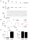
Figure 5. mTMEM16F Gives Rise to Small-Conductance Ca2+-Activated, Nonselective Cation Channels with Distinct Pharmacological Properties
(A) Noise analysis of mTMEM16F currents expressed in HEK293 cells. (i) Representative current traces recorded in the presence of various [Ca2+]i at +80 mV. (ii) 2 Hz high-pass-filtered current traces shown in (i). (iii) Enlarged high-pass-filtered current traces with 2 s duration under each [Ca2+]i. (B) The variance of the current traces shown in (A) was plotted against the current amplitudes and fitted with σ2 = IVmγ − I2/N. The obtained fitting parameters were γ = 0.47 pS and N = 67,006. The averaged channel conductance obtained from 15 different patches was γ = 0.45 ± 0.07 pS. (C) 300 μM intracellular niflumic acid (NFA) blocked most of the mTMEM16A Cl currents, but not the mTMEM16F Na+ currents. Inside-out patches from Axolotl oocytes were exposed to symmetrical 140 mM NaCl and 100 μM [Ca2+]i for mTMEM16A and symmetrical 140 mM NaMES and 100 μM [Ca2+]i for mTMEM16F. Currents were elicited by voltage pulses stepped from 380 to +80 mV. (D) Summary of the NFA effects shown in (C). INFA and IO are the maximum peak current amplitudes in the presence and absence of NFA, respectively. n = 7–8. ***p < 0.0001. Error bars represent SEM. (E) 2 mM free [AMP]i and [ATP]i had little effect on the mTMEM16F currents. Inside-out patches from Axolotl oocytes were exposed to symmetrical 140 mM NaMES, 100 μM [Ca2+]I, and AMP or ATP. IO is the maximum current amplitude in the absence of the nucleotides. n = 6–7. Error bar represents SEM. See also Figure S5.
Figure 6
Figure 6. A Residue in the Transmembrane Segment 5 Is Important for the Ion Selectivity of mTMEM16F-SCAN and mTMEM16A-CaCC Channels
(A) Sequence alignment of the transmembrane segment 5 (TM5) of human TMEM16A (h16A), murine TMEM16A (m16A), Xenopus TMEM16A (x16A), murine TMEM16B (m16B), and murine TMEM16F (m16F). (B) Representative macroscopic current traces of WT and Q559K mutant mTMEM16F channels expressed in HEK293 cell. The inside-out patches were exposed to 100 μM [Ca2+]i and symmetrical 140 mM NaCl. Testing potentials were from −80 mV to +180 mV with 20 mV increments. Both holding and repolarizing potentials were –80 mV. (C) Semi-log plots of mean activation time constants (τ) as a function of voltage for the WT and Q559K-mTMEM16F channels. t was derived from the single exponential fitting of the current traces shown in (B). n = 5–7. Error bar represents SEM. (D and E) Representative I-V relationships of the Q559K-mTMEM16F (D) and K584Q-mTMEM16A (E) mutant channels recorded from inside-out patches exposed to 14 and 140 mM [NaCl]i and 500 μM [Ca2+]i. (F and G) Summary of the reversal potential shift (ΔERev) (F) and relative ion permeability (G) for the WT and mutant channels when [NaCl]i switched from 140 mM to 14 mM. n = 8–19. ***p < 0.0001. Error bar represents SEM. All of these experiments were recordings from inside-out membrane patches from HEK293 cells.
Figure 7
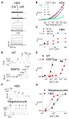
Figure 7. Gating of Heterologously Expressed mTMEM16F and Endogenous TMEM16F Channels in Megakaryocytes Is Ca2+ and Voltage Dependent
(A) Representative macroscopic current traces of an inside-out patch excised from a mTMEM16F-expressing HEK293 cell exposed to 1, 2, 7, and 100 μM [Ca2+]i. Testing potentials were from −80 mV to +180 mV with 20 mV increments. Both holding and repolarizing potentials were −80 mV. (B) Mean G-V relations of the mTMEM16F channels under different [Ca2+]i. Relative conductance was determined by measuring the amplitude of tail currents 400 μs after repolarization to a fixed membrane potential (−80 mV). The smooth curves represent Boltzmann fits I/IMax = 1/(1 + exp(−ze(VV1/2)/kT)) (see Table S1). IMax, tail current amplitude in response to deploarizaton to +180 mV in 100 μM [Ca2+]i. Error bar represents SEM. (C) Ca2+ dose-response of the mTMEM16F channel at +60, +120, and +180 mV. I/IMax values were from (B). The smooth curves represent the fits to the Hill equation: I/IMax = Amp/(1 + (KD/[Ca])H), wherein KD is the apparent dissociation constant, H is the Hill coefficient, and Amp is the maximum value of I/IMax at certain voltage. +60 mV: (KD = 10.8 ± 4.6 μM, H = 2.8); +120 mV: (KD = 5.4 ± 1.6 μM, H = 1.5); +180 mV: (KD = 3.4 ± 0.5 μM, H = 1.8). n = 5–21. Error bar represents SEM. (D) Representative inside-out patch-clamp recordings of WT (top) and E667Q mutant (bottom) mTMEM16F channels expressed in HEK293 cells. The membrane patches were voltage clamped at +60 mV and exposed to different [Ca2+]i. (E) Comparison of Ca2+ sensitivity between the WT and E667Q mutant TMEM16F channels at +60 mV. The smooth curves represent the fits to the Hill equation. WT: KD = 13.6 ± 1.8 μM, H = 2.2 ± 0.5; E667Q: KD = 2.8 ± 0.3 mM, H = 1.6 ± 0.3. n = 11–17. Error bar represents SEM. (F) Representative inside-out patch-clamp recordings from the mTMEM16F-expressing HEK293 cells (top) and megakaryocytes of WT mice (bottom). The membrane patches were voltage clamped at +60 mV and exposed to different [Ca2+]i. (G) Comparison of the Ca2+ sensitivity of the endogenous TMEM16F-SCAN current in megakaryocytes with that of the heterologously expressed TMEM16F channels in HEK293 cells at +60 mV. The smooth curves represent the fits to the Hill equation. For mTMEM16F currents in HEK293 cells, KD = 13.9 ± 2.9 μM, H = 1.6; for endogenous TMEM16F currents in megakaryocytes, KD = 5.1 ± 2.0 μM, H = 2.5. n = 6–12. Error bar represents SEM. All experiments were recordings from inside-out membrane patches in symmetrical 140 mM NaMES solutions except for the experiments shown in (D) and (E), which were done in symmetrical 140 mM NaCl solutions. See also Figure S6 and Table S1.

References

    1. Almaça J, Tian Y, Aldehni F, Ousingsawat J, Kongsuphol P, Rock JR, Harfe BD, Schreiber R, Kunzelmann K. TMEM16 proteins produce volume-regulated chloride currents that are reduced in mice lacking TMEM16A. J Biol Chem. 2009;284:28571–28578. - PMC - PubMed
    1. Ayoub C, Wasylyk C, Li Y, Thomas E, Marisa L, Robé A, Roux M, Abecassis J, de Reyniès A, Wasylyk B. ANO1 amplification and expression in HNSCC with a high propensity for future distant metastasis and its functions in HNSCC cell lines. Br J Cancer. 2010;103:715–726. - PMC - PubMed
    1. Bevers EM, Williamson PL. Phospholipid scramblase: an update. FEBS Lett. 2010;584:2724–2730. - PubMed
    1. Bevers EM, Comfurius P, Zwaal RF. Changes in membrane phospholipid distribution during platelet activation. Biochim Biophys Acta. 1983;736:57–66. - "VSports在线直播" PubMed
    1. Caputo A, Caci E, Ferrera L, Pedemonte N, Barsanti C, Sondo E, Pfeffer U, Ravazzolo R, Zegarra-Moran O, Galietta LJ. TMEM16A, a membrane protein associated with calcium-dependent chloride channel activity. Science. 2008;322:590–594. - "V体育官网入口" PubMed

Publication types

MeSH terms