<noscript lang="yzKP23u"></noscript><ins id="wa8JoINV"></ins> Skip to main page content (VSports)
U.S. flag

An official website of the United States government

Dot gov

The . gov means it’s official VSports app下载. Federal government websites often end in . gov or . mil. Before sharing sensitive information, make sure you’re on a federal government site. .

Https

The site is secure. The https:// ensures that you are connecting to the official website and that any information you provide is encrypted and transmitted securely. V体育官网.

. 2012 Sep 27;3(9):e393.
doi: 10.1038/cddis.2012.135.

Regulation of the tumor suppressor PTEN by SUMO

Affiliations

Regulation of the tumor suppressor PTEN by SUMO (VSports注册入口)

J González-Santamaría et al. Cell Death Dis. .

Abstract

The crucial function of the PTEN tumor suppressor in multiple cellular processes suggests that its activity must be tightly controlled. Both, membrane association and a variety of post-translational modifications, such as acetylation, phosphorylation, and mono- and polyubiquitination, have been reported to regulate PTEN activity. Here, we demonstrated that PTEN is also post-translationally modified by the small ubiquitin-like proteins, small ubiquitin-related modifier 1 (SUMO1) and SUMO2 VSports手机版. We identified lysine residue 266 and the major monoubiquitination site 289, both located within the C2 domain required for PTEN membrane association, as SUMO acceptors in PTEN. We demonstrated the existence of a crosstalk between PTEN SUMOylation and ubiquitination, with PTEN-SUMO1 showing a reduced capacity to form covalent interactions with monoubiquitin and accumulation of PTEN-SUMO2 conjugates after inhibition of the proteasome. Moreover, we found that virus infection induces PTEN SUMOylation and favors PTEN localization at the cell membrane. Finally, we demonstrated that SUMOylation contributes to the control of virus infection by PTEN. .


Figures

Figure 1
Figure 1
Covalent modification of PTEN by SUMO1 or SUMO2 in vitro and in vivo. (a) Recombinant PTEN protein (left panel) or in vitro translated [35S]methionine-labeled PTEN protein (right panel) was used as a substrate in an in vitro SUMOylation assay in the presence of SUMO1 or SUMO2. The reaction products were resolved on an 8% SDS-polyacrilamide gel and analyzed by western blot with anti-PTEN antibody (left panel) or dried for 1 h and exposed to X-ray film (right panel). (b) Deconjugation of SUMO1 from PTEN by SENP1. [35S]methionine-labeled PTEN-SUMO1 obtained in an in vitro SUMOylation reaction was incubated with GST-SENP1 as described in Materials and Methods. The reaction products were resolved on an 8% SDS-polyacrilamide gel, dried for 1 h, and exposed to X-ray film. (c) HEK-293 cells were co-transfected with HA-PTEN together with pcDNA, pcDNA-Ubc9 and pcDNA-His6-SUMO1 or pcDNA-Ub9 and pcDNA-His6-SUMO2. Total protein extracts and the Histidine-tagged proteins purified using nickel columns were then resolved on an 8% SDS-polyacrilamide gel and analyzed by western blot with anti-HA antibody. (d) HEK-293 cells were transfected with pcDNA or pcDNA-Ubc9 and pcDNA-His6-SUMO2. Total protein extracts and the Histidine-tagged proteins purified using nickel columns were then analyzed by western blot with anti-PTEN antibody
Figure 2
Figure 2
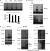
Identification of Lysines 266 and 289 as SUMO targets. (a) [35S]methionine-labeled wild-type or mutant PTEN proteins were used as substrates in an in vitro SUMOylation assay in the presence of SUMO1 as indicated. The reaction products were resolved on an 8% SDS-polyacrylamide gel, dried for 1 h, and exposed to an X-ray film. The intensity of SUMO-modified and -unmodified PTEN bands was quantified by densitometry using ImageJ software, and the ratio of SUMOylated to non-SUMOylated PTEN was normalized to that of the WT protein (right panel). The data represented in right panel are means ± S.D. of five independent experiments. *P<0.05; **P<0.005, by Student's t-test compared with PTEN WT. (b) Total protein extracts and Histidine-tagged purified proteins prepared from HEK-293 cells co-transfected with WT HA-PTEN or the different HA-PTEN mutants together with pcDNA or pcDNA-Ubc9 and pcDNA-His6-SUMO2, were resolved on an 8% SDS-polyacrilamide gel and analyzed by western blot with anti-HA antibody
Figure 3
Figure 3
Cytoplasmic localization of SUMOylation-deficient PTEN mutants. (a) MCF-7 cells were transfected with plasmids encoding WT or mutant HA-PTEN proteins and then stained with anti-HA antibody followed by Alexa 594-conjugated anti-mouse antibody and DAPI. Subcellular localization of the expressed proteins was analyzed under a confocal laser scanning microscope. Images were processed with Adobe Photoshop. Right panel shows quantification of the subcellular distribution of PTEN in at least 200 transfected cells per sample. (b) MCF-7 cells were transfected as described above and at 48 h after transfection, nuclear and cytoplasmic fractions were isolated, resolved on an 12% SDS-polyacrylamide gel and analyzed by western blot with the indicated antibodies. (c) PC3 cells were transfected with plasmids encoding WT or mutant HA-PTEN proteins, then stained with anti-PTEN antibody followed by Alexa 488-conjugated anti-mouse antibody and DAPI, and finally analyzed by confocal microscopy. Right panel shows quantification of the subcellular distribution of PTEN in at least 200 transfected cells per sample. **P<0.005, ***P<0.0005, by Student's t-test compared with PTEN WT
Figure 4
Figure 4
Crosstalk between SUMOylation and ubiquitination of PTEN. (a) [35S]methionine-labeled wild-type or mutant PTEN proteins were used as substrates in an in vitro ubiquitination assay in the presence of Ub-KO as indicated. The reaction products were resolved on an 8% SDS-polyacrylamide gel, dried for 1 h, and exposed to an X-ray film. The intensity of ubiquitin-modified and -unmodified PTEN bands was quantified by densitometry using ImageJ software, and the ratio of ubiquitinated to non-ubiquitinated PTEN was normalized to that of the WT protein (lower panel). The data represented are means±S.D. of at least three independent experiments. *P<0.05, by Student's t-test compared with PTEN WT. (b) Recombinant PTEN protein was used as a substrate in an in vitro SUMOylation assay in the presence or absence of SUMO1 and then the products of the SUMOylation reactions were used as substrates in an in vitro ubiquitination assay as indicated. The reaction products were then resolved on an 8% SDS-polyacrylamide gel and analyzed by western blot with anti-PTEN antibody. Membranes were subsequently washed and then incubated with anti-ubiquitin antibody. (c) HEK-293 cells were co-transfected with HA-PTEN together with pcDNA or Ubc9 and His6-SUMO2 and at 24 h after transfection cells were treated with MG132 (25 mM) for 12 h. Total protein extracts and the Histidine-tagged proteins purified using nickel columns were then resolved on an 8% SDS-polyacrylamide gel and analyzed by western blot with the indicated antibodies. Ub, ubiquitin
Figure 5
Figure 5
VSV infection promotes SUMOylation of PTEN. (a) MEFs WT (left panel) or MCF-7 cells transfected with HA-PTEN (right panel) were left uninfected (U) or infected with VSV for the indicated times and then cell lysates were resolved on an 8% SDS-polyacrylamide gel, and analyzed by western blot with anti-PTEN (left panel) or anti-HA antibody (right panel). (b) HEK-293 cells were co-transfected with HA-PTEN together with pcDNA, pcDNA-Ubc9 and pcDNA-His6-SUMO2 and 24 h after transfection cells were infected with VSV at MOI of 5 PFU/ml. Total protein extracts and the Histidine-tagged proteins purified using nickel columns at the indicated times after infection were resolved on an 8% SDS-polyacrylamide gel and analyzed by western blot with the indicated antibodies
Figure 6
Figure 6
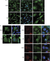
VSV infection promotes membrane localization of PTEN. (a) MEFs WT uninfected or infected with VSV for 4 h were analyzed by immunofluorescence staining for subcellular localization of PTEN. Cells were stained with anti-PTEN antibody followed by Alexa 488-conjugated anti-mouse antibody and DAPI, and finally analyzed by confocal microscopy. Digital zoom ( × 2) of the indicated area is shown in the right panels. (b) PTEN-null U251MG cells transfected with HA-PTEN, uninfected or infected with VSV for 4 h were analyzed by immunofluorescence staining for subcellular localization of PTEN. Cells were stained with anti-HA antibody followed by Alexa 488-conjugated anti-mouse antibody and DAPI, and finally analyzed by confocal microscopy. (c) Co-localization of PTEN and SUMO at the plasma membrane. MEFs were infected with VSV, fixed and stained with anti-PTEN and anti-SUMO1 or anti-SUMO2 antibodies and DAPI. The images were analyzed by confocal microscopy
Figure 7
Figure 7
Effect of PTEN WT or PTEN SUMOylation mutants on VSV replication. (a) U251MG cells were transfected in triplicate with the indicated plasmids and at 48 h after transfection, cells were infected with VSV at MOI of 5 and quantification of the virus yield after total destruction of the monolayer was assessed. Data represents means±S.E. for one experiment. *P<0.05, compared with pcDNA transfected cells, Student's t-test. Protein extracts from the transfected cells were loaded on an 8% SDS-polyacrylamide gel and revealed on western blot by the indicated antibodies. The lower panel represents the expression levels of PML-IV (positive control) and PTEN genes transfected in two wells of one experiment. (b) U251MG cells were transfected in triplicate with the indicated plasmids and at 48 h after transfection cells were infected with VSV at MOI of 5 and quantification of the virus yield after total destruction of the monolayer was assessed. Data represents means±S.E. for one experiment. *P<0.05, Student's t-test. Protein extracts from the transfected cells were loaded on an 8% SDS-polyacrylamide gel and revealed on western blot by PTEN antibody. (c) PTEN+/− MEFs were nucleofected with the indicated plasmids and at 48 h after transfection, cells were infected with VSV at MOI of 5. At the indicated times, protein extracts were loaded on a 12% SDS-polyacrylamide gel and revealed on western blot by antibodies against the M protein from VSV. The right panel represents the expression levels of PTEN genes transfected in the cells

"VSports" References

    1. Li DM, Sun H. TEP1, encoded by a candidate tumor suppressor locus, is a novel protein tyrosine phosphatase regulated by transforming growth factor beta. Cancer Res. 1997;57:2124–2129. - PubMed
    1. Li J, Yen C, Liaw D, Podsypanina K, Bose S, Wang SI, et al. PTEN, a putative protein tyrosine phosphatase gene mutated in human brain, breast, and prostate cancer. Science. 1997;275:1943–1947. - PubMed
    1. Steck PA, Pershouse MA, Jasser SA, Yung WK, Lin H, Ligon AH, et al. Identification of a candidate tumour suppressor gene, MMAC1, at chromosome 10q23.3 that is mutated in multiple advanced cancers. Nat Genet. 1997;15:356–362. - VSports app下载 - PubMed
    1. Cantley LC, Neel BG. New insights into tumor suppression: PTEN suppresses tumor formation by restraining the phosphoinositide 3-kinase/AKT pathway. Proc Natl Acad Sci USA. 1999;96:4240–4245. - "V体育安卓版" PMC - PubMed
    1. Di Cristofano A, Pandolfi PP. The multiple roles of PTEN in tumor suppression. Cell. 2000;100:387–390. - "V体育安卓版" PubMed

V体育平台登录 - Publication types

MeSH terms

LinkOut - more resources