<b dir="vogbXs"></b>
Skip to main page content
U.S. flag

An official website of the United States government

Dot gov

The . gov means it’s official. Federal government websites often end in . gov or . mil. Before sharing sensitive information, make sure you’re on a federal government site VSports app下载. .

Https

The site is secure V体育官网. The https:// ensures that you are connecting to the official website and that any information you provide is encrypted and transmitted securely. .

. 2012 Oct;122(10):3516-28.
doi: 10.1172/JCI63352. Epub 2012 Sep 24.

Pancreas-specific deletion of mouse Gata4 and Gata6 causes pancreatic agenesis

Affiliations

Pancreas-specific deletion of mouse Gata4 and Gata6 causes pancreatic agenesis

Shouhong Xuan et al. J Clin Invest. 2012 Oct.

Abstract

Pancreatic agenesis is a human disorder caused by defects in pancreas development. To date, only a few genes have been linked to pancreatic agenesis in humans, with mutations in pancreatic and duodenal homeobox 1 (PDX1) and pancreas-specific transcription factor 1a (PTF1A) reported in only 5 families with described cases. Recently, mutations in GATA6 have been identified in a large percentage of human cases, and a GATA4 mutant allele has been implicated in a single case. In the mouse, Gata4 and Gata6 are expressed in several endoderm-derived tissues, including the pancreas. To analyze the functions of GATA4 and/or GATA6 during mouse pancreatic development, we generated pancreas-specific deletions of Gata4 and Gata6. Surprisingly, loss of either Gata4 or Gata6 in the pancreas resulted in only mild pancreatic defects, which resolved postnatally. However, simultaneous deletion of both Gata4 and Gata6 in the pancreas caused severe pancreatic agenesis due to disruption of pancreatic progenitor cell proliferation, defects in branching morphogenesis, and a subsequent failure to induce the differentiation of progenitor cells expressing carboxypeptidase A1 (CPA1) and neurogenin 3 (NEUROG3). These studies address the conserved and nonconserved mechanisms underlying GATA4 and GATA6 function during pancreas development and provide a new mouse model to characterize the underlying developmental defects associated with pancreatic agenesis. VSports手机版.


Figures
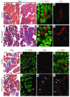
Figure 1
Figure 1. Pancreas-specific deletion of either Gata4 or Gata6 causes mild embryonic pancreas defects.
(AD) H&E staining of representative sections of E18.5 dorsal pancreas from control Gata4fl/fl (A and C) and Pdx1Cre;Gata4fl/fl embryos (B and D). (EH) Immunofluorescence staining for Sox9 (red) and Cpa1 (green) of E18.5 dorsal pancreas from control Gata4fl/fl (E and G) and Pdx1Cre;Gata4fl/fl embryos (F and H). Cpa1 is expressed in acinar cells (E and F). Sox9 is expressed in ductal cells and centroacinar cells in both control and Pdx1Cre;Gata4fl/fl embryos (E and F). In acinar cells, Sox9 is expressed in control pancreas (G), but is absent in most Gata4-depleted acinar cells of Pdx1Cre;Gata4fl/fl pancreas (H). Original magnification, ×200 (A and B); ×640 (CH). (I and J) H&E staining of representative sections of E18.5 control Gata6fl/+ (I) and Pdx1Cre;Gata6fl/fl (J) embryos. (KP) Immunofluorescence staining of E18.5 control Gata6fl/+; R26R-LacZ (K, M, and O) and Pdx1Cre;Gata6fl/fl (L, N, and P) embryos. Cpa1 (green) expression is in the acinar cells, and DBA (red) marks the epithelial ductal regions. Increased ductal tissues are observed in Pdx1Cre;Gata6fl/fl pancreas (arrows in J, N, and P). Original magnification, ×200 (IN); ×640 (O and P).
Figure 2
Figure 2. Mice retaining only 1 allele of Gata4 or 1 allele of Gata6 display dissimilar pancreatic defects.
Representative E18.5 pancreata from embryos retaining only 1 allele of Gata4 or Gata6 were analyzed in whole mounts (AC), H&E-stained sections (DF), and coimmunofluorescence staining with CK19 (green) and insulin (red) (GI). In comparison with control pancreata (A, D, and G), the pancreata in Pdx1Cre;Gata4fl/+;Gata6fl/fl mice display more ductal structures (E, H, and J) and have insulin-expressing cells residing within the ductal epithelium (arrows in J). In Pdx1Cre;Gata4fl/fl;Gata6fl/+ embryos, pancreas volume is significantly reduced due to decreased acinar tissue (C, F, and I). Original magnification, ×14 (A); ×20 (B and C); ×200 (DI); ×400 (J).
Figure 3
Figure 3. Pancreas-specific deletion of Gata4 and Gata6 causes pancreatic agenesis.
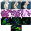
(A and B) Dissected pancreas, stomach, spleen, and intestine of newborn control (A) and DKO (B) pups. The DKO pancreas is predominantly made up of mesenchymal-like stromal tissue. (C and D) H&E staining of representative sections from E18.5 embryos shows pancreas tissue occupying an area from the body wall to the midline in the control (C), whereas at the same plane of section, there is no apparent pancreas in the DKO (D). (EG) LacZ staining of pancreatic progeny in whole E18.5 pancreas tissue shows fully developed pancreas in control embryos (E and F) and no apparent pancreatic tissue in the DKO embryos (G). Control pancreas from animals lacking the ROSA26:LacZ allele display endogenous LacZ staining in the gut tube (E). P, pancreas; Stm, stomach; Sp, spleen; Liv, liver; Kid, kidney. Pancreas or the region that should contain the pancreas is delineated by dashed lines in each panel. Original magnification, ×15 (A and B); ×25 (C and D); ×40 (E, F, and G).
Figure 4
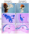
Figure 4. Pancreatic buds are specified in the E9.5 DKO embryos.
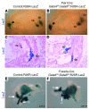
(A and B) Whole-mount LacZ labeling of Pdx1 progeny in E9.5 embryos shows distinctively formed pancreatic buds in both controls (A) and DKO embryos (B). A LacZ-positive region (arrowhead) was also detected in the hypothalamus region, as reported by Wicksteed et al. (53). (C and D) Representative sections of the pancreatic region from embryos shown in A and B confirm the presence of the dorsal and ventral buds in both control (C) and DKO embryos (D). (E and F) Whole-mount LacZ staining of Foxa3 lineage cells in control and embryos deleted for Gata4 and Gata6 in the Foxa3-Cre prepancreatic endodermal domain. Similar to the phenotype of embryos deleted for Gata4 and Gata6 in the later Pdx1 domain, dorsal and ventral pancreatic buds form in control E11.5 embryo (A) and in Foxa3-cre;Gata4fl/fl;Gata6fl/fl embryos (B). dp, dorsal pancreas; vp, ventral pancreas. Original magnification, ×40 (A and B); ×100 (C and D); ×80 (E and F).
Figure 5
Figure 5. At E10.5, fewer endoderm cells are differentiated into Pdx1+ pancreatic progenitor cells, due partially to the loss of Gata4 regulation of Pdx1 gene expression.
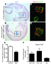
(AD) Representative sagittal sections from E10.5 LacZ-stained control (A) and DKO (B) embryos. The regions containing the pancreatic buds are highlighted by boxes. Pancreatic bud formation appears morphologically normal in the DKO embryo (boxed areas in A and B). (C and D) Immunofluorescence costaining using anti-Pdx1 and anti-Foxa1/2 antibodies shows that there is a smaller percentage of Pdx1+ pancreatic progenitor cells relative to the number of Foxa1/a2+ cells in the pancreatic endodermal region of DKO embryos (D vs. C). (E) Cell quantification of the relative number of Pdx1+ cells within the Foxa1/a2+ foregut endoderm (n = 4), p, pancreas; g, gut. ***P < 0.001. (F) Real-time qPCR analysis of DNA products immunoprecipitated by an anti-Gata4 antibody in 266-6 cells. Gata consensus sequences within Pdx1 enhancer area III (–1841 bp from the ATG) and Pdx1 enhancer area IV (–6010 bp from the ATG) are bound by the Gata4 protein. Gata4 does not bind the Arx promoter, which serves as a negative control. *P < 0.05. Original magnification, ×50 (A and B); ×200 (C and D).
Figure 6
Figure 6. Morphological and molecular defects in DKO pancreas are apparent during secondary transition.
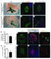
(A and B) Whole-mount LacZ lineage labeling of Pdx1 progeny in control (A) versus DKO (B) embryos indicates that by E11.5, the DKO embryos have smaller pancreatic buds. (C and D) Coimmunofluorescence staining of caspase 3 (red) and Pdx1 (green) in E11.5 sections of control (C) vs. DKO embryos (D) suggests that there is no obvious change in apoptosis in the DKO pancreas. Insets show positively caspase 3–labeled tissue from the same sections. (E and F) Coimmunofluorescence staining of BrdU (red) and Pdx1 (green) in E11.5 sections of control (E) vs. DKO embryos (F) indicates there are fewer proliferating Pdx1+ pancreatic cells. (G) Quantification of BrdU-labeled Pdx1+ cells indicates a significant (28%) reduction in the number of replicating Pdx1+ progenitor cells in DKO embryos. (H) FACS analysis of PI-stained cells to quantify overall cellular proliferation shows significant (38%) reduction of S phase cells in the DKO pancreatic buds. (I and J) Immunofluorescence staining of Sox9 (green) in the E12.5 ventral pancreatic bud. (K and L) Coimmunofluorescence staining of Cpa1 (green), Pdx1 (red), and Neurog3 (white) in the ventral pancreatic bud indicates that the DKO embryos are defective in pancreatic cell differentiation at E12.5, with the loss of multipotent progenitor marker Cpa1 and endocrine progenitor marker Neurog3 (n = 4; 20 sections evenly distributed through the entire pancreas were analyzed for each n). (M and N) Coimmunofluorescence staining of Ptf1a (red) and glucagon (green) in the E12.5 ventral pancreatic bud shows more centralized Pft1a distribution and much reduced glucagon-positive cells (N vs. M) *P < 0.05. Original magnification, ×60 (A and B); ×200 (CF); ×400 (IN, and insets).
Figure 7
Figure 7. Outgrowth of the pancreas during the secondary transition is defective in DKO embryos.
(AD) LacZ staining of Pdx1 progeny at E13.5. LacZ whole-mount staining shows the normal branched structure of the pancreas in a representative control embryo (A), whereas only a small group of labeled cells are present in the ventral bud region of DKO pancreas (B). Sections of the ventral pancreatic regions show extensive branching morphogenesis in the control pancreas (C) and an unbranched structure of tightly clustered cells in the DKO pancreas (D). Note the expansion of LacZ-labeled Pdx1 progeny cells into the proximal stomach (B). Sections of the stomachs from control (E) and DKO (F) embryos demonstrate that Pdx1 descendent cells are found in the anterior part of the stomach (arrows in F). Panc, pancreas; Liv, liver; Stm, stomach. Original magnification, ×40 (A and B); ×200 (C and D); ×100 (E and F).
Figure 8
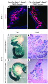
Figure 8. At E17.5, pancreatic lineage cells remain unbranched and undifferentiated.
(A and B) The Rosa26:Tomato reporter (red) labels the Pdx1+ lineage in control (A) and DKO (B) pancreatic epithelium at E15.5. By this stage of development, the Pdx1+ progenies are restricted to a relatively unbranched single-cell–layered epithelium in the DKO pancreas. (C and D) Whole-mount and sections of LacZ-stained E17.5 control (C and E) and DKO (D and F) abdominal organs show well-extended glandular structure in the control pancreas (C) and only a small pancreatic remnant in the DKO (D). Sections through the remaining (ventral) pancreatic remnant in the DKO show LacZ-labeled Pdx1 progenies present in unbranched cystic ductal structures, and there is no morphological evidence of differentiated pancreatic tissue (F) as compared with the control sections (E). Note the extension of Pdx1-lineage–labeled cells in the proximal stomach in the DKO embryo (arrows in D). Original magnification, ×200 (A and B); ×20 (C and D); ×200 (E and F).

Comment in

References (VSports在线直播)

    1. Pan FC, Wright C. Pancreas organogenesis: from bud to plexus to gland. Dev Dyn. 2011;240(3):530–565. doi: 10.1002/dvdy.22584. - DOI - PubMed
    1. Jorgensen MC, Ahnfelt-Ronne J, Hald J, Madsen OD, Serup P, Hecksher-Sorensen J. An illustrated review of early pancreas development in the mouse. Endocr Rev. 2007;28(6):685–705. doi: 10.1210/er.2007-0016. - DOI - PubMed
    1. Bort R, Martinez-Barbera JP, Beddington RS, Zaret KS. Hex homeobox gene-dependent tissue positioning is required for organogenesis of the ventral pancreas. Development. 2004;131(4):797–806. doi: 10.1242/dev.00965. - DOI - PubMed
    1. Wandzioch E, Zaret KS. Dynamic signaling network for the specification of embryonic pancreas and liver progenitors. Science. 2009;324(5935):1707–1710. doi: 10.1126/science.1174497. - DOI - PMC - PubMed
    1. Pictet R, Rutter WJ. Handbook of Physiology, Section 7: Endocrinology. 1972. Development of the embryonic endocrine pancreas. In: Steiner DF, Freinkel N, eds. pp. 25–66. Vol. 1. Washington, DC, USA: American Physiological Society;

VSports手机版 - Publication types

MeSH terms