Skip to main page content
U.S. flag

An official website of the United States government

Dot gov

The VSports app下载. gov means it’s official. Federal government websites often end in . gov or . mil. Before sharing sensitive information, make sure you’re on a federal government site. .

Https

The site is secure. The https:// ensures that you are connecting to the official website and that any information you provide is encrypted and transmitted securely. V体育官网.

. 2012 Sep 3:3:277.
doi: 10.3389/fimmu.2012.00277. eCollection 2012.

Monosodium urate crystals induce extracellular DNA traps in neutrophils, eosinophils, and basophils but not in mononuclear cells

Affiliations

Monosodium urate crystals induce extracellular DNA traps in neutrophils, eosinophils, and basophils but not in mononuclear cells

Christine Schorn et al. Front Immunol. .

Abstract (V体育平台登录)

Neutrophil extracellular traps (NETs) are fibers of extracellular DNA released from neutrophils due to overwhelming phagocytic stimuli. The function of NETs is to trap and kill microbes to avoid spreading of potential pathogens. NETs are formed after encounter with various gram-positive and -negative bacteria but also in response to mediators causing sterile inflammation like interleukin-8 (IL-8), tumor necrosis factor (TNF), and phorbol myristate acetate (PMA). Here we show the formation of NETs (NETting) in response to monosodium urate (MSU) crystals as further model for sterile inflammation. We identified monocytes, neutrophils, and eosinophils as MSU phagocytosing cells. Basophils did not take up the crystals, instead they upregulated their activation marker CD203c after contact with MSU. Nevertheless, MSU crystals induced extracellular trap formation also in basophils, like in eosinophils and neutrophils, which phagocytose the crystals. In contrast, monocytes do not form NETs despite uptake of the MSU crystals. In contrast to the canonical stimuli like bacteria and PMA, MSU-induced NETosis was not abrogated by plasma VSports手机版. Our data show that MSU crystals induce extracellular DNA trap formation in all three granulocytes lineages (NETs, EETs, and BETs) but not in monocytes, and DNA externalization does not necessitate the uptake of the crystals. .

Keywords: MSU; NETs; PMA; bacteria; granulocytes; inflammation; neutrophil extracellular traps. V体育安卓版.


Figures
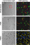
Figure 1
Figure 1
MSU crystals induce NET formation in PMN but not in PBMC. Whole blood cells (A), isolated PMN (B) and isolated PBMC (C) were incubated with MSU crystals. Cytospins were prepared and stained for nuclear and extranuclear DNA. The transmission microscopy (left) shows phagocytes which have ingested MSU crystals. The DNA staining (right) shows NETting granulocytes in whole blood cells (A, red square) and isolated PMN (B). No MSU-induced extracellular DNA was observed in isolated PBMC (C). In (A) and (B) erythrocytes and free MSU crystals were dissolved by acid treatment. (A–C) NETs were artificial colored in green, PMN in red, mononuclear cells in blue and MSU crystals in yellow. All experiments were performed at least 3 times. Scale bars, 100 μm.
Figure 2
Figure 2
MSU induced NETs contain histon H3. (A–D) Whole blood cells were incubated with MSU crystals. Cytospins were prepared and stained for nuclear and extranuclear DNA (B) and histon H3 (C). The merged picture (D) shows the co-localization of DNA and histon H3. Erythrocytes and free MSU crystals were dissolved by acid treatment. The MSU crystals were artificial colored in white. All experiments were performed at least 3 times. Scale bars, 100 μm.
Figure 3
Figure 3
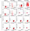
Monocytes, neutrophils and eosinophils ingest MSU crystals. (A–H) Whole blood cells were incubated with MSU crystals. After lysis of the erythrocytes the morphology (forward scatter, side scatter) of the leukocytes was characterized by flow cytometry. The individual leukocyte populations were defined by cell lineage-specific surface markers. The uptake of crystals was reflected by dramatic increases of the cells' SSc values after phagocytosis. Monocytes (A) neutrophils (B) and eosinophils (C) ingested MSU crystals. Basophils (D) did not take up the crystals, but upregulated their surface activation marker CD203c. T cells (E) B cells (F) NK cells (G) and pDCs (H) did not increase the side scatter in response to MSU crystals. SSc, side scatter; FSc, forward scatter; Mono, monocytes; neutro, neutrophils; eos, eosinophils; baso, basophils; NK, natural killer cells; pDC, plasmacytoid dendritic cells. All experiments were performed at least three times.
Figure 4
Figure 4
Neutrophils, eosinophils and basophils form extracellular traps after incubation with MSU. (A–C) Isolated neutrophils (A), eosinophils (B) or basophils (C) in plasma were incubated without (upper) or with MSU crystals (lower). Cytopsins were prepared and stained for extranuclear DNA. The DNA staining showed extracellular trap formation and clumping in neutrophils, eosinophils and basophils. (D) Cytokine/chemokine induction of eosinophils and neutrophils were analyzed after MSU incubation. Eosinophils produce traces of IL-6 and intermediate amounts of IL-8 after MSU incubation. Neutrophils release high amounts of IL-6 and IL-8 after incubation with MSU crystals. Values of mock-treated cells served as baseline. (E) Isolated eosinophils were incubated without (left), with MSU crystals (middle), or with silica crystals (right). The DNA staining shows extracellular trap formation (arrows) only in samples with MSU crystals. All experiments were performed at least three times. Scale bars, 100 μm; ***P < 0.001.
Figure 5
Figure 5
MSU induced NETting of neutrophils is not inhibited by plasma. (A–D) Isolated neutrophils were incubated without stimuli (A), with PMA (B), with bacteria (C), or with MSU crystals (D) in the presence of different concentrations of plasma (0% upper; 20% middle, 100% lower). High plasma concentrations inhibit NETting induced by PMA (B) and by bacteria (C). MSU induced NETting (D) is not inhibited and rather increased by high concentrations of plasma. All experiments were performed at least three times. Scale bars, 100 μm.

References

    1. Borregaard N., Cowland J. B. (1997). Granules of the human neutrophilic polymorphonuclear leukocyte. Blood 89, 3503–3521 - PubMed
    1. Brill A., Fuchs T. A., Savchenko A. S., Thomas G. M., Martinod K., De Meyer S. F., Bhandari A. A., Wagner D. D. (2012). Neutrophil extracellular traps promote deep vein thrombosis in mice. J. Thromb. Haemost. 10, 136–144 10.1111/j.1538-7836.2011.04544.x - DOI - PMC - PubMed
    1. Brinkmann V., Reichard U., Goosmann C., Fauler B., Uhlemann Y., Weiss D. S., Weinrauch Y., Zychlinsky A. (2004). Neutrophil extracellular traps kill bacteria. Science 303, 1532–1535 10.1007/978-1-61779-527-5_7 - DOI - PubMed
    1. Brinkmann V., Zychlinsky A. (2007). Beneficial suicide: why neutrophils die to make NETs. Nat. Rev. Microbiol. 5, 577–582 10.1038/nrmicro1710 - DOI - PubMed
    1. Burnett D., Chamba A., Stockley R. A., Murphy T. F., Hill S. L. (1993). Effects of recombinant GM-CSF and IgA opsonisation on neutrophil phagocytosis of latex beads coated with P6 outer membrane protein from Haemophilus influenzae. Thorax 48, 638–642 - PMC (VSports注册入口) - PubMed

LinkOut - more resources