Skip to main page content
U.S. flag

An official website of the United States government

Dot gov

The . gov means it’s official VSports app下载. Federal government websites often end in . gov or . mil. Before sharing sensitive information, make sure you’re on a federal government site. .

Https

The site is secure. The https:// ensures that you are connecting to the official website and that any information you provide is encrypted and transmitted securely. V体育官网.

. 2012 Oct;13(10):991-9.
doi: 10.1038/ni.2416. Epub 2012 Sep 9.

Induction and molecular signature of pathogenic TH17 cells

Affiliations

Induction and molecular signature of pathogenic TH17 cells

Youjin Lee et al. Nat Immunol. 2012 Oct.

Abstract

Interleukin 17 (IL-17)-producing helper T cells (T(H)17 cells) are often present at the sites of tissue inflammation in autoimmune diseases, which has led to the conclusion that T(H)17 cells are main drivers of autoimmune tissue injury. However, not all T(H)17 cells are pathogenic; in fact, T(H)17 cells generated with transforming growth factor-β1 (TGF-β1) and IL-6 produce IL-17 but do not readily induce autoimmune disease without further exposure to IL-23. Here we found that the production of TGF-β3 by developing T(H)17 cells was dependent on IL-23, which together with IL-6 induced very pathogenic T(H)17 cells. Moreover, TGF-β3-induced T(H)17 cells were functionally and molecularly distinct from TGF-β1-induced T(H)17 cells and had a molecular signature that defined pathogenic effector T(H)17 cells in autoimmune disease VSports手机版. .


Figures

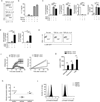
Figure 1
Figure 1. The induction of TGF-β3 in TH17 cells
(a) Mean clinical scores (disease incidence) in wild-type recipients 30 days after transfer of naïve CD4+ T cells (5×10⁶ cells) from 2D2 transgenic mice that were differentiated in vitro with TGF-β1-IL-6-IL-23 or TGF-β1-IL-6. (b) Quantitative RT-PCR analysis of Tgfb3 mRNA in naïve CD4+ T cells differentiated for four day in vitro with TGF-β1-IL-6-IL-23 or no cytokines. (c) Quantitative RT-PCR analysis of Tgfb3 mRNA in naïve CD4+ T cells from wild-type (WT) or Il23r−/− mice differentiated in vitro with TGF-β1-IL-6-IL-23 or no cytokines. (d) Flow cytometry analysis of TGF-β3-YFP expression in CD4+ T cells, CD8+ T cells, γδ T cells and B cells from the lymph nodes and spleen of TGF-β3crexRosa26yfp mice. (e) Intracellular cytokine staining showing co-expression of TGF-β3-YFP with either IL-17 or IFN-γ in naïve CD4+ T cells from MOG35–55+CFA immunized TGF-β3crexRosa26yfp mice stimulated with PMA+ionomycin four days post in vitro culture with or without IL-23. (f) Intracellular cytokine staining showing IL-17 and IFN-γ expression in PMA+ionomycin ex-vivo stimulated splenocytes from MOG35–55-immunized TGF-β3Cre+xRosaDTR and TGF-β3Cre-xRosaDTR littermate control (WT) mice treated with diptheria toxin on day 4 post-immunization. (g) IL-17 expression in naïve TGF-β3fl/fl CD4+ T cells cultured in vitro with IL-1β-IL-6-IL-23 (TH17) or no cytokines (TH0) then retrovirally transduced with Cre-GFP to delete TGF-β3. All data are a representative of more than three independent experiments with similar results. Statistical significance of *p<0.05, **p<0.01, or ***p<0.001 is indicated for the RT-PCR data. Error bars indicate mean ± s.d.
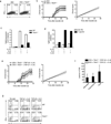
Figure 2
Figure 2. TGF-β3-induced TH17 cells are highly pathogenic in inducing autoimmunity
(a) Intracellular cytokine staining showing IL-17 expression of PMA+ionomycin stimulated naïve CD4+ T cells from C57BL/6 (wt) mice differentiated in vitro with TGF-β1-IL-6 or TGF-β3-IL-6 for four days. (b) ELISA showing IL-17 production from supernatants of naïve CD4+ T cells from C57BL/6 (wt) mice stimulated in vitro with TGF-β1, TGF-β3, TGF-β1-IL-6, TGF-β3-IL-6 for four days. (c,d) Quantitative RT-PCR of Rorc, IL-17a, IL-17f, IFNγ, Il23r and Il22 mRNA expression from wt naïve CD4+ T cells stimulated in vitro with TGF-β1-IL-6, TGF-β3-IL-6 or no cytokines for four days. (e) Flow cytometry analysis of IL-23R-GFP expression of naive CD4+ T cells from IL-23R-GFP knock-in mice differentiated in vitro with TGF-β1-IL-6, TGF-β3-IL-6 or no cytokines for four days (f) Mean clinical scores (disease incidence) in wild-type recipients 30 days after transfer of naïve CD4+ T cells (5×10⁶ cells) from 2D2 transgenic mice that were differentiated in vitro with TGF-β1-IL-6 or TGF-β3-IL-6. Statistical analysis by linear regression curve was performed and graphed including the 95% confidence band of the regression line (***p<0.001) (g) Quantification of CNS lesions in the meninges and parenchyma from recipient mice sacrificed on day 30 (**p<0.01). Data is pooled from three independent experiments (n=6). (h) TGF-β signaling PCR array analysis of Smad 1, 3, 5 from wt naïve CD4+ T cells differentiated with TGF-β1-IL-6 or TGF-β3-IL-6 for 24 hours. cDNA was prepared and processed according to manufacturer’s instruction (Sabioscience, CA). The graph illustrates the relative fold change in the Smads of TGF-β3-IL-6 condition compared to TGF-β1-IL-6. The dotted line represents the normalized expression of TGF-β1-IL-6 set at one for each of the Smads. The data is pooled from three independent replicates. (i) Flow cytometry analysis showing p-smad1&5 or p-smad3 expression from wt naïve CD4+ T cells stimulated with TGF-β1 or TGF-β3 for 30 minutes. Adoptive transfer data from (f) is a representative of four independent experiments. All data are a representative of more than three independent experiments, unless otherwise mentioned, with similar results. Statistical significance of *p<0.05, **p<0.01, or ***p<0.001 is indicated for the RT-PCR data. Error bars indicate mean ± s.d.
Figure 3
Figure 3. The pathogenicity of TGF-β3–IL-6 TH17 cells are highly comparable to IL-1β-IL-6-IL-23 TH17 cells
(a) Flow cytometry analysis showing IL-17-GFP expression in naïve CD4+ T cells from the IL-17-GFP knock-in mice differentiated in vitro with IL-1β-IL-6-IL-23, TGF-β3-IL-6, or no cytokines for four days. (b) Quantitative RT-PCR analysis of Il23r mRNA expression from naïve CD4+ T cells from wt mice differentiated in vitro with IL-1β-IL-6-IL-23, TGF-β3-IL-6, or no cytokines for four days. (c) Mean clinical scores (disease incidence) in wild-type recipients 30 days after transfer of naïve CD4+ T cells (5×10⁶ cells) from 2D2 transgenic or 2D2xIL-23R−/− mice that were differentiated in vitro with IL-1β-IL-6-IL-23 or TGF-β3-IL-6. Statistical analysis by linear regression curve was performed and graphed including the 95% confidence band of the regression line. (ns) denotes not significant. (d) Quantitative RT-PCR showing Tgfb3 mRNA expression of CNS-infiltrating CD4+ T cells isolated from MOG35–55+CFA+pertussis toxin (PT) immunized wt mice with either score 2–3 (peak of disease) or score 0 (no disease). Figure represents data of twelve individual mice pooled from three independent experiments. (e) Mean clinical scores (disease incidence) in wt mice immunized with MOG35–55+CFA+PT with either isotype control or anti-TGF-β3 neutralizing antibody in the emulsion then monitored for 21 days. Statistical analysis by linear regression curve was performed and graphed including the 95% confidence band of the regression line. Adoptive transfer data from (c) is pooled data from three independent experiments (n=8) and (e) is pooled data from two independent experiments (n=10). Cumulative data on the number of animals used are shown in Table 1. All in vitro experiments are a representative of at least three independent experiments unless specified differently. Error bars indicate mean ± s.d. (**p<0.01). (ns) denotes not significant.
Figure 4
Figure 4. Identification of transcriptional signature for pathogenic TH17 cells
(a) A heat map of the microarray analysis showing differential expression of 434 genes that were up- or down-regulated (>2 fold; compared with TH0 cells) from wt naïve CD4+ T cells differentiated in vitro with IL-1β-IL-6, IL-1β-IL-6-IL-23, TGF-β1-IL-6, TGF-β1-IL-6-IL-23, TGF-β3-IL-6, or TGF-b3-IL-6-IL-23 for 60 hours. (b) Bar graph representing analysis of 23 genes selected from a pool of 233 targets that had greater than 1.5 fold difference between TGF-β3-IL-6 and TGF-β1-IL-6 differentiated TH17 cells. Data is represented as a fold change of TGF-β3-induced TH17 cells over TGF-β1-induced TH17 cells. Up-regulated genes denote a “pathogenic signature” from TGF-β3-induced TH17 cells while down-regulated genes are “nonpathogenic signature” from TGF-β1-induced TH17 cells. (c) Quantitative RT-PCR analysis showing mRNA expression of several targets highlighted in (b) from wt naïve CD4+ T cells differentiated in vitro with TGF-β1-IL-6 or TGF-β3-IL-6 for four days. Error bars indicate mean ± s.d., *p<0.05, **p<0.01, ***p<0.001). Data is pooled from at least four independent experiments. (d,e) Principal component analysis (PCA) of the 23 selected genes (d) or 233 genes (e) that were differentially expressed and had greater than 1.5 fold difference between the TGF-β3-IL-6 and TGF-β1-IL-6 differentiated TH17 groups.
Figure 5
Figure 5. Exogenous TGF-β3 can overcome the absence of T-bet
(a) Intracellular cytokine staining showing IL-17A and IFN-γ expression in naïve CD4+ T cells from 2D2 or 2D2 Tbx21−/− mice stimulated with PMA+ionomycin after in vitro differentiation with TGF-β1-IL-6-IL-23 for four days. (b) Mean clinical scores (disease incidence) in wild-type recipients 30 days after transfer of naïve CD4+ T cells (5×10⁶ cells) from 2D2 transgenic or 2D2 Tbx21−/− mice that were differentiated in vitro with TGF-β1-IL-6-IL-23. Analysis by linear regression curve was performed and graphed including the 95% confidence band of the regression line. Data is pooled from two independent experiments (n=7). (c) Quantitative RT-PCR analysis of Tgfb3 mRNA expression from Tbx21−/− or wt naïve CD4+ T cells differentiated in vitro with IL-1β-IL-6-IL-23, IL-6-IL-23 or no cytokines for four days (d) Quantitative RT-PCR of Tgfb3 mRNA expression of wt CD4+ T cells differentiated in vitro with IL-1β-IL-6-IL-23, IL-6-IL-23 or no cytokines and retrovirally over-expressed with T-bet-RV or empty-vector-RV (both Thy1.1 marker) for four days. (e) Mean clinical scores (disease incidence) in wild-type recipients 30 days after transfer of naïve CD4+ T cells from 2D2 Tbx21−/− mice (5×10⁶ cells) that were differentiated in vitro with TGF-β1-IL-6 or TGF-β3-IL-6. Analysis by linear regression curve was performed and graphed including the 95% confidence band of the regression line. Data is pooled from three independent experiments (n=11). (f) Quantification of CNS lesions in the meninges and parenchyma from recipient mice sacrificed on day 30 (***p<0.001). Data was pooled from three independent experiments, n=4. (g) Intracellular cytokine staining of IFNγ and IL-17 expression from naïve wt and Tbx21−/− CD4 T cells differentiated in vitro with TGF-β1-IL-6, TGF-β3-IL-6, or no cytokines for four days. All in vitro experiments are representative of at least three experiments with similar results. Adoptive transfer experiments are pooled from two to three independent experiments and cumulative data on the number of animals used are shown in Table 1.

References (V体育2025版)

    1. Abbas AK, Murphy KM, Sher A. Functional diversity of helper T lymphocytes. Nature. 1996;383:787–793. - PubMed
    1. Szabo SJ, et al. A novel transcription factor, T-bet, directs Th1 lineage commitment. Cell. 2000;100:655–669. - PubMed
    1. Szabo SJ, Sullivan BM, Peng SL, Glimcher LH. Molecular mechanisms regulating Th1 immune responses. Annu Rev Immunol. 2003;21:713–758. - PubMed
    1. Zheng W, Flavell RA. The transcription factor GATA-3 is necessary and sufficient for Th2 cytokine gene expression in CD4 T cells. Cell. 1997;89:587–596. - PubMed
    1. Mosmann TR, Coffman RL. TH1 and TH2 cells: different patterns of lymphokine secretion lead to different functional properties. Annu Rev Immunol. 1989;7:145–173. - PubMed

Publication types

MeSH terms

Substances (V体育官网)

Associated data