Skip to main page content
U.S. flag

An official website of the United States government

Dot gov

The . gov means it’s official VSports app下载. Federal government websites often end in . gov or . mil. Before sharing sensitive information, make sure you’re on a federal government site. .

Https

The site is secure. The https:// ensures that you are connecting to the official website and that any information you provide is encrypted and transmitted securely V体育官网. .

. 2012 Oct;122(10):3603-17.
doi: 10.1172/JCI62229. Epub 2012 Sep 4.

HOXA9 promotes ovarian cancer growth by stimulating cancer-associated fibroblasts

Affiliations

HOXA9 promotes ovarian cancer growth by stimulating cancer-associated fibroblasts (VSports手机版)

Song Yi Ko et al. J Clin Invest. 2012 Oct.

Abstract (V体育平台登录)

Epithelial ovarian cancers (EOCs) often exhibit morphologic features of embryonic Müllerian duct-derived tissue lineages and colonize peritoneal surfaces that overlie connective and adipose tissues. However, the mechanisms that enable EOC cells to readily adapt to the peritoneal environment are poorly understood. In this study, we show that expression of HOXA9, a Müllerian-patterning gene, is strongly associated with poor outcomes in patients with EOC and in mouse xenograft models of EOC. Whereas HOXA9 promoted EOC growth in vivo, HOXA9 did not stimulate autonomous tumor cell growth in vitro. On the other hand, expression of HOXA9 in EOC cells induced normal peritoneal fibroblasts to express markers of cancer-associated fibroblasts (CAFs) and to stimulate growth of EOC and endothelial cells. Similarly, expression of HOXA9 in EOC cells induced normal adipose- and bone marrow-derived mesenchymal stem cells (MSCs) to acquire features of CAFs VSports手机版. These effects of HOXA9 were due in substantial part to its transcriptional activation of the gene encoding TGF-β2 that acted in a paracrine manner on peritoneal fibroblasts and MSCs to induce CXCL12, IL-6, and VEGF-A expression. These results indicate that HOXA9 expression in EOC cells promotes a microenvironment that is permissive for tumor growth. .


Figures

Figure 1
Figure 1. High HOXA9 expression is associated with reduced survival of patients with EOC.
Kaplan-Meier plot analysis of overall survival times of patients in the AOCS data set stratified by transcript levels of each indicated HOX gene in tumors. For each HOX gene, transcript levels were defined as High (≥ upper quartile) and Low (≤ lower quartile) (n = 72 cases per group). Significance values were determined by log-rank test.
Figure 2
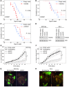
Figure 2. HOXA9 promotes tumor growth in mouse xenograft models of EOC.
(AC) Comparison of survival rates of female nude mice inoculated i.p. with vector-control and with (A) +HOXA9, (B) +HOXA10, and (C) +HOXA11 MOSEC lines (n = 10 per group). (D) Western blot of HOXA9 in SKOV3ip and ES-2 lines stably expressing empty pGFP-V-RS vector, nontargeting shRNA, and shRNAs targeting different sites of HOXA9 (shA9-A, shA9-B). Full uncut gels are shown in the Supplemental Material. (E and F) Growth rates of s.c. tumors derived from +HOXA9 control (Empty vector, Nontargeting) and HOXA9-knockdown (shA9-A, shA9-B) (E) SKOV3ip and (F) ES-2 lines (n = 5 per group). *P < 0.0005. (G and H) Mice were inoculated i.p. with GFP-expressing (G) SKOV3ip and (H) ES-2 lines and sacrificed at 4 weeks and 20 days, respectively. Implants were viewed under a fluorescence stereoscope. Omental implants in +HOXA9 control groups are indicated by arrows. Original magnification, ×0.8.
Figure 3
Figure 3. HOXA9 expression in EOC is associated with increased CAF abundance.
(A) Differences in transcript levels of ACTA2 (encoding αSMA) and FAP, between HOXA9-Low and HOXA9-High tumors in the AOCS data set, as estimated by Mann Whitney U test. In box-and-whisker plots, horizontal bars indicate the medians, boxes indicate 25th to 75th percentiles, and whiskers indicate minimum and maximum values. (BD) αSMA expression was evaluated in tumors of mice sacrificed at 20 days after inoculation with SKOV3ip and ES-2 lines. (B) The average number of αSMA+ cells per 1,000 tumor cells was calculated by scoring 5 random fields of stained tissue sections of each mouse (n = 5 mice per group). *P < 0.0001. Immunofluorescence staining of GFP-expressing tumor cells (green) and αSMA (red) in tumors of mice inoculated with (C) +HOXA9 control and HOXA9-knockdown SKOV3ip lines (scale bar: 100 μm) and (D) +HOXA9 control (nontargeting) SKOV3ip and ES-2 lines (scale bar: 50 μm). Nuclei were visualized by staining with DAPI (blue).
Figure 4
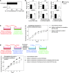
Figure 4. HOXA9 expression in EOC cells induces CAF features in normal omental fibroblasts.
(A) Diagram of fibroblast-priming assays. +HOXA9 control and HOXA9-knockdown SKOV3ip cells were cultured for 2 days to generate tumor-conditioned media (shown in pink). Normal omental fibroblasts were incubated for 5 days in SKOV3ip-conditioned medium (i.e., primed) or nonconditioned medium (i.e., unprimed) and then analyzed by Western blot and qRT-PCR. Fresh nonconditioned medium was added to washed fibroblasts. At 2 days thereafter, medium conditioned by fibroblasts (shown in light blue) was analyzed by ELISA and used for incubating control (nontargeting) SKOV3ip cells. (B) Western blot of αSMA levels in unprimed and primed fibroblasts. Full uncut gels are shown in the Supplemental Material. (C) Levels of growth factors in media conditioned by unprimed and primed fibroblasts. (D) Growth rates of control SKOV3ip cells incubated in fibroblast-conditioned medium. Average results of assays using 3 independent sets of each type of fibroblast-conditioned medium are shown in C and D. (E) Relative growth of control SKOV3ip cells at 6 days after incubation in medium conditioned by fibroblasts that were initially primed in +HOXA9 control SKOV3ip-conditioned medium, where fibroblast-conditioned medium was left untreated, treated with IgG, or depleted by IP with Abs to CXCL12 and IL-6. *P < 0.001; #P < 0.005.
Figure 5
Figure 5. HOXA9 expression in EOC cells promotes the ability of fibroblasts to stimulate endothelial cell growth.
(A) The average number of microvessels per 104 tumor cells was calculated in tumors derived from +HOXA9 control (nontargeting) and HOXA9-knockdown (shA9-B) SKOV3ip lines by scoring 5 random fields of CD34-stained tissue sections of each mouse (n = 5 mice per group). *P < 0.005. (B) Relative mRNA levels of IL6 and VEGFA in cultured SKOV3ip cells and of IL6 and VEGFA (in human EOC cells) and Il6 and Vegfa (in mouse host cells) in omental tumors of mice that were inoculated with SKOV3ip lines (n = 5 mice per group). *P = 0.03; P = 0.007. P values > 0.05 were considered not significant. Evaluation of specificity of human- and mouse- specific qRT-PCR primers is shown in Supplemental Figure 6B. (C) Growth rates of mouse endothelial cells incubated in nonconditioned medium and in SKOV3ip-conditioned media. **P < 0.005. (D) Normal omental fibroblasts were left unprimed or primed with SKOV3ip-conditioned media (shown in pink) for 5 days. Fresh nonconditioned medium was added to washed fibroblasts. Two days thereafter, medium conditioned by fibroblasts (shown in light blue) was collected. Growth rates of endothelial cells incubated in fibroblast-conditioned medium were measured. *P < 0.005. Average results of assays using 3 independent sets of each type of conditioned medium are shown in C and D.
Figure 6
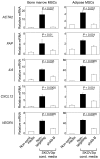
Figure 6. HOXA9 expression in EOC cells induces CAF features in bone marrow– and adipose-derived MSCs.
Levels of ACTA2, FAP, IL6, CXCL12, and VEGFA transcripts were assayed in bone marrow MSCs and adipose MSCs at 5 days after incubation in media conditioned by +HOXA9 control (Nontargeting) and HOXA9-knockdown (shA9-B) SKOV3ip cells. The mRNA level of each gene, assayed by qRT-PCR, is expressed relative to its level in MSCs incubated in nonconditioned medium.
Figure 7
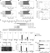
Figure 7. HOXA9 induces TGF-β2 expression in EOC cells.
(A) Relative TGFB1 and TGFB2 mRNA levels in SKOV3ip lines. *P < 0.001; **P < 0.005. (B) TGF-β1 and TGF-β2 levels in media conditioned by SKOV3ip lines. *P < 0.001. (C) Relative HOXA9, TGFB1, and TGFB2 mRNA levels in primary ovarian tumors of 33 cases. Correlations were determined by Spearman test. (D) Representation of the mouse Tgfb2 promoter. Locations of 5 putative HOXA9-binding sites evaluated by chromatin IP (S1 to S5) and regions evaluated in luciferase reporter assays (pA, pB, pC, pC-mtS4) relative to the transcription start site (TSS) are indicated. Wild-type sequences within the S4 site (in pA and pC) and mutant sequences (in pC-mtS4) are shown. (E) Chromatin IP analysis of interactions of FLAG-tagged HOXA9 in MOSEC cells with sites S1 to S5. Immunoprecipitated DNA was assayed by qPCR and is expressed as a percentage of total chromatin input. (F) Chromatin IP analysis of interactions of endogenous HOXA9 in SKOV3ip cells with conserved sites S4 and S5 in the human TGFB2 promoter. The input corresponds to 1% of chromatin solution before IP. IP using cells expressing FLAG-tag alone or cells expressing HOXA9 shRNA (shA9-B) as well as IP with IgG and amplification of Gapdh and GAPDH as irrelevant genes are included as negative controls in E and F. Full uncut gels are shown in the Supplemental Material. (G) qPCR analysis of immunoprecipitated DNA from assays in F, expressed as a percentage of total chromatin input. (H) Activity of Tgfb2 promoter regions shown in D was assayed in +HOXA9 control (black bar) and HOXA9-knockdown (white bar) SKOV3ip cells. Average relative luciferase activities of 3 independent assays are shown.
Figure 8
Figure 8. TGF-β2 induces expression of TGF-β ligands and CAF markers in omental fibroblasts.
(A) Levels of TGF-β1 and TGF-β2 released by omental fibroblasts that were primed in SKOV3ip-conditioned medium or left unprimed, as in Figure 4C. Average results of ELISAs of 3 independent sets of each type of fibroblast-conditioned medium are shown. *P < 0.005; #P < 0.001. (B) Relative levels of TGFB1 and TGFB2 mRNAs (in human EOC cells) and Tgfb1 and Tgfb2 mRNAs (in mouse host cells) in omental tumors of mice that were inoculated with +HOXA9 control (Nontargeting) and HOXA9-knockdown (shA9-B) SKOV3ip lines (n = 5 mice per group). Specificity of human- and mouse-specific qRT-PCR primers is shown in Supplemental Figure 6B. *P = 0.025; P = 0.018. (C and D) Relative TGFB1, TGFB2, ACTA2, and FAP mRNA levels in omental fibroblasts at 5 days after incubation with (C) recombinant TGF-β1 and TGF-β2 at the indicated concentrations and (D) media conditioned by +HOXA9 control SKOV3ip cells, where SKOV3ip-conditioned medium was left untreated, treated with IgG, or depleted by IP with Ab to TGF-β2.
Figure 9
Figure 9. Effects of HOXA9 on growth of EOC xenografts are mediated via its induction of tumor-derived TGF-β2.
(A) TGF-β2 levels released by control (Nontargeting), TGF-β2–knockdown (shTGF-β2), and HOXA9-knockdown (shA9-B) SKOV3ip lines and a HOXA9-knockdown line stably expressing TGF-β2 (shA9-B + TGF-β2). *P < 0.001. (B and C) Female nude mice were inoculated i.p. with SKOV3ip lines and sacrificed at 4 weeks thereafter. (B) Implants viewed under a fluorescence stereoscope. The arrow indicates the omental implant in the control group. Original magnification, ×0.7. (C) The average number of Ki-67+ tumor cells, αSMA+ cells, and microvessels in tumors was calculated by scoring 5 random fields of stained tumor tissue sections of each mouse (n = 5 mice per group). *P < 0.005; P < 0.01. (D) Survival rates of mice inoculated i.p. with SKOV3ip lines (n = 10 per group). Significance values for each group as compared with the Nontargeting control group are indicated. (E) Survival rates of patients in the AOCS data set stratified by TGFB2 expression in tumors, where TGFB2 transcript levels were defined as High (≥ upper quartile) and Low (≤ lower quartile) (n = 72 cases per group).
Figure 10
Figure 10. Bidirectional signaling between EOC cells and CAFs.
Control of growth factor expression and interactions among EOC cells, CAFs, and endothelial cells by HOXA9-induced, tumor-derived TGF-β2.

References

    1. Lengyel E. Ovarian cancer development and metastasis. Am J Pathol. 2010;177(3):1053–1064. doi: 10.2353/ajpath.2010.100105. - DOI - PMC - PubMed
    1. Naora H, Montell DJ. Ovarian cancer metastasis: Integrating insights from disparate model organisms. Nat Rev Cancer. 2005;5(5):355–366. doi: 10.1038/nrc1611. - "VSports" DOI - PubMed
    1. Clements PB, Young RH. Atlas of Gynecologic Surgical Pathology. Philadelphia, Pennsylvania, USA: WB Saunders; 2000.
    1. Pearson JC, Lemons D, McGinnis W. Modulating HOX gene functions during animal body patterning. . Nat Rev Genet. 2005;6(12):893–904. - PubMed
    1. Abate-Shen C. Deregulated homeobox gene expression in cancer: cause or consequence? Nat Rev Cancer. 2002;2(10):777–785. doi: 10.1038/nrc907. - DOI - PubMed

Publication types

MeSH terms