"V体育官网入口" Skip to main page content
U.S. flag

An official website of the United States government

Dot gov

The . gov means it’s official. Federal government websites often end in . gov or VSports app下载. mil. Before sharing sensitive information, make sure you’re on a federal government site. .

Https

The site is secure. The https:// ensures that you are connecting to the official website and that any information you provide is encrypted and transmitted securely V体育官网. .

. 2012 Nov;92(5):999-1009.
doi: 10.1189/jlb.0512242. Epub 2012 Aug 31.

"V体育官网" Immature murine NKT cells pass through a stage of developmentally programmed innate IL-4 secretion

Affiliations

Immature murine NKT cells pass through a stage of developmentally programmed innate IL-4 secretion

Nina Dickgreber (VSports) et al. J Leukoc Biol. 2012 Nov.

Abstract

We assessed the production of the canonical Th2 cytokine IL-4 by NKT cells directly in vivo using IL-4-substituting strains of reporter mice that provide faithful and sensitive readouts of cytokine production without the confounding effects of in vitro stimulation. Analysis in naïve animals revealed an "innate" phase of IL-4 secretion that did not need to be triggered by administration of a known NKT cell ligand. This secretion was by immature NKT cells spanning Stage 1 of the maturation process in the thymus (CD4(+) CD44(lo) NK1. 1(-) cells) and Stage 2 (CD4(+) CD44(hi) NK1 VSports手机版. 1(-) cells) in the spleen. Like ligand-induced IL-4 production by mature cells, this innate activity was independent of an initial source of IL-4 protein and did not require STAT6 signaling. A more sustained level of innate IL-4 production was observed in animals on a BALB/c background compared with a C57BL/6 background, suggesting a level of genetic regulation that may contribute to the "Th2-prone" phenotype in BALB/c animals. These observations indicate a regulated pattern of IL-4 expression by maturing NKT cells, which may endow these cells with a capacity to influence the development of surrounding cells in the thymus. .


Figures

Figure 1.
Figure 1.. i.v. administration of α-GalCer induces IL-4 reporter expression in NKT cell in G4 mice.
(A) Representative dot plots showing expression of the IL-4 reporter on gated NKT cells (CD1d Tetr-binding cells, as shown in Supplemental Fig. 1) from spleen and liver of heterozygous G4 mice on a BALB/c background administered 200 ng α-GalCer, relative to nontransgenic and untreated control animals. (B) Mean percentages of IL-4 reporter-positive cells (±sem) within the CD4+ and CD4 NKT cell subsets of the indicated tissues over time after α-GalCer administration (n=4–6 animals/group). Significant differences between the subsets were determined by two-way ANOVA with Bonferroni post-test. Tissues were also examined from similarly treated homozygous G4 mice and mice homozygous for the G4 reporter and STAT6 deficiency. A graph showing levels of IL-4 protein in the serum is also presented. (C) Representative dot plots from a similar analysis in G4 mice on a C57BL/6 background.
Figure 2.
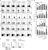
Figure 2.. Immature thymic NKT cells pass through a stage of innate IL-4 secretion.
(A) Flow cytometry was used to analyze GFP as a surrogate for IL-4 expression in thymocytes from naïve heterozygous G4 mice on C57BL/6 and BALB/c backgrounds. Dot plots show gating of NKT cells and analysis of GFP, CD4, CD44, and NK1.1 expression. (B) Mean fluorescence intensity of binding of CD4, CD44, and NK1.1 antibodies and CD1d Tetr on GFP+ (IL-4+) and GFP (IL-4) NKT cells; bars represent mean ± sem from four to six mice/age group analyzed (two-way ANOVA with Bonferroni post-test).
Figure 3.
Figure 3.. Strong binding of CD1d Tetr is associated with an immature NKT cell phenotype in a BALB/c strain congenic for NK1.1.
Increased levels of CD1d Tetr binding are correlated with an immature CD4+CD44hiNK1.1 phenotype in thymic NKT cells from naïve BALB.B6-Cmv1r mice. The gating strategy used to identify NKT cells is shown together with representative FACS plots of CD4, CD44, and NK1.1 expression on (A) CD1d Tetrhi, (B) CD1d Tetrint, and (C) CD1d Tetrlo cells. Results are representative of five different mice.
Figure 4.
Figure 4.. Analysis of huCD2 as a surrogate for IL-4 expression in KN2 mice.
Expression of the reporter huCD2 was examined in thymic NKT cells from naïve homozygous KN2 mice compared with BALB/c controls. The gating strategy used to identify NKT cells is shown together with representative FACS plots of CD4 and CD44 on (A) CD1d Tetrhi, (B) CD1d Tetrint, and (C) CD1d Tetrlo cells. Dot plots are representative of three mice/group.
Figure 5.
Figure 5.. IL-4 protein is detected in thymic cultures.
Mean levels of IL-4 protein in cell suspensions prepared from thymic tissue from mice from the indicated mouse strains cultured for 24 h; NKT cells were removed from some cultures, as indicated, using PE-labeled CD1d Tetr and anti-PE-coated magnetic beads. KO, Knockout.
Figure 6.
Figure 6.. Innate expression from the Il4 locus in NKT cells is independent of an initial source IL-4 and STAT6 signaling.
Analysis of IL-4 reporter expression in thymic NKT cells from groups (n=4–6) of naïve heterozygous G4 mice (BALB/c IL-4+/G4; IL-4-sufficient), homozygous G4 mice (BALB/c IL-4G4/G4; IL-4−/−), and mice homozygous for the G4 reporter and STAT6 deficiency (BALB/c IL-4G4/G4 STAT6−/−). Mean percentages of GFP reporter+ cells in CD4+ and CD4 NKT cells are presented ±sem.
Figure 7.
Figure 7.. Proportion of thymic NKT cells exhibiting innate expression from the Il4 locus is determined by age and genetic background.
Numbers of IL-4 reporter-positive thymic NKT cells (IL-4+ NKT cells) were determined in different age groups of naïve heterozygous G4 mice on C57BL/6 and BALB/c backgrounds. Right panels show mean percentage of GFP reporter+ of NKT cells/mouse. All graphs show mean values ± sem/group (n=4–6).
Figure 8.
Figure 8.. Splenic NKT cells exhibit innate expression of IL-4.
(A) Representative dot plots of analysis of IL-4 reporter expression in NKT cells from the spleen of naïve heterozygous G4 mice on C57BL/6 and BALB/c backgrounds. (B) Mean fluorescence intensity of binding of CD4, CD44, and NK1.1 antibodies and CD1d Tetr on GFP+ (IL-4+) and GFP (IL-4) NKT cells; bars represent mean ± sem from four to six mice/age group analyzed (two-way ANOVA with Bonferroni post-test). (C) Expression of the IL-4 reporter in splenic NKT cells from groups (n=4–6) of naïve, heterozygous G4 mice, homozygous G4 mice, and mice homozygous for G4 and STAT6 deficiency. Mean percentages of GFP reporter+ cells in CD4+ and CD4 NKT cells are presented ±sem.

References

    1. Bendelac A., Savage P. B., Teyton L. (2007) The biology of NKT cells. Annu. Rev. Immunol. 25, 297–336 - PubMed
    1. Godfrey D. I., MacDonald H. R., Kronenberg M., Smyth M. J., Van Kaer L. (2004) NKT cells: what's in a name? Nat. Rev. Immunol. 4, 231–237 - PubMed
    1. Zajonc D. M., Kronenberg M. (2007) CD1 mediated T cell recognition of glycolipids. Curr. Opin. Struct. Biol. 17, 521–529 - PMC - PubMed
    1. Godfrey D. I., Kronenberg M. (2004) Going both ways: immune regulation via CD1d-dependent NKT cells. J. Clin. Invest. 114, 1379–1388 - "V体育官网入口" PMC - PubMed
    1. Mohrs M., Shinkai K., Mohrs K., Locksley R. M. (2001) Analysis of type 2 immunity in vivo with a bicistronic IL-4 reporter. Immunity 15, 303–311 - PubMed

Publication types

Substances