Skip to main page content
U.S. flag

An official website of the United States government

Dot gov

The . gov means it’s official. Federal government websites often end in . gov or VSports app下载. mil. Before sharing sensitive information, make sure you’re on a federal government site. .

Https

The site is secure V体育官网. The https:// ensures that you are connecting to the official website and that any information you provide is encrypted and transmitted securely. .

. 2012 Aug 24;37(2):223-34.
doi: 10.1016/j.immuni.2012.04.015.

TBK-1 promotes autophagy-mediated antimicrobial defense by controlling autophagosome maturation

Affiliations

TBK-1 promotes autophagy-mediated antimicrobial defense by controlling autophagosome maturation

"VSports注册入口" Manohar Pilli et al. Immunity. .

Abstract (V体育官网)

Autophagy is a fundamental biological process of the eukaryotic cell contributing to diverse cellular and physiological functions including cell-autonomous defense against intracellular pathogens. Here, we screened the Rab family of membrane trafficking regulators for effects on autophagic elimination of Mycobacterium tuberculosis var. bovis BCG and found that Rab8b and its downstream interacting partner, innate immunity regulator TBK-1, are required for autophagic elimination of mycobacteria in macrophages VSports手机版. TBK-1 was necessary for autophagic maturation. TBK-1 coordinated assembly and function of the autophagic machinery and phosphorylated the autophagic adaptor p62 (sequestosome 1) on Ser-403, a residue essential for its role in autophagic clearance. A key proinflammatory cytokine, IL-1β, induced autophagy leading to autophagic killing of mycobacteria in macrophages, and this IL-1β activity was dependent on TBK-1. Thus, TBK-1 is a key regulator of immunological autophagy and is responsible for the maturation of autophagosomes into lytic bactericidal organelles. .


Figures

Fig. 1
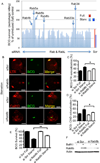
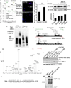
Fig. 1. Analysis of murine Rab factors in cell-autonomous autophagic elimination of mycobacteria and the role of Rab8b
A. Sixty-two Rab or Rablike (RabL) factors encoded by the Mus musculus genome were knocked down by siRNA in RAW264.7 macrophages (details and identity of each bar in Suppl. Table S1), macrophages infected with M. tuberculosis var. bovis BCG, autophagy induced by starvation (Starv), and autophagic killing of BCG quantified by colony forming unit (CFU) counting. Scr, scrambled (control) siRNA. Bars, BCG survival (relative to Scr siRNA) following induction (4 h) of autophagy by starvation for 4 h. B–D. Effect of Rab8b knockdown on maturation of BCG phagosomes into autophagolysosomes. LTR, Lysotracker Red (acidotropic dye); CathD, cathepsin D. Images: LTR (red fluorescence), BCG (green fluorescence) and merged fields (yellow, colocalization of LTR and BCG). Bar graphs, quantitative colocalization analysis of images. (% of total green profiles showing red fluoresecence) E,F. Validation (E, % survival measured and represented as in A); F, Rab8b knockodown analysis by immunoblotting).of the role for Rab8b in autophagic killing of BCG. BCG survival, % of BCG CFU recovered from RAW 264.7 macrophages pretreated with siRNAs; si Scr, control scrambled siRNA; si Rab8b, Rab8b siRNA. Full, full medium; Starve, autophagy induced by starvation. [Data, means ± se (n≥3; †, p≥0.05*, p<0.05; **, p<0.01; ANOVA).
Fig. 2
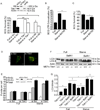
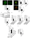
Fig. 2. Role of TBK-1 in autophagic maturation
A. RAW 264.7 macrophages were pretreated with siRNAs, infected with BCG, and autophagy induced by starvation (Starve). BCG survival (% of BCG) was analyzed and represented as in Fig. 1. Full, full medium (control conditions); si Scr, scrambled siRNA (control siRNA) si TBK-1, siRNA to TBK-1. Si Htt, siRNA to huntingtin (Htt). Immunoblot, analysis of TBK-1 and Htt knockdowns. B. Effect of TBK-1 inhibitor BX795 on acidification of BCG-containing organelles following induction of autophagy. RAW 264.7 macrophages were pretreated with 10 nM BX795, infected, and induced for autophagy by starvation. C. Autophagic killing of BCG in RAW 264.7 macrophages pretreated with BX795. BCG survival, % CFU recovered from RAW 264.7 cells. D,E. RAW 264.7 expressing RFP-GFP-LC-3 and pretreated with scrambled or TBK-1 siRNAs were untreated (Full) or induced (Starv) for autophagy. Puncta; R+G+ (RFP+ GFP+), early autophagic organelles; R+G- (RFP+ GFP-), late autophagic organelles. Images, merged red and green channels. Inset in E, immunoblot analysis of TBK-1 knockdown. F,G. Effects of TBK-1 on LC3-II levels and degradation during autophagic maturation. Tbk-1−/− and Tbk-1+/+ MEFs were uninduced and induced for autophagy, treated or not treated with bafilomycin A1 (BafA1) to inhibit autophagic degradation of LC3-II. Data, means ± se (n≥3; †, p≥0.05*, p<0.05; **, p<0.01; ANOVA).
Fig. 3
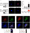
Fig. 3. Rab8b and TBK-1 interact and colocalize with autophagic organelles
A. HEK 293T cell extracts, transiently transfected with control (EGFP), GFPRab8b (Wt, wild type) and GFP-Rab8b Q67L (Q67L, constitutively active) expression constructs were immunoprecipitated with anti-GFP antibody; immunoblots of immune complexes and inputs were probed with anti-TBK-1 and anti-GFP antibodies. B. Proximity ligation in situ assay (P-LISA) for proteinprotein interactions in murine primary bone marrow macrophages (BMM). BMMs were fixed, incubated with primary antibodies against two proteins tested (Ab1 and Ab2), and P-LISA oligonucleotides added. After nucleic acid ligation, rolling circle amplification, and hybridization with fluorescent oligonucleotides (and counterstaining of nuclei with DAPI), red fluorescent dots (see scheme for distance requirements between probes for positive signal) were imaged by confocal microscopy. Control, no primary antibodies. TBK-1-Rab8b, antibody 1 (Ab1): anti-TBK-1, antibody 2 (Ab2): anti-Rab8b. Rab8b-LC3, Ab1: anti-Rab8b, Ab2: anti-LC3. TBK-1-NDP52, Ab1: anti-TBK-1, Ab2: anti-NDP52. Graph, quantitative analysis of P-LISA red-fluorescent dots (indicating close proximity of proteins recognized by Ab1 and Ab2). Cells (≤ 25 per sample) with positive red fluorescing dots were morphometrically analyzed and the mean number of red fluorescing dots per cell quantified. Up to 10% of the cells in any given sample were positive for red dots. Values, means ± sd; * p<0.05 (t-test). C. Colocalization analysis of Rab8b and its downstream effector, TBK-1 with the basal autophagic machinery factor LC3. Fluorescence; endogenous TBK-1 (red, Alexa568), Rab8B (green, Alexa488), LC3 (blue, Alexa633). Cells (BMM) were induced for autophagy by starvation in the presence of bafilomycin A1 to inhibit autophagic maturation and degradation. Arrows, colocalization of Rab8b, TBK-1, and LC3. D. Representative line tracing of three fluorescence channels in images in A. E. Analysis of TBK-1 localization relative to the autophagic adaptor sequestosome 1/p62 and LC3. Cells (BMM), treatments, labels and graphs as in C and D. Arrows, colocalization of TBK-1, p62 and LC3. Cells (BMM) were induced for autophagy by starvation in the presence of bafilomycin A1 to inhibit autophagic maturation. F. Representative line tracing of fluorescence channels in images in C. Data, representative of ≥3 independent experiments.
Fig. 4
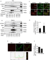
Fig. 4. TBK-1 co-fractionates and colocalizes with intracellular membranous organelles containing autophagic adaptors and machinery
A,B. Analysis by subcellular fractionation of Rab8b, TBK-1 and components of autophagic machinery (UVRAG, p62, LC3-II) in resting cells (Full, full medium) or upon induction of autophagy by starvation (Starve). Membranous organelles from RAW 264.7 macrophages uninduced (A) or induced (B) for autophagy were subjected to subcellular fractionation by isopycnic sucrose density gradient centrifugation, fractions collected, and proteins analyzed by immunolotting. PNS, postnuclear supernatant. 1–4, pooled fractions. Rectangle over fraction 9, convergence in autophagic organelles (LC3-II) of: Rab8b, TBK-1, UVRAG (Beclin 1 interacting protein specific for autophagosomal maturation), and autophagic adaptor p62. Refractive indexes below the lanes reflect sucrose density of each fraction. C. Images; confocal microscopy analysis of endogenous UVRAG (Alexa568) and endogenous TBK-1 (Alexa488). Cells, BMM, uninduced (Full) and induced (Starvation) for autophagy. Arrows, coloclaization; insets, enlarged areas. D. Pearson’s coefficient for TBK-1 and UVRAG colocalization (Starv, starvation). E. Analysis of TBK-1 localization relative to autophagic adaptor p62 in BMM. Images: endogenous TBK-1 (Alexa568; red), p62 (Alexa488; green) and merged. Line tracing, analysis of colocalization of TBK-1 (red tracing) and p62 (green tracing). F. Pearson’s colocalization coefficients for TBK-1-p62. (Starv, starvation). Data, means ± se (n=3, three independent experiments with at least 5 images analyzed per experiment; †, p≥0.05; **, p<0.01; ANOVA).
Fig. 5
Fig. 5. TBK-1 controls p62 phosphorylation, and affects autophagic clearance of p62 and its cargo capture, delivery and degradation
A–C. High content imaging analysis (using Cellomics high-content microscopy system) of p62 puncta (endogenous, revealed by imunofluorescence) in BMM with or without treatment with TBK-1 inhibitor BX795. Panel A shows output from Cellomics high-content microscopy and analysis software comparing the number of p62 puncta between TBK-1 inhibitor-treated (BX795) and control (DMSO) BMM. Vertical axis denotes the mean number of p62 puncta per cell and horizontal axis denotes the position of the well (B, BX795 series; C, control series) on the plate. Between 754 and 2395 cells were analyzed per well. Panel C shows t test (data, means ± se; **, p<0.01) from cumulative data treating only whole wells as independent samples (n=4). D. Analysis of the effects of TBK-1 pharmacological inhibitor, BX795 on p62 levels. Tbk-1+/+ MEFs were treated with BX795; bafilomycin A1 (BafA1) to inhibit autophagic degradation. Densitometric analyses of p62 levels normalized against actin levels were plotted (n=2; error bars, range). E. Analysis of TBK-1 role in autophagic clearance of polyubiquitinated proteins. Cell lysates from Tbk1+/+ and Tbk1−/− MEFs uninduced and induced for autophagy by starvation were incubated with TUBE2 agarose beads and bound material pulled down. Protein blots were probed for K63 polyubiquitin chains. F–H. Identification of TBK-1-dependent S303 phosphorylation of the UBA domain of p62. In vivo phosphorylation of p62 UBA domain following cotransfection of GFP-p62D69A (D69A mutation prevents oligomerization with endogenous p62) and expression constructs of TBK-1 wild type or kinase defective form. Immunoprecipitated (GFP-p62) material was subjected to tandem mass spectrometry. A triply charged ion with the mass 857.01 was selected for fragmentation. This ion was identified as the phosphorylated LIESLSQMLpSMGFSDEGGWLTR peptide (shown in panel H) from p62. Panel G shows MS spectra from LC-MS, showing the phosphopeptide of 857.01 m/z observed in p62 phosphorylated by TBK-1. The peptide was not observed when GFP-p62 was co-transfected with the kinase-defective K38D mutant of TBK-1. Spectra are taken from the same retention time in both runs, confirmed by the unspecific peaks observed in both spectra. I. HEK293 cells transfected with vector control, myc-TBK-1 or myc-TBK-1 K38D were left untreated or were treated for 2 h with 1 µM BX795. Cell extracts were immunoblotted with antibodies against phospho-p62 (S303), p62, myc and actin. Abbreviations: end. p62, endogenous p62. J. MBP or MBP-tagged p62 proteins were expressed and affinity-purified from E. coli. TBK-1 mediated phosphorylation was assessed by incubating recombinant MBP, MBP-p62 or MBP-p62 S303A with recombinant active TBK-1 in the presence of [γ-32P] ATP for 10 min at 30°C. The reaction products were analyzed by autoradiography (AR). CBB, Coomassie Brilliant Blue staining.
Fig. 6
Fig. 6. IL-1β induces autophagy in macrophages
A. RAW264.7 macrophages were transiently transfected with EGFP-LC3 and treated with 10 ng/ml murine IL- 1β for 2 h, and assayed for LC3 puncta formation by confocal microscopy (only puncta ≥ 1µm were scored as positive). B. RAW264.7 macrophages transfected with mRFP-GFP-LC3 tandem probe, treated with 10 ng/ml IL-1β for 2 h were scored for number (per transfected cell) of RFP+GFP+ puncta (R+G+; early autophagosomes), RFP+GFP (R+Gβ− ; autolysosomes), and total LC3 puncta. C. Immunoblot analysis of endogenous LC3 conversion to lipidated form (LC3-II) in RAW 264.7 cells upon treatment with 10 ng/ml IL-1β for 2 h, in the absence or presence of bafilomycin A1. Graph, ratio of LC3-II to actin intensity in immunoblots from bafilomycin A1-treated samples. D. RAW264.7 murine macrophages were co-transfected with tandem mRFP-GFP-LC3 probe and expression constructs containing either wild-type MyD88 (MyD88-WT) or a dominant-negative mutant of MyD88 (MyD88-DN). Following stimulation with 10 ng/ml IL-1β for 2 h, LC3 puncta were quantified as in B. E. Induction of autophagy in response to IL-1β is abrogated in bone marrow-derived macrophages (BMM) from MyD88-deficient (Myd88−/−) mice, measured by ratios of LC3-II band relative to actin following treatments of BMMs and immunoblotting of cellular extracts. F. Proteolysis of stable proteins (radiolabeled by a pulsechase protocol) upon stimulation of RAW264.7 cells with 10 ng/ml IL-1β for 2 h (Full + IL-1β) relative to control (Full) or starvation-induced autophagy (Starve).. Data, means ± se, except in E where data are means ± sd (n≥3; †, p≥0.05; *, p<0.05; **, p<0.01; ANOVA).
Fig. 7
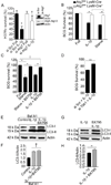
Fig. 7. Requirement for TBK-1 in IL-1β mediated autophagic killing of mycobacteria
A. RAW264.7 macrophages were knocked down for Atg7 (by Atg7 siRNA transfection 48 h prior to infection), infected with M. tuberculosis H37Rv for 1h, washed and then left untreated or treated with 10 ng/ml recombinant murine IL-1β for 2 h after which they were lysed and plated for CFU determination, and survival expressed relative to sample transfected with control scrambled siRNA and not treated with IL-1β. Immunoblots, Atg7 knockdown and levels of Atg5-Atg12 complexes. B. BCG survival, % CFU of M. tuberculosis var. bovis BCG recovered from bone marrow macrophages derived from Atg7fl/fl LysM-Cre and Atg7fl/fl LysM-Cre+ mice treated with 50 ng/ml IL-1β (16 h preinfection + 4 h post-infection). Data, means ± se (n=3, *, p<0.05; t-test. C. BCG survival, % CFU recovered from RAW264.7 macrophages treated with IL-1β with and without 10 nM BX795. D. BCG survival in infected RAW264.7 (and knocked down or not for TBK-1) macrophages stimulated with IL-1β. E,F. RAW264.7 macrophages were incubated in full medium (Control) or induced by adding IL-1β to full medium. Cells were pretreated with 10 nM BX795 where indicated. Macrophages were treated with or without bafilomycin A1 (BafA1) to inhibit autophagic degradation of LC3-II, and cellular extracts analyzed by immunoblotting. Graphs, densitometric analyses of LC3-II levels normalized to actin levels (LC3-II/Actin). G,H. Cells, treatments and analysis as in E and F but in the absence of bafilomycin A1. Data, means ± se (n=3; †, p≥0.05; **, p<0.01; ANOVA).

"VSports手机版" References

    1. Alonso S, Pethe K, Russell DG, Purdy GE. Lysosomal killing of Mycobacterium mediated by ubiquitin-derived peptides is enhanced by autophagy. Proc Natl Acad Sci U S A. 2007;104:6031–6036. - "V体育官网入口" PMC - PubMed
    1. Bjorkoy G, Lamark T, Brech A, Outzen H, Perander M, Overvatn A, Stenmark H, Johansen T. p62/SQSTM1 forms protein aggregates degraded by autophagy and has a protective effect on huntingtin-induced cell death. J Cell Biol. 2005;171:603–614. - PMC - PubMed
    1. Bjorkqvist M, Wild EJ, Thiele J, Silvestroni A, Andre R, Lahiri N, Raibon E, Lee RV, Benn CL, Soulet D, et al. A novel pathogenic pathway of immune activation detectable before clinical onset in Huntington's disease. J Exp Med. 2008;205:1869–1877. - PMC - PubMed
    1. Clark K, Plater L, Peggie M, Cohen P. Use of the pharmacological inhibitor BX795 to study the regulation and physiological roles of TBK1 and IkappaB kinase epsilon: a distinct upstream kinase mediates Ser-172 phosphorylation and activation. J Biol Chem. 2009;284:14136–14146. - VSports最新版本 - PMC - PubMed
    1. Criollo A, Niso-Santano M, Malik SA, Michaud M, Morselli E, Marino G, Lachkar S, Arkhipenko AV, Harper F, Pierron G, et al. Inhibition of autophagy by TAB2 and TAB3. The EMBO journal. 2011;30:4908–4920. - PMC - PubMed

VSports最新版本 - Publication types

MeSH terms

Substances (V体育官网入口)