Skip to main page content
U.S. flag

An official website of the United States government

Dot gov

The . gov means it’s official. Federal government websites often end in . gov or VSports app下载. mil. Before sharing sensitive information, make sure you’re on a federal government site. .

Https

The site is secure. The https:// ensures that you are connecting to the official website and that any information you provide is encrypted and transmitted securely V体育官网. .

. 2012 Oct;40(19):9661-74.
doi: 10.1093/nar/gks745. Epub 2012 Aug 16.

Cockayne Syndrome group B protein interacts with TRF2 and regulates telomere length and stability

Affiliations

VSports app下载 - Cockayne Syndrome group B protein interacts with TRF2 and regulates telomere length and stability

"VSports" Nicole L Batenburg et al. Nucleic Acids Res. 2012 Oct.

Abstract

The majority of Cockayne syndrome (CS) patients carry a mutation in Cockayne Syndrome group B (CSB), a large nuclear protein implicated in DNA repair, transcription and chromatin remodeling. However, whether CSB may play a role in telomere metabolism has not yet been characterized. Here, we report that CSB physically interacts with TRF2, a duplex telomeric DNA binding protein essential for telomere protection. We find that CSB localizes at a small subset of human telomeres and that it is required for preventing the formation of telomere dysfunction-induced foci (TIF) in CS cells. We find that CS cells or CSB knockdown cells accumulate telomere doublets, the suppression of which requires CSB. We find that overexpression of CSB in CS cells promotes telomerase-dependent telomere lengthening, a phenotype that is associated with a decrease in the amount of telomere-bound TRF1, a negative mediator of telomere length maintenance. Furthermore, we show that CS cells or CSB knockdown cells exhibit misregulation of TERRA, a large non-coding telomere repeat-containing RNA important for telomere maintenance. Taken together, these results suggest that CSB is required for maintaining the homeostatic level of TERRA, telomere length and integrity. These results further imply that CS patients carrying CSB mutations may be defective in telomere maintenance VSports手机版. .


VSports在线直播 - Figures
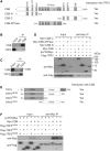
Figure 1.
Figure 1.
CSB interacts physically with TRF2. (A) Schematic diagram of CSB. NLS: nuclear localization sequence; NTB: nucleotide binding domain; and UBD: ubiquitin binding domain. (B) Co-immunoprecipitation with HeLa cell extracts and anti-CSB antibody. Anti-IgG IP was used as a negative control. Immunoblotting was carried out with anti-CSB or anti-TRF2 antibody. (C) Co-IP with HeLa nuclear extracts and anti-TRF2 antibody. Anti-IgG IP was used as a negative control. Immunoblotting was carried out with anti-CSB or anti-TRF2 antibody. (D) IP with anti-Myc antibody was carried out with protein extracts from 293T cells coexpressing Flag-TRF2 in conjunction with either the vector alone, Myc-CSB, Myc-CSB-N, Myc-CSB-ATPase or Myc-CSB-C. Immunoblotting was performed with anti-Myc or anti-Flag antibody. (E) Schematic diagram of TRF2. B stands for basic domain. (F) IP with anti-Myc antibody was carried out with protein extracts from 293T cells coexpressing the vector or Myc-CSB in conjunction with Flag-TRF2linker, Flag-TRF2TRFH or Flag-TRF2ΔBΔM. Immunoblotting was performed with anti-Myc or anti-Flag antibody.
Figure 2.
Figure 2.
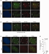
CSB localizes at a small subset of human telomeres and prevents the formation of TIFs in CS cells. (A) Analysis of indirect immunofluorescence (IF) on HeLaII and CSB-complemented hTERT-GM10905 cells. IF was perfromed with mouse anti-CSB (red) in conjunction with rabbit anti-hRap1 (green). Cells were extracted with detergent prior to fixation by paraformaldehyde to remove soluble proteins. Cell nuclei were stained with DAPI shown in blue. Arrowheads indicate the overlap between anti-CSB and anti-hRap1 staining. (B) Analysis of IF-FISH on GM16095 cells expressing Myc-CSB. IF–FISH analyses were performed with anti-Myc antibody (red) in conjunction with a FITC-conjugated (CCCTAA)3-containing PNA probe (green). Cell nuclei were stained with DAPI shown in blue. Arrowheads indicate the colocalization of CSB with telomeric DNA. (C) Indirect immunofluorescence using anti-hRap1 in conjunction with anti-γ-H2AX was performed with fixed hTERT-GM10905 cells expressing either the vector alone or wild-type CSB. Arrowheads indicate sites of colocalization of γH2AX and hRap1. (D) Quantification of percentage of cells with five or more TIFs. For each cell line, a total of 300 cells from three independent experiments were scored. Standard deviations from three independent experiments are indicated.
Figure 3.
Figure 3.
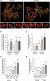
CS primary fibroblasts carrying CSB mutations accumulate telomere doublets. (A) Analysis of metaphase chromosomes from GM10901 and GM10905. Chromosomes were stained with DAPI and false colored in red. Telomeric DNA was detected by FISH using a FITC-conjugated (CCCTAA)3-containing PNA probe (green). Open arrows represent telomere doublets whereas asterisks indicate telomere loss. Enlarged images of chromosomes with telomere doublets or telomere loss are shown at the bottom. (B–E) Quantification of telomere loss or telomere doublets from indicated cell lines. For each cell line, a total of 2410–2699 chromosomes from 60 metaphase cells were scored in a blind manner for the presence of telomere loss (B and D) as well as telomere doublets in (C and E). Standard deviations derived from three independent experiments are indicated. Passage numbers of cell lines used are indicated above the bars.
Figure 4.
Figure 4.
CSB is required to prevent the formation of telomere doublets. (A) Quantification of telomere loss from indicated cell lines. For each cell line, a total of at least 2649–2668 chromosomes from 60 metaphase cells were scored in a blind manner. Standard deviations derived from three independent experiments are indicated. (B) Quantification of telomere doublets from indicated cell lines. For each cell line, a total of 2649–2668 chromosomes from 60 metaphase cells were scored in a blind manner. Standard deviations derived from three independent experiments are indicated. (C) Quantification of telomere doublets from hTERT-GM10905 cells expressing indicated constructs. For each cell line, a total of 2707–2754 chromosomes from 60 metaphase cells were scored in a blind manner. Standard deviations derived from three independent experiments are indicated. (D) Quantification of telomere doublets from GM16095 cells expressing indicated constructs. For each cell line, a total of 4774–4923 chromosomes from 60 metaphase cells were scored in a blind manner. Standard deviations derived from three independent experiments are indicated. (E) Western analysis of CSB expression. CSB was stably knocked down in HeLaI.2.11 cells. Immunoblotting was performed with anti-CSB or anti-γ-tubulin antibody. The latter was used as a loading control. (F) Quantification of telomere doublets from HeLaI.2.11 cells expressing the vector alone or pRS-shCSB. For each cell line, a total of 2678–2961 chromosomes from at least 43 metaphase cells were scored in a blind manner. Standard deviations derived from three independent experiments are indicated. (G) Quantification of telomere doublets from GM16095 cells expressing indicated constructs. Cells were treated with DMSO or aphidicolin (0.2 µM) for 16 h. For each cell line, a total of 3879–4321 chromosomes from 51 to 53 metaphase cells were scored in a blind manner. Standard deviations derived from three independent experiments are indicated.
Figure 5.
Figure 5.
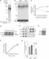
CSB is required for telomere length maintenance. (A) Genomic blot of telomeric restriction fragments from hTERT-immortalized GM10901 and GM10905 cells. About 3 µg of RsaI/HinfI-digested genomic DNA from each sample was used for gel electrophoresis. DNA molecular weight markers are shown on the left of the blot. Median telomere length of indicated cell lines are shown on the bottom of the blot. (B) Genomic blots of telomeric restriction fragments from hTERT-GM10905 cells expressing either the vector alone or wild-type CSB as indicated above the lanes. About 3 µg of RsaI/HinfI-digested genomic DNA from each sample was used for gel electrophoresis. PDs are indicated above the lanes whereas DNA molecular weight markers are shown on the left of the blots. The bottom panel, taken from an ethidium bromide-stained agarose gel, is used as a loading control. (C) Median telomere length of indicated cell lines was plotted against PDs. (D) Western analysis of CSB expression in hTERT-GM10905 cells. Immunoblotting was performed with anti-CSB or anti-γ-tubulin antibody. The indicated CSB-PiggyBac fusion protein is a product of alternative splicing involving the first five exons of CSB and a conserved PGBD3 located within the intron 5 of the CSB gene (52). (E) Growth curve of hTERT-GM10905 cells expressing various constructs as indicated. The number of PDs was plotted against days in culture. (F) Dot blots of ChIPs with anti-TRF1 or anti-TRF2 antibody. ChIPs were performed with lysates from hTERT-GM10905 cells expressing either the vector alone or wild-type CSB. Anti-IgG ChIP was used as a control. (G) Quantification of ChIPs from (E). The signals from dot blots were quantified by ImageQuant (IQ) analysis. Standard deviations from three independent experiments are shown. (H) Western analysis of protein expression. Immunoblotting was carried out with anti-TRF1, anti-TRF2 or anti-γ-tubulin antibody.
Figure 6.
Figure 6.
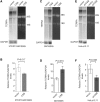
CSB is required for maintaining the homeostatic level of TERRA. (A) Analysis of TERRA expression from hTERT-GM10905 cells expressing the vector alone or CSB. Northern blotting was performed with a 32P-labeled telomeric DNA-containing probe shown on the left top panel. The northern blot of GAPDH shown on the left bottom panel was used as a loading control. The right panel was taken from the ethidium bromide-stained agarose gel. The position of 28S or 18S ribosomal RNA is indicated. (B) Quantification of relative TERRA levels from (A). The signals from northern blots were quantified by ImageQuant analysis. The TERRA signal from each lane was normalized to the GAPDH signal in the corresponding lane, giving rise to the relative level of TERRA to GAPDH. (C) Northern analysis of TERRA expression from GM16095 cells expressing the vector alone or wild-type CSB. The northern blot of GAPDH shown on the left bottom panel was used as a loading control. The right panel was taken from the ethidium bromide-stained agarose gel. The position of 28S or 18S ribosomal RNA is indicated. (D) Quantification of relative TERRA levels from (C). Quantification was performed as described in (B). (E) Northern analysis of TERRA expression from HeLaI.2.11 cells stably expressing the vector alone or pRS-shCSB. The northern blot of GAPDH shown on the left bottom panel was used as a loading control. The right panel was taken from the ethidium bromide-stained agarose gel. The position of 28S or 18S ribosomal RNA is indicated. (F) Quantification of relative TERRA levels from (E). Quantification was performed as described in (B).

References

    1. de Lange T. Shelterin: the protein complex that shapes and safeguards human telomeres. Genes Dev. 2005;19:2100–2110. - PubMed
    1. Palm W, de Lange T. How shelterin protects Mammalian telomeres. Annu. Rev. Genet. 2008;42:301–334. - PubMed
    1. Liu D, O'Connor MS, Qin J, Songyang Z. Telosome, a mammalian telomere-associated complex formed by multiple telomeric proteins. J. Biol. Chem. 2004;279:51338–51342. - PubMed
    1. Walker JR, Zhu XD. Post-translational modification of TRF1 and TRF2 and their roles in telomere maintenance. Mech. Ageing Dev. 2012;133:421–434. - PubMed
    1. Azzalin CM, Reichenbach P, Khoriauli L, Giulotto E, Lingner J. Telomeric repeat containing RNA and RNA surveillance factors at mammalian chromosome ends. Science. 2007;318:798–801. - PubMed

Publication types (VSports最新版本)

MeSH terms

Substances