Skip to main page content
U.S. flag

An official website of the United States government

Dot gov

The . gov means it’s official. Federal government websites often end in . gov or . mil. Before sharing sensitive information, make sure you’re on a federal government site. VSports app下载.

Https

The site is secure V体育官网. The https:// ensures that you are connecting to the official website and that any information you provide is encrypted and transmitted securely. .

. 2012 Aug 17;150(4):764-79.
doi: 10.1016/j.cell.2012.06.035.

"VSports" The BMP inhibitor Coco reactivates breast cancer cells at lung metastatic sites

Affiliations

The BMP inhibitor Coco reactivates breast cancer cells at lung metastatic sites

Hua Gao et al. Cell. .

Erratum in

  • Cell. 2012 Dec 7;151(6):1386-8

Abstract

The mechanistic underpinnings of metastatic dormancy and reactivation are poorly understood. A gain-of-function cDNA screen reveals that Coco, a secreted antagonist of TGF-β ligands, induces dormant breast cancer cells to undergo reactivation in the lung VSports手机版. Mechanistic studies indicate that Coco exerts this effect by blocking lung-derived BMP ligands. Whereas Coco enhances the manifestation of traits associated with cancer stem cells, BMP signaling suppresses it. Coco induces a discrete gene expression signature, which is strongly associated with metastatic relapse to the lung, but not to the bone or brain in patients. Experiments in mouse models suggest that these latter organs contain niches devoid of bioactive BMP. These findings reveal that metastasis-initiating cells need to overcome organ-specific antimetastatic signals in order to undergo reactivation. .

PubMed Disclaimer (V体育安卓版)

Figures

Figure 1
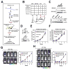
Figure 1. Coco Mediates Lung Colonization
(A) Gain-of-function retroviral cDNA screen. (B) Metastatic capabilities of the cell lines and results of the first screen. (C) Proteins precipitated from supernatants (4 ml) or extracted from cell layers (30 μg) were subjected to immunoblotting (top). Cells were treated with or without trypsin for 30 minutes before immunoblotting (bottom). (D) 4TO7-TGL cells transduced with myc-Coco or empty vector (top) and 4T1 cells expressing a control shRNA (sh-Control) or 2 shRNAs targeting Coco (sh #1 and #3) were subjected to immunoblotting. (E, F) 4TO7-TGL cells expressing Coco or not (E) and control and Coco-silenced 4T1-TGL cells (F) were inoculated orthotopically in syngeneic mice. The graphs show mean tumor volumes. (G, H) 4TO7-TGL cells expressing Coco or not (G) and control and Coco-silenced 4T1-TGL cells (H) were inoculated i.v.. Lung metastasis was measured by bioluminescent imaging. The panels show representative images (left) and the graph shows the normalized photon flux (right). In all panels, error bars represent SD. See also Figure S1.
Figure 2
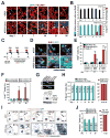
Figure 2. Coco Induces Reactivation in the Lung
(A) Mice were injected i.v. with 4TO7-TGL cells expressing Coco or not and sacrificed at the indicated times. Lung sections were stained as indicated. (B) The graph shows the number of GFP-positive solitary tumor cells per microscopic field (left vertical axis) and of metastatic lesions per lung section (right vertical axis) at the indicated times. Metastases were detected only in the lungs of mice injected with 4TO7-Coco cells (red bars; P < 5.6E-4 at 21 days and < 3.1E-05 at 35 days). (C) Outline of the EdU incorporation experiment. (D) Representative images of lung sections stained as indicated. (E) The graph shows the percentage of EdU+ cells at the indicated times. Cells expressing Coco were categorized as indicated. **p<0.01. (F) Control (sh-Control) and Coco-silenced (sh-Coco #1 and #3) 4T1-TGL cells were inoculated i.v.. Lung sections were stained with anti-Ki-67 and anti-GFP. The graph shows the percentage of Ki-67+ tumor cells at the indicated times. Control cells were categorized as solitary (Sol.) or outgrowing (Out.). **p<0.01. (G) Semiquantitative RT-PCR analysis of BMP receptors (top). Cells were left untreated or exposed to 1 ng/ml BMP4 for 30 minutes and subjected to immunoblotting (bottom). (H, I) 4TO7 and 4TO7-Coco cells were inoculated i.v. and lung sections were stained with anti-P-Smad 1, 5, 8 and anti-GFP. The graph on the left shows the percentage of P-Smad solitary tumor cells and the graph on the right the percentage of P-Smad metastatic outgrowths at the indicated times. Metastases were only detected in mice injected with 4TO7-Coco cells (red bars) (H). The panels show the positive staining pattern generated by anti-P-Smad antibodies on virtually all dormant 4TO7 cells and the lack of reactivity of the same antibodies with a subpopulation of dormant 4TO7-Coco cells. Metastases arising in mice injected with 4TO7-Coco were counterstained with hematoxylin (bottom right). (J) Control and Coco-silenced 4T1 cells were inoculated i.v. and lung sections were stained with anti-P-Smad and anti-GFP. The graph shows the percentage of P-Smad cells at the indicated times. Control cells were categorized as solitary (Sol.) or outgrowing (Out.). In all panels, error bars represent SD. See also Figure S2.
Figure 3
Figure 3. Coco Promotes Cancer Stem Cell Traits
(A) The graph shows the number of tumor spheres at each of three subsequent passages per 103 cells seeded. (B) Coomassie Blue staining of recombinant Coco (left) and number of tumor spheres formed by cells plated with the indicated concentrations (right). (C, D) Cells were plated with the indicated concentrations of Coco and BMP. The graph shows the efficiency of tumor sphere formation as compared to control cells. ***p<0.001 (C). Representative images (Coco: 0.9 μg/ml, BMP4: 10 ng/ml) (D). (E, F) Outline of PKH-26 staining and serial tumor sphere assay. ErbB2-transformed cells were stained with PKH-26 (left) and subjected to tumor sphere assay. The primary spheres were dissociated and their constituent cells were sorted according to staining intensity (PKHHIGH, PKHLOW, and PKHNEG) (center) and subjected to secondary tumor sphere assays. Finally, the PKHHIGH cells from secondary tumor spheres were subjected to the same protocol to derive tertiary tumor spheres (E). The graph show the results obtained with control and Coco-silenced ErbB2-transformed cells. *p<0.05, **p<0.01, ***p<0.001, NS-not significant. (G, H) 4TO7 and 4TO7-Coco cells (G) and control and Coco-silenced 4T1 cells (H) were inoculated orthotopically at the indicated doses. The graphs show mean tumor volumes. (I, J) Q-PCR of indicated genes in 4TO7 and 4TO7-Coco cells (I) and in 4TO7 cells left untreated or exposed to 10 ng/ml BMP4 for 2 days (J). In panel A, B, C and F, error bars represent SD. In panel G, H, I and J, error bars represent SEM. See also Figure S3 and Table S1, S2.
Figure 4
Figure 4. Inhibition of BMP Signaling Enhances Cancer Stem Cell Traits and Lung Colonization
(A) Cells were transduced with the indicated constructs were subjected to immunoblotting (left) and tumor sphere assay (right). (B) Representative images. (C) 4TO7 cells expressing the indicated constructs were inoculated i.v.. The panels show representative images (top) and the graph the normalized photon flux at the indicated times (bottom). (D) 4T1 cells transduced as indicated were injected orthotopically at the indicated doses. The graph shows the mean tumor volumes. (E) Cells were inoculated i.v. and lung metastasis was assessed as in C. In all panels, error bars represent SD. See also Figure S4.
Figure 5
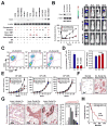
Figure 5. Coco Promotes Human Breast Cancer Metastasis
(A) Human breast cancer cells classified as indicated (Supplementary Experimental Procedures) were subjected to immunoblotting (Bo: bone only; L*: predominantly lung; BaA and BaB: basal A and B; Lu: luminal). (B) Control and Coco-silenced MDA-MB-231 cells were subjected to immunoblotting (top left) and lung colonization assay. The graph shows the normalized photon flux (bottom left, ±SD) and the panels show representative images (right). (C, D) Control and Coco-silenced MDA-MB-231 cells were subjected to FACS (C) and tumor sphere assay (D, left, ±SD). Viability at the end of the assay was measured by MTT staining (D, right, ±SEM). (E) Control and Coco-silenced MDA-MB-231 cells were inoculated orthotopically at the indicated doses in NOD/SCID/IL2Rγ−/− mice. The graphs show mean tumor volumes (± SEM. *p<0.05). **p<0.01, n.s.-not significant. (F) Normal breast tissue and a case of invasive ductal carcinoma were stained with monospecific antibodies to Coco and counterstained with Hematoxylin. Note the positivity of nests of tumor cells and scattered stromal cells (high magnification inset). (G) TMAs comprising 126 primary breast tumors were stained with anti-Coco. The pictures show images of cases exhibiting varying levels of positivity. The graph shows the distribution of staining intensity across samples. (H) Kaplan-Meier analysis of overall survival based on Coco expression in primary tumors. See also Figure S5 and Table S3, S4.
Figure 6
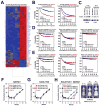
Figure 6. Coco Predicts Specifically Relapse to the Lung
(A) Hierarchical clustering of genes upregulated or downregulated (> 2 fold) in Coco-silenced MDA-MB231 cells as compared to control cells. NDRG1 and KIAA119 are underlined. (B) Kaplan-Meier analysis of metastasis-free survival in the NKI295 (left) or the indicated combined dataset (right). Patients were divided according to expression of the 14-gene Coco signature (red line, positive; blue line, negative). (C) MDA-MB231 cells and their lung and bone metastatic derivatives were subjected to immunoblotting. (D, E) Kaplan-Meier analysis of lung-only metastasis-free survival (left), bone-only metastasis-free survival (middle), and brain-only metastasis-free survival (right) in the MSK82, EMC192 and EMC286 combined dataset. Patients were divided according to expression of the 14-gene (D) or 2-gene Coco signature (E). (F, G) MDA-MB-231 cells expressing a control sh-RNA (sh-Control) and two sh-RNAs targeting NDRG1 (sh-NDRG #3 and #5) (F) or two sh-RNAs targeting KIAA1199 (sh-KIAA #1 and #5) (G) were subjected to lung colonization assay. (H) MDA-MB-231 cells expressing a control sh-RNA (sh-Control) or sh-RNAs targeting NDRG1 and KIAA1199 (sh-KIAA #1/ NDRG #3 and KIAA #5/ NDRG #5) were subjected to lung colonization assay. The graph shows the normalized photon flux (left) and the panels show representative images (right). In all panels, error bars represent SD. See also Figure S6 and Table S4-S7.
Figure 7
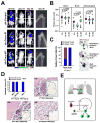
Figure 7. Coco-mediated Blockage of BMP is Not Required for Metastasis to the Bone or Brain
(A, B) Mice inoculated i.c. with control or Coco-silenced 4T1-TGL cells were subjected to bioluminescent imaging. Bone metastases were confirmed by X-ray radiography. The panels show representative images. Yellow dotted lines delineate the boundaries of osteolytic lesions in the radiograms (A). The graph shows the photon flux signal of metastases in the hind limbs, brains and adrenal glands of mice (B, ± SEM). (C) Mice were inoculated i.c. with 4T1-TGL cells and sacrificed 7 days later. Bone and brain sections were subjected to staining with anti-P-Smad and anti-GFP. The graph shows the percentage of P-Smad+ and P-Smad tumor cells in the bone and the brain. The images show representative images. (D) Mice were inoculated i.c. with 4T1 and 4TO7 cells and sacrificed at the indicated times. Bone sections were stained with H&E and anti-P-Smad. The graph shows the percentage of P-Smad+ and P-Smad tumor cells in micrometastases and macroscopic metastases generated by 4T1 cells and in micrometastatic clusters produced by 4TO7 cells (top left). The pictures show representative images. (E) Model of Coco’s function. See also Figure S7.

Comment in

  • Metastasis: Recharging with COCO.
    Seton-Rogers S. Seton-Rogers S. Nat Rev Cancer. 2012 Oct;12(10):655. doi: 10.1038/nrc3361. Epub 2012 Sep 21. Nat Rev Cancer. 2012. PMID: 22996601 No abstract available.

References

    1. Al-Hajj M, Wicha MS, Benito-Hernandez A, Morrison SJ, Clarke MF. Prospective identification of tumorigenic breast cancer cells. Proc Natl Acad Sci USA. 2003;100:3983–3988. - PMC - PubMed
    1. Aslakson CJ, Miller FR. Selective events in the metastatic process defined by analysis of the sequential dissemination of subpopulations of a mouse mammary tumor. Cancer Res. 1992;52:1399–1405. - PubMed
    1. Bell E, Munoz-Sanjuan I, Altmann CR, Vonica A, Brivanlou AH. Cell fate specification and competence by Coco, a maternal BMP, TGFβ and Wnt inhibitor. Development. 2003;130:1381–1389. - "V体育安卓版" PubMed
    1. Ben-Porath I, Thomson MW, Carey VJ, Ge R, Bell GW, Regev A, Weinberg RA. An embryonic stem cell-like gene expression signature in poorly differentiated aggressive human tumors. Nat Genetics. 2008;40:499–507. - PMC - PubMed
    1. Birkenkamp-Demtroder K, Maghnouj A, Mansilla’ F, Thorsen K, Andersen CL, Øster B, Hahn S, Ørntoft TF. Repression of KIAA1199 attenuates Wnt-signalling and decreases the proliferation of colon cancer cells. Brit J Cancer. 2011;105:552–561. - PMC - PubMed

"V体育安卓版" Publication types

Substances

VSports在线直播 - Associated data