"VSports在线直播" Skip to main page content
U.S. flag

An official website of the United States government

Dot gov

The . gov means it’s official VSports app下载. Federal government websites often end in . gov or . mil. Before sharing sensitive information, make sure you’re on a federal government site. .

Https

The site is secure. The https:// ensures that you are connecting to the official website and that any information you provide is encrypted and transmitted securely. V体育官网.

. 2012 Sep;8(9):1333-41.
doi: 10.4161/auto.20792. Epub 2012 Aug 14.

"VSports注册入口" ATG7 deficiency suppresses apoptosis and cell death induced by lysosomal photodamage

Affiliations

ATG7 deficiency suppresses apoptosis and cell death induced by lysosomal photodamage (VSports最新版本)

David H Kessel (VSports) et al. Autophagy. 2012 Sep.

Abstract (V体育2025版)

Photodynamic therapy (PDT) involves photosensitizing agents that, in the presence of oxygen and light, initiate formation of cytotoxic reactive oxygen species (ROS). PDT commonly induces both apoptosis and autophagy. Previous studies with murine hepatoma 1c1c7 cells indicated that loss of autophagy-related protein 7 (ATG7) inhibited autophagy and enhanced the cytotoxicity of photosensitizers that mediate photodamage to mitochondria or the endoplasmic reticulum. In this study, we examined two photosensitizing agents that target lysosomes: the chlorin NPe6 and the palladium bacteriopheophorbide WST11. Irradiation of wild-type 1c1c7 cultures loaded with either photosensitizer induced apoptosis and autophagy, with a blockage of autophagic flux. An ATG7- or ATG5-deficiency suppressed the induction of autophagy in PDT protocols using either photosensitizer. Whereas ATG5-deficient cells were quantitatively similar to wild-type cultures in their response to NPe6 and WST11 PDT, an ATG7-deficiency suppressed the apoptotic response (as monitored by analyses of chromatin condensation and procaspase-3/7 activation) and increased the LD(50) light dose by > 5-fold (as monitored by colony-forming assays). An ATG7-deficiency did not prevent immediate lysosomal photodamage, as indicated by loss of the lysosomal pH gradient. However, unlike wild-type and ATG5-deficient cells, the lysosomes of ATG7-deficient cells recovered this gradient within 4 h of irradiation, and never underwent permeabilization (monitored as release of endocytosed 10-kDa dextran polymers). We propose that the efficacy of lysosomal photosensitizers is in part due to both promotion of autophagic stress and suppression of autophagic prosurvival functions. In addition, an effect of ATG7 unrelated to autophagy appears to modulate lysosomal photodamage VSports手机版. .


Figures
None
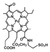
Figure 1. Structure of WST11.
None
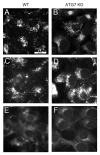
Figure 2. Subcellular localization of WST11 and effects on LTR fluorescence. Fluorescence localization patterns of WST11 in WT (A) or ATG7 KD (B) cultures after a 16 h incubation with 1 μM drug. (C–F) Effects of photodamage on the pattern of LTR fluorescence. Wild-type (C and E) or ATG7 KD cultures (D and F) were photosensitized with 1 μM WST11 for 16 h, then treated with LTR for 10 min before (C and D) or directly after (E and F) irradiation (90 mJ/cm2). White bar in (A): 20 μm. Photographs are representative of observations for three independent experiments.
None
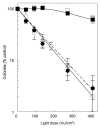
Figure 3. Effects of WST11 and NPe6 on 1c1c7 survival. Cultures of wild type (●, ○), ATG7 KD (■, □), and ATG5 KD (△) 1c1c7 cells were sensitized with 1 μM WST11 for 16 h (●, ■, △) or 20 μM NPe6 for 1 h (○, □) prior to being irradiated for different lengths of time to achieve the indicated light doses. Results with ATG5 KD cells are indicated by a dashed line. Colonies of 30 or more cells were determined 5 d later. Data are normalized to no-treatment controls and represent mean ± SD of three determinations. Similar results were obtained in a second independent experiment.
None
Figure 4. WST11 induced vacuolization. Cultures of wild-type (A–C), ATG7 KD (D–F) and ATG5 KD (G–I) 1c1c7 cells were incubated with 1 μM WST11 for 16 h prior to irradiation with light doses of 0 (A–G), 90 mJ/cm2 (B,E and H), or 405 mJ/cm2 (C, F and I). Images were captured 2 h after irradiation. Bar in (A): 20 μm. Images are representative of three independent experiments.
None
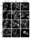
Figure 5. WST11 induction of autophagy. Cultures of GFP-LC3-expressing wild-type 1c1c7 cells were incubated with 1 μM WST11 for 16 h prior to being imaged by phase and fluorescence microscopy immediately prior to (A and B), 1 h (C and D) or 4 h (E and F) after irradiation (405 mJ/cm2). White bars: (A and B) 20 μm; (C–F) 15 μm. (G) Electron micrograph of representative autophagosomes found in WT 1c1c7 cultures loaded with 1 μM WST11 for 16 h and fixed for processing 4 h after irradiation with 405 mJ/cm2. White bar: 0.2 μm. (H) Western blot analysis of LC3 and actin in untreated WT (lane 1) or ATG7 KD (lane 2) cells. Alternatively, WT (lanes 3 and 4) and ATG7 KD cells (lanes 5 and 6) were incubated for 16 h with WST11 prior to exposure to a 405-J/cm2 light dose. NH4Cl (50 mM) was added to some cultures immediately after irradiation (lanes 4 and 6). All cultures were harvested 2 h after irradiation. Similar results were obtained in a second independent study.
None
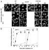
Figure 6. Effects of WST11 PDT on nuclear morphology and procaspase activation. (A) Cultures of wild-type (a–g) and ATG7 KD (h–n) 1c1c7 cells were loaded with 1 μM WST11 for 16 h prior to no treatment, or irradiation (90 or 405 mJ/cm2). Cultures were subsequently incubated with HO33342 for 10 min prior to being imaged 1, 4 or 24 h after irradiation. With the exceptions of (g) and the right half of (f), all other images are of adherent cells. (g) and the right half of (f) are images of HO33342 stained non-adherent floating cells which constitute ~50% and > 95% of all the cells in the plates, respectively. Arrows in (e) point to nuclei with condensed chromatin. Bar in (a): 20 μm. (B) Cultures of wild-type (●, ○), ATG7 KD (▲) or ATG5 KD (■) 1c1c7 cells were treated with 1 μM WST11 for 16 h or with 20 μM NPe6 for 1 h prior to irradiation with either 90 mJ/cm2 (○) or 405 mJ/cm2 (●,▲, ■). Cultures were harvested at specified times after irradiation for analyses of DEVDase activity. Data represent means ± SD of three analyses at each time point for all treatment groups. Similar results were obtained in a second independent study.
None
Figure 7. Effects of WST11 PDT on LTR labeling. WT, ATG5 KD and ATG7 KD cultures were left untreated (a, e and i) or incubated with 1 µM WST11 for 16 h prior to being irradiated (405 mJ/cm2), and labeled with LTR immediately (b ,f and j), 4 h (c, g and k) or 16 h (d, h and l) after irradiation. Bar in (a): 20 μm.
None
Figure 8. Analyses of lysosomal permeabilization following NPe6 PDT. Cultures of WT (A–F) and ATG7 KD (G–L) cells were preloaded with fluorescein-conjugated 10-kDa dextran polymers as described in Materials and Methods prior to being sensitized with 20 μM NPe6 and irradiated (270 mJ/cm2). LTR was added to the cultures 10 min prior to imaging. LTR (A–C and G–I) and dextran (D–F and J–L) fluorescence were captured 10 min and 4 h after irradiation. Similar results were obtained in a second study.

References

    1. Dougherty TJ, Gomer CJ, Henderson BW, Jori G, Kessel D, Korbelik M, et al. Photodynamic therapy. J Natl Cancer Inst. 1998;90:889–905. doi: 10.1093/jnci/90.12.889. - V体育安卓版 - DOI - PMC - PubMed
    1. Agostinis P, Berg K, Cengel KA, Foster TH, Girotti AW, Gollnick SO, et al. Photodynamic therapy of cancer: an update. CA Cancer J Clin. 2011;61:250–81. doi: 10.3322/caac.20114. - DOI - PMC - PubMed
    1. Agarwal ML, Clay ME, Harvey EJ, Evans HH, Antunez AR, Oleinick NL. Photodynamic therapy induces rapid cell death by apoptosis in L5178Y mouse lymphoma cells. Cancer Res. 1991;51:5993–6. - V体育官网 - PubMed
    1. Kim HR, Luo Y, Li G, Kessel D. Enhanced apoptotic response to photodynamic therapy after bcl-2 transfection. Cancer Res. 1999;59:3429–32. - PMC - PubMed
    1. Xue LY, Chiu SM, Oleinick NL. Photochemical destruction of the Bcl-2 oncoprotein during photodynamic therapy with the phthalocyanine photosensitizer Pc 4. Oncogene. 2001;20:3420–7. doi: 10.1038/sj.onc.1204441. - DOI - PubMed

V体育ios版 - Publication types

"V体育安卓版" MeSH terms