VSports在线直播 - Skip to main page content
U.S. flag

An official website of the United States government

Dot gov

The . gov means it’s official. Federal government websites often end in . gov or VSports app下载. mil. Before sharing sensitive information, make sure you’re on a federal government site. .

Https

The site is secure V体育官网. The https:// ensures that you are connecting to the official website and that any information you provide is encrypted and transmitted securely. .

. 2012 Aug 3;150(3):549-62.
doi: 10.1016/j.cell.2012.06.031.

HSF1 drives a transcriptional program distinct from heat shock to support highly malignant human cancers

Affiliations

"V体育平台登录" HSF1 drives a transcriptional program distinct from heat shock to support highly malignant human cancers

Marc L Mendillo et al. Cell. .

Abstract

Heat-Shock Factor 1 (HSF1), master regulator of the heat-shock response, facilitates malignant transformation, cancer cell survival, and proliferation in model systems. The common assumption is that these effects are mediated through regulation of heat-shock protein (HSP) expression. However, the transcriptional network that HSF1 coordinates directly in malignancy and its relationship to the heat-shock response have never been defined. By comparing cells with high and low malignant potential alongside their nontransformed counterparts, we identify an HSF1-regulated transcriptional program specific to highly malignant cells and distinct from heat shock VSports手机版. Cancer-specific genes in this program support oncogenic processes: cell-cycle regulation, signaling, metabolism, adhesion and translation. HSP genes are integral to this program, however, many are uniquely regulated in malignancy. This HSF1 cancer program is active in breast, colon and lung tumors isolated directly from human patients and is strongly associated with metastasis and death. Thus, HSF1 rewires the transcriptome in tumorigenesis, with prognostic and therapeutic implications. .


Figures

Figure 1
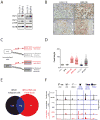
Figure 1. HSF1 is activated in metastatic and highly tumorigenic human mammary epithelial cell lines
A. Equal amounts of total protein were immunoblotted with the indicated antibody. B. IHC staining with anti-HSF1 antibody of the indicated tumors xenografted in mice. Upper panels show regions of viable tumor (high mag, scale bar 20 μm) and lower panels show the viable tumor/necrotic interface (low mag, scale bar 50 μm) C. Schematic of experimental groups analyzed by HSF1 ChIP-Seq. D. Graph of ChIP-Seq peak heights for each region of HSF1 occupancy, normalized by the number of reads in the dataset. E. Overlap of genes bound in malignant cells (BPLER, 37°C) and immortalized, non-tumorigenic cells after heat shock (BPE or HME cells, 42°C). F. Representative genes bound in BPLER cells (CKS2, LY6K, RBM23) and bound in both BPLER cells and heat-shocked HME and BPE cells (HSPA6, HSPA8, PROM2). X-axis: from −2kb from the transcription start site (TSS) to either +5, +6 or +10kb from the TSS for each gene; genes diagrams drawn to scale. See also Figure S1 and Tables S1 and S2.
Figure 2
Figure 2. The expression of HSF1-bound genes is altered by HSF1 depletion
A. Relative gene expression levels following shRNA-mediated knockdown of HSF1 in indicated cells. Scr and GFP were negative control shRNA. B. Graph showing the number of genes positively regulated (reduced expression upon HSF1 depletion) or negatively regulated (increased expression upon HSF1 depletion) by HSF1 relative to site of gene occupancy by HSF1 (promoter versus distal). See also Figure S2 and Table S3.
Figure 3
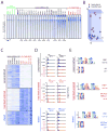
Figure 3. Genome-wide patterns of DNA occupancy by HSF1 across a broad range of common human cancer cell lines
A. Heat map of ChIP-Seq read density for all HSF1 target regions (union of all HSF1-bound regions in all datasets). Genomic regions from −1kb to +1kb relative to the peak of HSF1 binding are shown. Regions are ordered the same in all datasets. Read density is depicted for non-tumorigenic cells at 37°C (green), cancer lines at 37°C (black) and non-tumorigenic (nt) lines following heat shock at 42°C (red). Asterisks indicate datasets also used for the analysis in Figure 1E. B. Principal component analysis (PCA) of HSF1 binding in heat-shocked parental lines (red) and cancer lines (black). C. ChIP-Seq density heat map of genomic regions differentially bound by HSF1 in cancer lines at 37°C, heat-shocked non-tumorigenic lines, and regions shared under both conditions. D. HSF1 binding of representative genes in cancer lines at 37°C (black) and heat-shocked non-tumorigenic lines (red). Examples of genes with distinct patterns of binding are presented: Enriched in cancer lines, heat-shocked non-tumorigenic lines, or both. E. Motif analysis of 100bp regions surrounding HSF1 binding peaks for genes enriched in cancer lines (BT20, NCIH838 and SKBR3), heat-shocked non-tumorigenic lines (HME, BPE, MCF10A) and both cancer and heat-shocked non-tumorigenic lines. See also Figure S3 and Table S1.
Figure 4
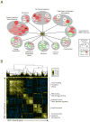
Figure 4. Distinct, coordinately-regulated modules of HSF1-bound genes
A. Graphical representation of the HSF1 cancer program integrating information on gene binding, regulation and function. The peak height is reflected in the diameter of the circle (log2 peak height: range ~3 to 9) and color intensity reflects gene regulation (average of log2 fold change in BPLER and MCF7 cells upon HSF1 knockdown; red - positively regulated; green – negatively regulated; gray – no data available). Well-bound, differentially regulated genes as well as several genes of biological interest are displayed. B. Gene-gene expression correlation matrix of HSF1-bound genes. Pair-wise correlation map is presented of the genes that were bound by HSF1 in at least two of the three cancer cell lines (BT20, NCIH38, and SKBR3). The Pearson correlation coefficient relating normalized mRNA expression data for each gene pair was assessed in nearly 12,000 expression profiles. Enriched GO (gene-ontology) categories for each module are shown. See also Figure S4.
Figure 5
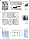
Figure 5. HSF1 is activated in a broad range of human tumors
A. IHC shows strong nuclear HSF1 staining in human breast tumor cells (top) with adjacent normal breast epithelial cells (bottom) showing a lack of nuclear HSF1. B. Images of HSF1 IHC on breast cancer tissue microarray (TMA) cores. Heat map shows scoring of three TMAs. The top panel depicts data from two TMAs (BRC1501 and BRC1502), containing 138 breast tumors of all major breast cancer subtypes. Progesterone receptor (PR), ER, and HER2 were also evaluated. The middle panel shows data from 161 triple negative (TN) breast cancer cases. The bottom panel shows the lack of HSF1 nuclear expression in 16 normal mammary tissue sections. A summary is provided in the bar graph (right). C. HSF1 IHC showing high level nuclear staining in indicated tumors; T, Tumor; N, Normal adjacent tissue. A summary is provided in the bar graph. D. ChIP-Seq analysis of human breast and colon cancer surgical resection specimens (patient tumors). Heat map depicting ChIP-Seq read density for all HSF1 target regions defined in Figure 3A. For reference, the binding profiles for cancer cell lines in culture (black; average across BT20, NCIH838 and SKBR3) and parental heat-shocked cell lines (red) are included. HSF1 expression was evaluated by IHC in the same patient tumors used for ChIP-Seq (see Figure S5C) and scored as in Panel B. E. HSF1 binding in cell lines compared to patient tumors. Average binding across cancer cell lines in cell culture (black; average across BT20, NCIH838 and SKBR3), parental heat-shocked cell lines (red), and patient tumors (cyan) are depicted for the representative target genes indicated. F. PCA of HSF1 binding in heat-shocked parental cell lines (red), cancer cells lines (black) and patient tumors (cyan). See also Figure S5 and Table S1.
Figure 6
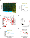
Figure 6. An HSF1-cancer signature is associated with reduced survival in patients with breast cancer
A. Representative dataset (Pawitan et al., 2005) is shown from a meta-analysis of 10 publicly available mRNA expression datasets (Table S5) derived from human breast tumors with known clinical outcome and representing a total of 1594 patients. Each column corresponds to a tumor, and each row corresponds to a microarray probe for an HSF1-cancer signature (HSF1-CaSig) gene. Median levels of expression are depicted in black, increased expression in yellow, and decreased expression in blue. Tumors are ordered by average level of expression of the HSF1-cancer signature, from low (blue) to high (yellow). Red bars indicate deaths. Kaplan-Meier (KM) analysis of the tumors with high expression of the HSF1-cancer signature (top 25%, “High HSF1-CaSig”, yellow) versus low expressors (bottom 75%, “Low HSF1-CaSig”, blue) is shown. B. Log-rank p-values for each of the indicated classifiers were calculated for each dataset; results are displayed as a heat map. Corresponding KM curves are provided in Figure S6. C. Random gene signature analysis of a representative dataset (Pawitan et al., 2005). KM analysis on the dataset to evaluate associations between 10,000 individual randomly generated gene signatures and patient outcome. The random signatures are binned and ordered from least significant to most significant by the KM-generated test statistic. The Red arrow indicates the test statistic of the HSF1-CaSig. For reference, black arrows indicate the test statistic of the random signature with the median test statistic (5000th) and the random signature with the 95th percentile test statistic. D. KM analysis of individuals with ER+/Lymph node negative tumors (Wang et al., 2005) with Low HSF1-CaSig (blue) or High HSF1-CaSig (yellow). E. KM analysis of 947 individuals from the NHS with ER+, lymph-node negative tumors expressing no, low or high nuclear HSF1 as measured by IHC. Data are from the NHS (1976–1997). Log-rank p-values are shown. See also Figure S6 and Tables S4, S5 and S6.
Figure 7
Figure 7. An HSF1-cancer signature is associated with reduced survival in patients with colon or lung cancers
A. KM analysis of survival in patients with colon or lung cancer based on Low HSF1-CaSig (blue) or High HSF1-CaSig (yellow). Log-rank p-values are shown. B. Heat map of log-rank p-values for each of the indicated classifiers in four datasets is shown. Corresponding KM curves are in Figure S7. See also Tables S4 and S5.

References

    1. Anckar J, Sistonen L. Regulation of HSF1 Function in the Heat Stress Response: Implications in Aging and Disease. Annu Rev Biochem. 2011;80:1089–1115. - PubMed
    1. Chiang WC, Ching TT, Lee HC, Mousigian C, Hsu AL. HSF-1 regulators DDL-1/2 link insulin-like signaling to heat-shock responses and modulation of longevity. Cell. 2012;148:322–334. - PMC - PubMed
    1. Christians ES, Yan LJ, Benjamin IJ. Heat shock factor 1 and heat shock proteins: Critical partners in protection against acute cell injury. Crit Care Med. 2002;30:S43–S50. - V体育官网入口 - PubMed
    1. Ciocca DR, Calderwood SK. Heat shock proteins in cancer: diagnostic, prognostic, predictive, and treatment implications. Cell Stress Chaperones. 2005;10:86–103. - PMC - PubMed
    1. Dai C, Whitesell L, Rogers AB, Lindquist S. Heat shock factor 1 is a powerful multifaceted modifier of carcinogenesis. Cell. 2007;130:1005–1018. - PMC - PubMed

Publication types (V体育2025版)

MeSH terms

"V体育2025版" Substances