<acronym id="x5nAtPft"></acronym>
Skip to main page content
U.S. flag

An official website of the United States government

Dot gov

The . gov means it’s official. Federal government websites often end in . gov or . mil VSports app下载. Before sharing sensitive information, make sure you’re on a federal government site. .

Https

The site is secure. The https:// ensures that you are connecting to the official website and that any information you provide is encrypted and transmitted securely V体育官网. .

. 2012;7(7):e41945.
doi: 10.1371/journal.pone.0041945. Epub 2012 Jul 31.

Staurosporine induces necroptotic cell death under caspase-compromised conditions in U937 cells

Affiliations

Staurosporine induces necroptotic cell death under caspase-compromised conditions in U937 cells

Zsuzsanna A Dunai et al. PLoS One. 2012.

Abstract

For a long time necrosis was thought to be an uncontrolled process but evidences recently have revealed that necrosis can also occur in a regulated manner. Necroptosis, a type of programmed necrosis is defined as a death receptor-initiated process under caspase-compromised conditions. The process requires the kinase activity of receptor-interacting protein kinase 1 and 3 (RIPK1 and RIPK3) and mixed lineage kinase domain-like protein (MLKL), as a substrate of RIPK3. The further downstream events remain elusive. We applied known inhibitors to characterize the contributing enzymes in necroptosis and their effect on cell viability and different cellular functions were detected mainly by flow cytometry VSports手机版. Here we report that staurosporine, the classical inducer of intrinsic apoptotic pathway can induce necroptosis under caspase-compromised conditions in U937 cell line. This process could be hampered at least partially by the RIPK1 inhibitor necrotstin-1 and by the heat shock protein 90 kDa inhibitor geldanamycin. Moreover both the staurosporine-triggered and the classical death ligand-induced necroptotic pathway can be effectively arrested by a lysosomal enzyme inhibitor CA-074-OMe and the recently discovered MLKL inhibitor necrosulfonamide. We also confirmed that the enzymatic role of poly(ADP-ribose)polymerase (PARP) is dispensable in necroptosis but it contributes to membrane disruption in secondary necrosis. In conclusion, we identified a novel way of necroptosis induction that can facilitate our understanding of the molecular mechanisms of necroptosis. Our results shed light on alternative application of staurosporine, as a possible anticancer therapeutic agent. Furthermore, we showed that the CA-074-OMe has a target in the signaling pathway leading to necroptosis. Finally, we could differentiate necroptotic and secondary necrotic processes based on participation of PARP enzyme. .


Conflict of interest statement

Competing Interests: The authors have read the journal's policy and have the following conflicts: IP is an employee of KPS Medical Biotechnology and Healthcare Services Ltd. KPS Medical Biotechnology did not support our studies at all. KPS Medical Biotechnology provides molecular pathology services to detect predictive biomarkers in tumor samples for personalized targeted therapies V体育安卓版. The company focuses on the diseases of the gastrointestinal tract and on lung cancer. This does not alter the authors' adherence to all the PLoS ONE policies on sharing data and materials.

Figures

Figure 1
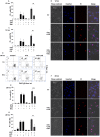
Figure 1. TRAIL induces necroptosis in the presence of caspase inhibitor.
TRAIL-induced cell death in U937 cells. Cells were treated as indicated for 20 hrs. (A) Cytospins were stained with hematoxylin-eosin. Blue arrows show the apoptotic, green ones the necrotic cells (400x) (representative of n = 2). Scale bar on the first subfigure applies to all the figures in the panel. (B) Western blot analysis of fragmented PARP-1 protein. Full length PARP-1 is 116 kDa, cleaved PARP-1 fragment is 89 kDa (representative of n = 2). (C) TRAIL-induced caspase activity in U937 cells. U937 cells were exposed to 50 ng/mL hr-TRAIL (114–281 aa) in the presence or absence of zVD (5 µM) for the indicated period of time. The ordinate shows the slope of the measured DEVDase activity curves of a representative experiment carried out in triplicates. (D) FACS analysis of treated cells. Plasma membrane integrity was analyzed after PI staining of cells. Inserted values on histograms show the percentage of the marked population (representative of n = 7). (E-F) Nec and GA protected U937 cells from TRAIL+zVD-induced necroptosis. U937 cell were exposed to TRAIL (50 ng/mL) in the presence or absence of zVD (5 µM) and (E) Nec (10 µM) or (F) GA (1 µM) for 20 hrs. PI stained cells were analyzed for membrane permeability. Percentages of PI positive cells were determined (n = 7 for Nec and n = 4 for GA). Values are mean±SD. *, P<0.05, **, P<0.01 and ***, P<0.001 calculated by Student’s t-probe.
Figure 2
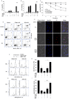
Figure 2. STS induces primary necrosis in the presence of caspase inhibitor.
(A-B) Nec (10 µM) and (C) GA (1 µM) significantly inhibited the STS-triggered necroptosis. Cells were exposed to STS (1 µM) in the presence or absence of zVD (5 µM) for 12 hrs. Percentage of PI positive cells was determined by flow cytometry (A, C). (n = 4) and by Hoechst/PI double staining technique (B) (representative of n = 2), (400x). Scale bar on the first subfigure applies to all the figures in the panel. (D) Nec (10 µM) arrested the STS-induced (1 µM) necroptosis in the presence of zVD (5 µM) after 20 hrs incubation. The mitochondrial transmembrane potential and plasma membrane integrity is shown in representative dot plots of DiOC6(3) and PI stained, unfixed cells. The values indicate the percentage of cells in the marked regions (n = 13). (E-G) Nec (10 µM) (E, F) and GA (1 µM) (G) partially inhibited the STS-triggered necroptosis. Cells were exposed to STS (1 µM) in the presence or absence of zVD (5 µM) for 20 hrs. Percentage of PI positive cells was determined by flow cytometric analysis (E, G) (n = 13 for Nec and n = 4 for GA), and by Hoechst/PI double staining technique (F) (n = 2). Scale bar on the first subfigure applies to all the figures in the panel. Values are mean±SD. *, P<0.05, **, P<0.01 and ***, P<0.001 calculated by Student’s t-probe.
Figure 3
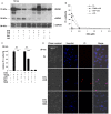
Figure 3. STS induces RIPK1 and MLKL-dependent necroptosis.
(A) zVD hampered RIPK1 fragmentation triggered by STS for 12 hrs. Western blot analysis was performed for the detection of RIPK1 protein level and presence of cleaved fragment due to caspase activity (representative of n = 2). (B) NSA reduced TRAIL and STS-induced necroptosis in a concentration-dependent manner after 20 hrs incubation time – representative experiment. U937 ells were exposed to STS (1 µM) and TRAIL (50 ng/mL) and varying concentrations of NSA (0–2.5 µM) in the presence of zVD (5 µM) for 20 hrs. Percentage of PI positive cells was determined. (C-D) NSA (0.5 µM) significantly hampered both the TRAIL and STS-triggered necroptosis. (C) Relative percentage of PI positive cells was determined by FACS analysis (n = 3). Values are mean±SD. *, P<0.05, **, P<0.01 and ***, P<0.001 calculated by Student’s t-probe. (D) Morphological signs of apoptosis and necrosis are shown in representative fluorescent microscopic images (400x) of Hoechst/PI double stained U937 cells (n = 2). Scale bar on the first subfigure applies to all the figures in the panel.
Figure 4
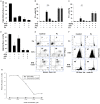
Figure 4. CA inhibits either the TRAIL or STS-induced necroptosis in presence of caspase inhibitor.
U937 cells were treated either with STS (1 µM) or with TRAIL (50 ng/mL) in the presence or absence of zVD (5 µM) for 20 hrs. Nec (10 µM) or CA (10 µM) were added 1 hr before cell death was induced. (A-B) CA (10 µM) considerably inhibited the (A) TRAIL (n = 4) or (B) STS-triggered necroptosis (n = 3). Percentage of PI positive cells was determined. (C) Time course analysis of cells with depolarized mitochondria is shown after DiOC6(3) staining of, unfixed cells for STS treatment combined with the indicated inhibitors (n = 2). Values are mean±SD. *, P<0.05, **, P<0.01 and ***, P<0.001 calculated by Student’s t-probe. (D) PS distribution in the plasma membrane is shown in representative dot plots of Annexin V-FITC and PI stained, unfixed cells analyzed by flow cytometry. The values indicate the percentage of cells in the marked regions (n = 2). (E) Morphological signs of apoptosis and necrosis are shown in representative fluorescent microscopic images (400x) of Hoechst/PI double stained U937 cells (n = 2). Scale bar on the first subfigure applies to all the figures in the panel. (F) Distribution of cells with various volumes of acidic compartments (endo-lysosomes) is shown in representative overlay histograms of AO stained cells analyzed by flow cytometry. (G) Column diagram of percentage of cells with high AO red fluorescence intensity (n = 2). (H) Distribution of cells with various mitochondrial transmembrane potential is shown in representative overlay histograms of DiOC6(3) stained cells analyzed by flow cytometry. (I) Column diagram of percentage of cells with high DiOC6(3) fluorescence intensity (n = 3). Values are mean±SD. *, P<0.05, **, P<0.01 and ***, P<0.001 calculated by Student’s t-probe.
Figure 5
Figure 5. PJ3-4 does not arrest either the TRAIL or STS-induced necroptosis in presence of caspase inhibitor.
U937 cells were treated either with STS (1 µM) or with TRAIL (50 ng/mL) in the presence or absence of zVD (5 µM) for 20 hrs. PJ-34 (1 µM) was added 1 hr before cell death was induced. (A) Column diagram of percentage of cells with high DiOC6(3) fluorescence intensity (n = 3). (B-C) PJ-34 (1 µM) considerably inhibited the (B) STS (n = 8) or (C) TRAIL-triggered (n = 8) secondary necrosis but not the necroptosis. Percentage of PI positive cells was determined. Values are mean±SD. *, P<0.05, **, P<0.01 and ***, P<0.001 calculated by Student’s t-probe. (D) Column diagram of percentage of cells with high AO fluorescence intensity (n = 2). (E) Dot plot distribution of cells stained with Annexin V-FITC and PI analyzed by flow cytometry. The values indicate the percentage of cells in the marked regions (representative of n = 2). (F) Representative histograms of distribution of PI stained, ethanol-fixed (sub-G1) cells were analyzed by flow cytometry (n = 8). The numbers indicate the percentage of cells in the marked regions. (G) PJ-34 concentration dependently reduced the proportion of PI positive cells for STS treatment for 20 hrs – representative experiment.
Figure 6
Figure 6. Schematic diagram about the action of inhibitors on TRAIL and STS- induced cell death pathways.
Both TRAIL and STS triggered necroptosis in caspase depleted U937 cells upon prolonged incubation time. RIPK1 inhibitors Nec and GA could completely inhibit the TRAIL-evoked necroptosis, but had partial inhibitory potential to the STS-provoked process. Contrarily CA and NSA abolished both the TRAIL and STS-induced necroptosis. In absence of caspase inhibitor TRAIL and STS induced apoptosis which is followed by secondary necrosis. PJ-34 delayed the necrotic plasma membrane disruption during secondary necrosis, but failed to inhibit necroptosis.

References

    1. Yuan J (2009) Neuroprotective strategies targeting apoptotic and necrotic cell death for stroke. Apoptosis 14: 469–477. - PMC - PubMed
    1. Mareninova OA, Sung KF, Hong P, Lugea A, Pandol SJ, et al. (2006) Cell death in pancreatitis: Caspases protect from necrotizing pancreatitis. Journal of Biological Chemistry 281: 3370–3381. - V体育平台登录 - PubMed
    1. Zhang X, Chen Y, Jenkins LW, Kochanek PM, Clark RSB (2005) Bench-to-bedside review: Apoptosis/programmed cell death triggered by traumatic brain injury. Critical Care 9: 66–75. - PMC - PubMed
    1. McCully JD, Wakiyama H, Hsieh YJ, Jones M, Levitsky S (2004) Differential contribution of necrosis and apoptosis in myocardial ischemia-reperfusion injury. American Journal of Physiology - Heart and Circulatory Physiology 286: H1923–H1935. - "V体育ios版" PubMed
    1. West T, Atzeva M, Holtzman DM (2006) Caspase-3 deficiency during development increases vulnerability to hypoxic-ischemic injury through caspase-3-independent pathways. Neurobiology of Disease 22: 523–537. - "VSports" PubMed

Publication types