V体育官网入口 - Skip to main page content
U.S. flag

An official website of the United States government

Dot gov

The . gov means it’s official VSports app下载. Federal government websites often end in . gov or . mil. Before sharing sensitive information, make sure you’re on a federal government site. .

Https

The site is secure. The https:// ensures that you are connecting to the official website and that any information you provide is encrypted and transmitted securely V体育官网. .

. 2012 Aug;9(8):1076-87.
doi: 10.4161/rna.21089. Epub 2012 Aug 1.

V体育官网 - Loss of the abundant nuclear non-coding RNA MALAT1 is compatible with life and development

Affiliations

Loss of the abundant nuclear non-coding RNA MALAT1 is compatible with life and development (VSports最新版本)

"VSports注册入口" Moritz Eißmann et al. RNA Biol. 2012 Aug.

Abstract

The metastasis-associated lung adenocarcinoma transcript 1, MALAT1, is a long non-coding RNA (lncRNA) that has been discovered as a marker for lung cancer metastasis VSports手机版. It is highly abundant, its expression is strongly regulated in many tumor entities including lung adenocarcinoma and hepatocellular carcinoma as well as physiological processes, and it is associated with many RNA binding proteins and highly conserved throughout evolution. The nuclear transcript MALAT-1 has been functionally associated with gene regulation and alternative splicing and its regulation has been shown to impact proliferation, apoptosis, migration and invasion. Here, we have developed a human and a mouse knockout system to study the loss-of-function phenotypes of this important ncRNA. In human tumor cells, MALAT1 expression was abrogated using Zinc Finger Nucleases. Unexpectedly, the quantitative loss of MALAT1 did neither affect proliferation nor cell cycle progression nor nuclear architecture in human lung or liver cancer cells. Moreover, genetic loss of Malat1 in a knockout mouse model did not give rise to any obvious phenotype or histological abnormalities in Malat1-null compared with wild-type animals. Thus, loss of the abundant nuclear long ncRNA MALAT1 is compatible with cell viability and normal development. .


Figures
None
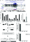
Figure 1. Expression and depletion of MALAT1 in human cells. (A) The evolutionary conservation of MALAT1 over its entire length is depicted using the University of California, Santa Cruz (UCSC) genome browser. (B) MALAT1 expression was determined in a panel of 11 human cell lines representing different tissues of origin. Shown is the expression relative to RPLP0 mRNA (B, left panel). On average, MALAT1 shows a 2.5-fold higher expression (p < 0.001) in these cell lines compared with the abundant housekeeping gene RPLP0 (B, right panel). (C) To deplete the highly abundant lncRNA MALAT1, a knockout approach was recently developed that is schematically explained here. (D) This method allowed the generation of A549 (lung) and HLE (liver) single cell clones that showed a ~1000-fold or ~200-fold reduction of full-length MALAT1, respectively. Given is the average expression measured in three independent experiments +SEM.
None
Figure 2. Proliferation and cell cycle progression of MALAT1 KO cells. The proliferative phenotype of A549 and HLE MALAT1 WT and KO cells was analyzed. (A+C) For both cell lines, relative proliferation rates were determined using a bromodeoxyuridine (BrdU) proliferation assay. Results were normalized to the parental cell line in each case. Mean values of three independent experiments +SEM are presented. (B+D) Cell cycle profiles were obtained from exponentially growing cells and the average percentages of cells in G1-, S- and G2/M-phase from at least two independent experiments are depicted.
None
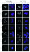
Figure 3. Nuclear morphology of human A549 wild-type and MALAT1 knockout cells. CLSM optical sections were acquired for different fluorescently labeled nuclear components in the A549 lung cancer cell line for wild-type and MALAT1 knockout cells. RNA was labeled via incorporated 5-ethynyl-uridine, and the proteins were visualized by immunofluorescence. The merged images show the indicated nuclear components in red and a DNA DAPI staining in blue. Scale bar, 10 µm. (A) RNA labeled. (B) Active RNA polymerase II. (C) Lamin A, a component of the nuclear envelope. (D) UBF, a marker for active ribosomal genes. (E) Splicing speckles visualized via the SC35 protein. (F) HP1α, enriched in pericentric heterochromatin. (G) TRF2, localizing to the telomeric shelterin complex.
None
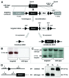
Figure 4. Generation and confirmation of the Malat1 knockout mouse line. (A) Schematic presentation of the strategy for deletion of Malat1 by homologous recombination in the mouse genome. LoxP (Cre recombination) and FRT (Flip recombination) sites are indicated and allow individual removal of the complete Malat1 locus (Cre) and the Neomycin (neo) resistance gene (Flip), respectively. Probe 2: 3′external probe (located outside of the targeting vector) for Southern Blot analysis. (B) PCR analysis of genomic DNA prepared from single electroporated ES cell clones. With the forward primer located in the neo resistance gene and the reverse primer hybridizing to the wild-type sequence at the 3′-end outside of the targeting construct, several ES cell clones with the desired homozygous recombination were identified via a 2420 bp PCR fragment which is absent in clones without correct targeting. (C) Southern Blot analysis with genomic DNA prepared from single electroporated ES cell clones and digested with the restriction enzymes BamHI and XhoI. Probe 2 recognized a 4.1 kb XhoI/BamHI fragment in the correctly targeted locus, while the wild-type allele was identified via a 10.6 kb BamHI/BamHI fragment. All relevant restriction sites are indicated in (A). (D) To confirm Cre-mediated deletion of Malat1 and to distinguish wild-type, heterozygous and homozygous Malat1 constitutive knockout mice, a three-primer-PCR-strategy was developed, resulting in two fragments with different lengths for the wild-type (120 bp) and the targeted (204 bp) locus.
None
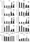
Figure 5.Malat1 and Neat1 expression. Malat1 and Neat1 expression were determined in eight different mouse tissues (A) and two human cancer cell lines (B) by qRT-PCR. (A) Malat1 was quantified with three independent amplicons and Neat1 was quantified using two independent amplicons in lung, spleen, small intestine, kidney, brain, colon, liver and heart of four wild-type (WT) and three Malat1 knockout mice (KO). Gapdh served as reference gene and data was normalized to expression in normal lung which displayed the highest Malat1 and Neat1 expression. Depicted is the average expression +SEM (B) NEAT1 was quantified in A549 and HLE cancer cell lines in the parental cell line, two wild-type clones (WT) and three MALAT1 knockout clones (KO) each. RN7SL1 served as reference gene, and data was normalized to expression in the wild-type cells. Depicted is the average expression +SEM.
None
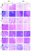
Figure 6. Depletion of Malat1 does not alter organ histomorphology. H&E staining of brain (A), thymus (B), lung (C), liver (D), pancreas (E), kidney (F) and genitals (G) from wild-type Malat1+/+ (left panel; ♀,♂) and knockout Malat1−/− (right panel; ♀,♂) mice. No histomorphogical differences were observed between wild-type and knockout mice in all tissues examined.

"V体育安卓版" References

    1. Birney E, Stamatoyannopoulos JA, Dutta A, Guigó R, Gingeras TR, Margulies EH, et al. ENCODE Project Consortium. NISC Comparative Sequencing Program. Baylor College of Medicine Human Genome Sequencing Center. Washington University Genome Sequencing Center. Broad Institute. Children’s Hospital Oakland Research Institute Identification and analysis of functional elements in 1% of the human genome by the ENCODE pilot project. Nature. 2007;447:799–816. doi: 10.1038/nature05874. - DOI - PMC - PubMed
    1. Cabili MN, Trapnell C, Goff L, Koziol M, Tazon-Vega B, Regev A, et al. Integrative annotation of human large intergenic noncoding RNAs reveals global properties and specific subclasses. Genes Dev. 2011;25:1915–27. doi: 10.1101/gad.17446611. - DOI - PMC - PubMed
    1. Carninci P, Kasukawa T, Katayama S, Gough J, Frith MC, Maeda N, et al. FANTOM Consortium. RIKEN Genome Exploration Research Group and Genome Science Group (Genome Network Project Core Group) The transcriptional landscape of the mammalian genome. Science. 2005;309:1559–63. doi: 10.1126/science.1112014. - "V体育2025版" DOI - PubMed
    1. Clark MB, Amaral PP, Schlesinger FJ, Dinger ME, Taft RJ, Rinn JL, et al. The reality of pervasive transcription. PLoS Biol. 2011;9:e1000625–, discussion e1001102. doi: 10.1371/journal.pbio.1000625. - DOI - PMC - PubMed
    1. Winter J, Jung S, Keller S, Gregory RI, Diederichs S. Many roads to maturity: microRNA biogenesis pathways and their regulation. Nat Cell Biol. 2009;11:228–34. doi: 10.1038/ncb0309-228. - DOI - PubMed

Publication types

Substances (V体育ios版)