<u date-time="3VVWu"></u><sub date-time="xQVNA1K"></sub><sub date-time="3Rd4H"><small dropzone="7TUChK"></small></sub> Skip to main page content
U.S. flag

An official website of the United States government

Dot gov

The . gov means it’s official. Federal government websites often end in . gov or . mil. Before sharing sensitive information, make sure you’re on a federal government site. VSports app下载.

Https

The site is secure. The https:// ensures that you are connecting to the official website and that any information you provide is encrypted and transmitted securely. V体育官网.

. 2012 Sep 15;303(6):F831-44.
doi: 10.1152/ajprenal.00441.2011. Epub 2012 Jul 11.

Human TSC-associated renal angiomyolipoma cells are hypersensitive to ER stress

Affiliations

Human TSC-associated renal angiomyolipoma cells are hypersensitive to ER stress

Brian J Siroky et al. Am J Physiol Renal Physiol. .

"V体育安卓版" Abstract

Tuberous sclerosis complex (TSC), an inherited tumor predisposition syndrome associated with mutations in TSC1 or TSC2, affects ∼1 in 6,000 individuals. Eighty percent of TSC patients develop renal angiomyolipomas, and renal involvement is a major contributor to patient morbidity and mortality. Recent work has shown that mammalian target of rapamycin complex 1 (mTORC1) inhibition caused angiomyolipoma shrinkage but that this treatment may cause cytostatic not a cytotoxic effect. Endoplasmic reticulum (ER) stress can develop in TSC-associated cells due to mTORC1-driven protein translation. We hypothesized that renal angiomyolipoma cells experience ER stress that can be leveraged to result in targeted cytotoxicity VSports手机版. We used immortalized human angiomyolipoma cells stably transfected with empty vector or TSC2 (encoding tuberin). Using cell number quantification and cell death assays, we found that mTORC1 inhibition with RAD001 suppressed angiomyolipoma cell proliferation in a cytostatic manner. Angiomyolipoma cells exhibited enhanced sensitivity to proteasome inhibitor-induced ER stress compared with TSC2-rescued cells. After proteasome inhibition with MG-132, Western blot analyses showed greater induction of C/EBP-homologous protein (CHOP) and more poly (ADP-ribose) polymerase (PARP) and caspase-3 cleavage, supporting ER stress-induced apoptosis. Live cell numbers also were decreased and cell death increased by MG-132 in angiomyolipoma cells compared with TSC2 rescued. Intriguingly, while pretreatment of angiomyolipoma cells with RAD001 attenuated CHOP and BiP induction, apoptotic markers cleaved PARP and caspase-3 and eukaryotic translation initiation factor 2α phosphorylation were increased, along with evidence of increased autophagy. These results suggest that human angiomyolipoma cells are uniquely susceptible to agents that exacerbate ER stress and that additional synergy may be achievable with targeted combination therapy. .


Figures
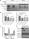
Fig. 1.
Fig. 1.
A: patient-derived AML cell line that had been transfected with vector alone (TRI102), or the vector containing the tuberous sclerosis complex 2 (TSC2) gene (TRI103) were cultured with and without 10% FBS, and treated with or without RAD001 (20 nM, 24 h). Lysates were prepared, and tuberin, total- and phospho-S6 ribosomal proteins, and α-smooth muscle actin (α-SMA) were detected by Western blot. B: cell numbers were quantified under serum free conditions by crystal violet DNA dye binding in TRI102 and TRI103 cells treated with or without RAD001 (20 nM) for 24 h (n = 6), or 72 h (n = 10). C: cell death was measured under serum free conditions by propidium iodide uptake assay in TRI102 and TRI103 cells treated with or without RAD001 (20 nM) for 72 h or stuarosporine (ST; 250 nM) for 24 h (n = 3; **P < 0.01; ***P < 0.001). D: Western blot of apoptosis and cell cycle markers in TRI102 and TRI103 cells treated without (“C”) or with (“R”) RAD001 (20 nM) for 24 h. Results are represent 3 separate experiments. PARP, poly (ADP-ribose) polymerase; F.L., full length; Cl., cleaved.
Fig. 2.
Fig. 2.
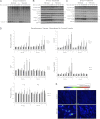
A: Western blot for ubiquitin in TRI102 and TRI 103 cells following 4-h treatment with MG-132 (doses ranging from 0 to 500 nM). B and C: Western blots for phospho-SAPK/JNK, phospho-eukaryotic initiation factor 2α (eIF2α), BiP, C/EBP-homologous protein (CHOP), BiP, full-length and cleaved PARP, and GAPDH in TRI102 and TRI103 cells following 4-h (B) and 8-h (C) treatment with MG-132 (doses ranging from 0 to 500 nM) in serum free conditions (“s.e.,” short exposure; “l.e.,” long exposure). D: graphs depicting densitometric analysis of phospho-SAPK/JNK, CHOP, phospho-eIF2α, BiP, and cleaved PARP levels normalized to untreated control values (n = 4). E: immunofluorescence images of TRI102 and TRI103 cells labeled with anti-CHOP 1° and Alexa 488-conjugated 2° antibodies with and without MG-132 treatment (500 nM, 8 h, serum free conditions). Image acquisition settings remained constant. Results represent 3 separate experiments.
Fig. 3.
Fig. 3.
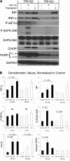
A: Western blots for BiP, inositol-requiring enzyme-1α (IRE1α), phospho (P)-eIF2α, phospho-SAPK/JNK, SAPK/JNK, CHOP, full-length and cleaved PARP, and GAPDH in TRI102 and TRI103 cells given no treatment, 500 nM MG-132, or 1 μM thapsigargin (Tg) for 8 h in serum free conditions. B: graphs depicting densitometric analysis of BiP, IRE1α, phospho-eIF2α, phospho-SAPK/JNK, CHOP, and cleaved PARP levels normalized to untreated control values (n = 4).
Fig. 4.
Fig. 4.
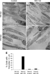
Electron micrographs of TRI102 (A and B) and TRI103 (C and D) cells untreated or treated with MG-132 (500 nM, 8 h). Note the pronounced vacuolization in MG-132 treated TRI102 cells (B) compared with other groups. E: graph depicting the percentage of cells containing vacuoles >4 μm in longest diameter. A minimum of 50 cells were counted for each group.
Fig. 5.
Fig. 5.
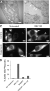
A and B: electron micrographs of MG-132-treated TRI102 cells demonstrating the presence of ribosomes on the edges of large vacuoles. White arrows indicate examples of ribosomes. White bars = 500 nm. C: fluorescence images of 621–101 human renal angiomyolipoma cells expressing an endoplasmic retituclum (ER)-localized RFP-containing an ER targeting and retention sequence. A diffuse pattern of fluorescence consistent with ER can be seen in untreated control (top left). Pronounced vacuolization is evident with MG-132 treatment (500 nM, 8 h; top right). Small, bright ER-derived vacuoles can clearly be seen (white arrow) and also very large vacuoles with a weaker, more diffused RFP signal due to increased size (arrowhead). Rapamycin suppressed vacuole formation by MG-132 (bottom right). White bars = 10 μm. D: graph depicting the percent of vacuolated TRI102 cells treated as follows in serum free conditions: no treatment, 500 nM MG-132 (8 h), 20 nM RAD001 (72 h), or RAD001 + MG-132 (as previous). Vacuolated cells were determined from immunofluorescence imaging of cells labeled with anti-LC3 (see Fig. 8). Importantly, LC3 did not appear to directly label the numerous large vacuoles, but vacuoles were clearly discernable inside cells in these images (see Fig. 8E).
Fig. 6.
Fig. 6.
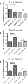
A: graph depicting cell number quantification measured by crystal violet DNA dye binding assay in TRI102 and TRI103 cells without and with 500 nM MG-132 treatment for 8 h in serum free conditions (n = 5). B and C: graph depicting cell death measured by propidium iodide (PI) uptake assay in TRI102 and TRI103 cells following 8-h (B) and 24-h (C) treatment with 500 nM MG-132 in serum free conditions (n = 3, each).
Fig. 7.
Fig. 7.
A: Western blots for phospho-S6, CHOP, BiP, phospho-eIF2α, cleaved caspase-3, full-length and cleaved PARP, and β-tubulin in TRI102 cells given no treatment, or 500 nM MG-132 (8 h) with and without 20 nM RAD001 pretreatment (72 h; n = 7). B: graphs depicting densitometric analysis (values normalized to untreated control levels). *P < 0.05; **P < 0.01; ***P < 0.001. C: graphs depicting caspase-3 activity measured in TRI102 and TRI103 cells given no treatment, or 500 nM MG-132 (8 h) with and without 20 nM RAD001 pretreatment (72 h; n = 4). **P < 0.01.
Fig. 8.
Fig. 8.
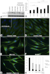
A: Western blot of LC3 I and II in TRI102 cells given no treatment, 100 nM or 500 nM MG-132 (8 h) with and without 20 nM RAD001 pretreatment (72 h) in serum free conditions (n = 7). B: graph depicting densitometric analysis. CG: immunofluorescence images of TRI102 cells stained for LC3. C: control with FBS; inset: no primary antibody negative control. D: control, serum free for 72 h. E: 500 nM MG-132 for 8 h, serum free 72 h. F: 20 nM RAD001 pretreatment for 72 h, serum free 72 h. G: 20 nM RAD001 pretreatment for 72 h, 500 nM MG-132 for final 8 h, serum free 72 h. H: graph depicting percentage of TRI102 cells with LC3 staining patterns of “None,” “Light/Moderate,” or “Strong/Punctate” given the aforementioned treatments; 175 to 260 individual cells in randomly selected fields from 6 separate experiments were evaluated separately by 2 investigators (B. J. Siroky, unblinded and J. J. Bissler, blinded) with a correlation coefficient of 0.992 between data sets.
Fig. 9.
Fig. 9.
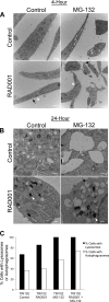
Electron micrographs of TRI102 cells treated with RAD001 (20 nM) and/or MG-132 (500 nM) for 4 h (A) or 24 h (B). A: images are lower magnification (white bars = 2 μm) and demonstrate vacuole formation with MG-132 treatment that is attenuated with RAD001 pretreatment. Electron dense lysosomes (white arrowheads) and autophagosomes surrounding partially digested cellular material (black arrows) are present in RAD001-treated cells, but appear more abundant in RAD001/MG-132 treated groups. B: images are higher magnification (white bars = 500 nm) and demonstrate similar findings. C: graph depicting the percentage of TRI102 cells expressing lysosomes and autophagosomes after 24 h of each indicated treatment. For each group, ≥50 cells were examined from 3 different experiments.
Fig. 10.
Fig. 10.
Immunohistochemical analysis of BiP (AC) and phospho-eIF2α (DF) in de-identified human renal angiomyolipoma tissue from two TSC patients. A: spindle cells displaying strong interspersed BiP staining. B: epithelioid cells with weakly BiP-positive and scattered strongly BiP-positive large cell populations. C: adipocyte-like cells with positive BiP staining in cytoplasm and weakly positive epithelioid cells interspersed. D: spindle cells with strong phospho-eIF2α staining. E: epithelioid cells displaying strong phospho-eIF2α staining. F: adipocyte-like cells with strong cytoplasmic phospho-eIF2α staining.
Fig. 11.
Fig. 11.
A: immunohistochemical analysis of BiP expression in renal cell carcinomas (RCC) and RCC cyst (lumen marked with *) tissue from a patient with Birt-Hogg-Dubé syndrome. Lower magnification (B) and higher magnification (C) immunohistochemical images of normal renal tissue (obtained via core needle biopsy) stained for BiP.
Fig. 12.
Fig. 12.
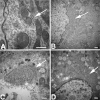
Electron microscopy demonstrating expanded endoplasmic reticulum in angiomyolipoma cells from different TSC patients compared with non-TSC associated kidney tissue. Arrows denote endoplasmic reticulum. A: normal renal tubule cell. B: angiomyolipoma spindle-morphology cell with significantly expanded endoplasmic reticulum. C: expanded endoplasmic reticulum in epithelioid cell angiomyolipoma. D: expanded endoplasmic reticulum in epithelioid cell with lipid droplets. White bar = 500 nm.

References

    1. Babcock JT, Quilliam LA. Rheb/mTOR activation and regulation in cancer: novel treatment strategies beyond rapamycin. Curr Drug Targets. - PubMed
    1. Bissler JJ, McCormack FX, Young LR, Elwing JM, Chuck G, Leonard JM, Schmithorst VJ, Laor T, Brody AS, Bean J, Salisbury S, Franz DN. Sirolimus for angiomyolipoma in tuberous sclerosis complex or lymphangioleiomyomatosis. N Engl J Med 358: 140–151, 2008 - PMC - PubMed
    1. Catley L, Weisberg E, Kiziltepe T, Tai YT, Hideshima T, Neri P, Tassone P, Atadja P, Chauhan D, Munshi NC, Anderson KC. Aggresome induction by proteasome inhibitor bortezomib and alpha-tubulin hyperacetylation by tubulin deacetylase (TDAC) inhibitor LBH589 are synergistic in myeloma cells. Blood 108: 3441–3449, 2006 - PMC - PubMed
    1. Cocciolone RA, Crotty KA, Andrews L, Haass NK, Moloney FJ. Multiple desmoplastic melanomas in Birt-Hogg-Dube syndrome and a proposed signaling link between folliculin, the mTOR pathway, and melanoma susceptibility. Arch Dermatol 146: 1316–1318 - PubMed (VSports)
    1. Davenport EL, Moore HE, Dunlop AS, Sharp SY, Workman P, Morgan GJ, Davies FE. Heat shock protein inhibition is associated with activation of the unfolded protein response pathway in myeloma plasma cells. Blood 110: 2641–2649, 2007 - "VSports在线直播" PubMed

Publication types

MeSH terms

"V体育官网" Substances