Skip to main page content
U.S. flag

An official website of the United States government

Dot gov

The . gov means it’s official. Federal government websites often end in . gov or VSports app下载. mil. Before sharing sensitive information, make sure you’re on a federal government site. .

Https

The site is secure V体育官网. The https:// ensures that you are connecting to the official website and that any information you provide is encrypted and transmitted securely. .

. 2012 Dec;19(12):1928-38.
doi: 10.1038/cdd.2012.71. Epub 2012 Jun 15.

V体育平台登录 - Disruption of the VDAC2-Bak interaction by Bcl-x(S) mediates efficient induction of apoptosis in melanoma cells

Affiliations

Disruption of the VDAC2-Bak interaction by Bcl-x(S) mediates efficient induction of apoptosis in melanoma cells

"VSports最新版本" M Plötz et al. Cell Death Differ. 2012 Dec.

Abstract

The proapoptotic B-cell lymphoma (Bcl)-2 protein Bcl-x(S) encloses the Bcl-2 homology (BH) domains BH3 and BH4 and triggers apoptosis via the multidomain protein Bak, however, the mechanism remained elusive. For investigating Bcl-x(S) efficacy and pathways, an adenoviral vector was constructed with its cDNA under tetracycline-off control. Bcl-x(S) overexpression resulted in efficient apoptosis induction and caspase activation in melanoma cells. Indicative of mitochondrial apoptosis pathways, Bcl-x(S) translocated to the mitochondria, disrupted the mitochondrial membrane potential and induced release of cytochrome c, apoptosis-inducing factor and second mitochondria-derived activator of caspases. In melanoma cells, Bcl-x(S) resulted in significant Bak activation, and Bak knockdown as well as Bcl-x(L) overexpression abrogated Bcl-x(S)-induced apoptosis, whereas Mcl-1 (myeloid cell leukemia-1) knockdown resulted in a sensitization. With regard to the particular role of voltage-dependent anion channel 2 (VDAC2) for inhibition of Bak, we identified here a notable interaction between Bcl-x(S) and VDAC2 in melanoma cells, which was proven in reciprocal coimmunoprecipitation analyses. On the other hand, Bcl-x(S) showed no direct interaction with Bak, and its binding to VDAC2 appeared as also independent of Bak expression. Suggesting a new proapoptotic mechanism, Bcl-x(S) overexpression resulted in disruption of the VDAC2-Bak interaction leading to release of Bak. Further supporting this pathway, overexpression of VDAC2 strongly decreased apoptosis by Bcl-x(S) VSports手机版. New proapoptotic pathways are of principle interest for overcoming apoptosis deficiency of melanoma cells. .


Figures
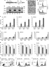
Figure 1
Figure 1
Apoptosis induction by strong and tightly controlled expression of Bcl-xS. (a) The structure of AdV-XS is shown. The Bcl-xS cDNA driven by a tetracyclin-controlled promoter (PTRE) was subcloned into the Ad5 E1 region, and E3 had been replaced by the tetracyclin-suppressed transactivator (tTA) driven by a CMV promoter (PCMV). The tTA mediates Tet-off regulation. Striped boxed indicate poly(A) regions. (b) Inducible Bcl-xS expression is shown by western blotting in A-375, Mel-HO and Mel-2a at 48 h after transduction of AdV-XS (MOI=50). Doxycycline was given for suppression (off condition) or was omitted (on condition). Equal protein loading was confirmed by β-actin, and Bcl-xL-transfected SK-Mel-13 cells (SKM13-Bcl-xL, C) are shown as control for Bcl-xL expression. (c) Rounded cells indicating apoptosis are shown of Mel-2a at 48 h after transduction with AdV-XS under off and on conditions. (d) Examples of cell cycle analysis after PI staining indicating sub-G1 apoptotic cell populations in Mel-2a at 72 h of transduction. (eg) Time course analyses of apoptosis (e, flow cytometry after PI staining), cytotoxicity (f, LDH release) and cell numbers (g, WST-1 assay) are shown for A-375, Mel-HO and Mel-2a cells at 24, 48 and 72 h after transduction with AdV-XS (50 MOI, +Dox=off, −Dox=on). As positive controls for induced cytotoxicity, cell lines were completely lysed by triton X-100 (T=100%) or were treated with doxorubicin (D, 500 nM, 72 h). WST-1 values are expressed as percent of non-treated controls (=100%). A luciferase-encoding adenovirus (Ad5-CMV-Luc) applied at the same MOI served as mock control (M). Mean values±S.Ds of at least six individual values are shown, and statistical significance (P<0.05) is indicated by asterisks. (hi) In Mel-2a at 48 h of Bcl-xs induction, apoptotic cells were determined by flow cytometric measurement of annexin V/PI staining (h) or by annexin V single staining (i), and numbers of viable cells were determined according to calcein staining (j). A shift to the right indicates annexin V-positive cells (i) and a shift to the left indicates calcein-negative (nonviable) cells (j)
Figure 2
Figure 2
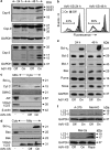
Activation of Csps and mitochondria. (a) Processing of Csps -8, -9 and -3 is shown in Mel-2a at 24 and 48 h after transduction of AdV-XS (MOI=50; on/off conditions). Equal protein loading (20 μg/lane) was confirmed by glyceraldehyde 3-phosphate dehydrogenase (GAPDH). Two independent experiments revealed comparable results. (b) Decrease of the mitochondrial membrane potential (Δψm) is shown for Mel-2a at 24 and 48 h after transduction of AdV-XS. Overlays of on conditions (open graphs) and off conditions (gray) are shown. Two independent experiments revealed highly comparable results. (c and d) Cytosolic fractions (cyto) and mitochondrial fractions (mito) were isolated from Mel-2a cells at 48 h after transduction with AdV-XS (MOI=50) and culturing under off and on conditions. Non-transfected cells (−) served as controls. (c) Cytosolic extracts demonstrate release of mitochondrial factors. Mitochondrial extracts served as control for mitochondrial proteins, and β-actin served as loading control. An antibody for mitochondrial VDAC proteins (VDAC1/2/3) was used for ruling out any contaminations of cytosolic extracts with mitochondria. (d) Mitochondrial extracts demonstrate mitochondrial translocation of Bcl-xS, and cytosolic extracts served as controls. Here, expression of VDAC proteins confirmed equal mitochondrial protein loading. The whole experiment was performed two times, resulting in highly comparable results. (e) Expression levels of Bcl-2 proteins in total protein extracts were determined by western blotting in Mel-2a at 24 and 48 h after transduction with AdV-XS (on/off conditions). Equal protein loading (20 μg/lane) was confirmed by GAPDH. (f) Expression of LC3 was determined by western blotting in A-375, Mel-HO and Mel-2a at 48 h after transduction of AdV-XS (MOI=50). Rapamycin-treated Mel-2a cells (Rapa, 10 μM, 24 h) served as positive control
Figure 3
Figure 3
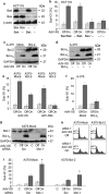
Dependency on Bak and inhibition by antiapoptotic Bcl-2 proteins. (a) Expression of Bax and Bak is shown in HCT116 parental cells and in subclones derived by Bak knockdown (Bax/−), Bax knockout (−/Bak) or double knockdown (−/−). Loading control: β-actin. (b) HCT116 cells were transduced with AdV-XS, cultured under off or on conditions, and percentages of apoptotic cell populations (sub-G1) were determined after 24 h. (c and d) Expression levels of Bcl-2, Bcl-xL and Bcl-xS were determined by western blotting in A-375-Mock and A-375-Bcl-2 (c) and in A-375 parental cells transiently transfected with Bcl-xL (d). Proteins were isolated at 64 h after transfection and at 48 h after transduction with AdV-XS (50 MOI) and culturing under off or on conditions. Equal protein loading was confirmed by glyceraldehyde 3-phosphate dehydrogenase (GAPDH). (e) Percentages of apoptotic cell populations (sub-G1) were determined at 48 h of transduction with AdV-XS in subclones of A-375 stably transfected with Bcl-2 (A-375-Bcl-2) or mock-transfected (A-375-Mock). (f) Percentages of apoptotic cell populations (sub-G1) were determined in A-375 after transient transfection with a Bcl-xL expression plasmid at 48 h after transduction with AdV-XS and growth under on and off conditions. (g) Expression of Mcl-1 and Bcl-xS is shown in A-375-Mock cells at 64 h after siRNA transfection against Mcl-1 and at 48 h after transduction with AdV-XS as well as culturing under on or off conditions. Scramble siRNA-transfected cells as well as non-transduced and non-transfected cells are shown as controls (C). A parallel experiment with A-375-Bcl-2 showed a highly similar result (data not shown). (h) Representative histograms displaying sub-G1 cell populations (apoptotic cells) at 48 h after transduction are shown. (i) Quantification of apoptosis in A-375-Bcl-2 and A-375-Mock cells after siRNA transfection (Mcl-1 or scramble) at 48 h after transduction with AdV-XS and growth under on and off conditions. Non-transduced and non-transfected cells served as controls (C). Experiments were performed at least two times, which showed highly comparable results. Bar charts, mean values±S.Ds of at least six individual values are shown. Statistical significance is indicated by asterisks
Figure 4
Figure 4
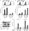
Activation of Bak by Bcl-xS. (a and b) Activation of Bax and Bak was determined by flow cytometry after staining with conformation-specific antibodies (Bax-NT, Bak-NT) in Mel-2a at 24 h and 48 after transduction of 50 MOI of AdV-XS (a) and of AdV-Nbk (b). Bars in the upper panels indicate the populations with activated Bax and Bak, respectively. (c) Expression of Bak and Bcl-xS is shown in A-375 cells at 64 h after transfection of anti-Bak siRNA or scramble siRNA (Scr) and at 48 h after transduction with AdV-XS and culturing under on or off conditions. (d) Apoptosis induction was determined by quantification of sub-G1 cells in A-375 after AdV-XS transduction and anti-Bak siRNA treatment. Bar charts, mean values±S.Ds of at least six individual values are shown, and statistical significance is indicated by asterisks
Figure 5
Figure 5
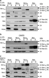
Immunoprecipitation analyses of Myc-tagged Bcl-2 proteins. (a) A-375 cells were transiently transfected with Myc-tagged Bcl-xL, Bcl-xS or Bax. Immunoprecipitates with anti-Myc antibody were analyzed by western blotting for Bcl-2 proteins. Mock controls were transfected with pcDNA3. Myc-tagged proteins with a higher molecular weight (M) are distinguished from endogenously expressed proteins (E). Supernatants not bound to the columns (S) were compared with the immunoprecipitated pellet fractions (P). (b) Binding to Bax and Bak was also analyzed, when cells were lysed without triton-X100 in CHAPS-containing cell lysis buffer. (c) Another experiment was performed with SK-Mel-13 cells stably transfected for Bcl-xL overexpression (SKM13-Bcl-xL). For further control, cells were here also transfected with Myc-Nbk. Each two independent experiments (ac) gave highly comparable results
Figure 6
Figure 6
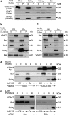
Bcl-xS disrupts the VDAC2–Bak interaction. (a) Immunoprecipitates with anti-Myc antibody were generated from A-375 cells transfected with Myc-tagged copies of Bcl-xL, Bcl-xS or Bax. Controls were mock-transfected (pcDNA3). Coimmunoprecipitation of any of the three VDAC proteins with Bcl-xL or Bcl-xS was proven by western blotting with a non-specific antibody (VDAC 1/2/3), and specific binding of VDAC2 was proven by a selective VDAC2 antibody. Cell lysis was performed with either triton X-100 or CHAPS-containing lysis buffer, as indicated. Interactions are shown by protein bands in immunoprecipitated pellet (P) fractions. (b) Mel-2a cells transduced with AdV-XS were cultured for 24 h under off or on conditions. Lysates were immunoprecipitated with anti-VDAC2 antibody, and immune complexes were analyzed by western blotting for Bcl-xS and for endogenous VDAC2, Bax, Bcl-2 and Bak. Supernatant (S) and immunoprecipitated pellet (P) fractions were compared. (c) A reciprocal experiment is shown based on immunoprecipitation with anti-Bak antibody. (d) A coimmunoprecipitation analysis was performed after transfection of a Myc-tagged VDAC2 cDNA (plasmid pcDNA3-VDAC2-Myc). Mock-transfected cells received pcDNA3 empty plasmid. Selective VDAC2 immunoprecipitates were performed with an anti-Myc antibody. (e) VDAC2 immunoprecipitates were generated of A-375 cells after siRNA-mediated Bak knockdown or sramble siRNA treatment (Scr), and binding of Bcl-xS was determined (ae). For all immunoprecipations, at least two independent experiments were performed, which always revealed the same result
Figure 7
Figure 7
Model for Bcl-xS-induced apoptosis. (a) Percentages of apoptotic cell populations (sub-G1) were determined in A-375 after transient transfection with a VDAC2 expression plasmid (pCMV-Sport6-VDAC2) and after transduction with AdV-XS (MOI=50) and growth under on and off conditions for 24 h and at 48 h. Transduction started 16 h after transient transfection. Overexpression of VDAC2 and Bcl-xS as determined by western blotting at 48 h after transduction is shown in the right panel. (b) Apoptosis induction (sub-G1 cells) is shown in A-375 after siRNA-mediated VDAC2 knockdown as well as at 24 and 48 h after Bcl-xS induction. Transfection with siRNA was always 16 h before transduction. Expression of VDAC2 and Bcl-xS is shown at 48 h after transduction in the right panel. Scramble siRNA-transfected cells (Scr) are shown as controls. Mean values±S.Ds of at least six individual values are shown, and statistical significance (P<0.05) is indicated by asterisks. (c) A proposed model, according to which Bcl-xS activates Bak through neutralization of the antiapoptotic VDAC2 protein. Bcl-xS-induced apoptosis is negatively controlled by Bcl-xL and Mcl-1, whereas Bcl-2 controls Bax

References

    1. Eberle J, Kurbanov BM, Hossini AM, Trefzer U, Fecker LF. Overcoming apoptosis deficiency of melanoma-hope for new therapeutic approaches. Drug Resist Updat. 2007;10:218–234. - PubMed
    1. Vogler M, Weber K, Dinsdale D, Schmitz I, Schulze-Osthoff K, Dyer MJ, et al. Different forms of cell death induced by putative BCL2 inhibitors. Cell Death Differ. 2009;16:1030–1039. - "VSports最新版本" PubMed
    1. Garbe C, Leiter U. Melanoma epidemiology and trends. Clin Dermatol. 2009;27:3–9. - PubMed
    1. Krammer PH, Arnold R, Lavrik IN. Life and death in peripheral T cells. Nat Rev Immunol. 2007;7:532–542. - PubMed
    1. Fulda S, Galluzzi L, Kroemer G. Targeting mitochondria for cancer therapy. Nat Rev Drug Discov. 2010;9:447–464. - PubMed

"V体育官网入口" Publication types

V体育平台登录 - MeSH terms

Substances