Skip to main page content
U.S. flag

An official website of the United States government

Dot gov

The . gov means it’s official. Federal government websites often end in . gov or . mil. Before sharing sensitive information, make sure you’re on a federal government site. VSports app下载.

Https

The site is secure V体育官网. The https:// ensures that you are connecting to the official website and that any information you provide is encrypted and transmitted securely. .

. 2012 Jun 26;109(26):10480-5.
doi: 10.1073/pnas.1201836109. Epub 2012 Jun 13.

NLRP3 inflammasome induces chemotactic immune cell migration to the CNS in experimental autoimmune encephalomyelitis

Affiliations

NLRP3 inflammasome induces chemotactic immune cell migration to the CNS in experimental autoimmune encephalomyelitis

"VSports最新版本" Makoto Inoue et al. Proc Natl Acad Sci U S A. .

Abstract

The NLRP3 inflammasome is a multiprotein complex consisting of three kinds of proteins, NLRP3, ASC, and pro-caspase-1, and plays a role in sensing pathogens and danger signals in the innate immune system. The NLRP3 inflammasome is thought to be involved in the development of experimental autoimmune encephalomyelitis (EAE), an animal model of multiple sclerosis (MS). However, the mechanism by which the NLRP3 inflammasome induces EAE is not clear VSports手机版. In this study, we found that the NLRP3 inflammasome played a critical role in inducing T-helper cell migration into the CNS. To gain migratory ability, CD4(+) T cells need to be primed by NLRP3 inflammasome-sufficient antigen-presenting cells to up-regulate chemotaxis-related proteins, such as osteopontin, CCR2, and CXCR6. In the presence of the NLRP3 inflammasome, dendritic cells and macrophages also induce chemotactic ability and up-regulate chemotaxis-related proteins, such as α4β1 integrin, CCL7, CCL8, and CXCL16. On the other hand, reduced Th17 cell population size in immunized Nlrp3(-/-) and Asc(-/-) mice is not a determinative factor for their resistance to EAE. As currently applied in clinical interventions of MS, targeting immune cell migration molecules may be an effective approach in treating MS accompanied by NLRP3 inflammasome activation. .


Conflict of interest statement

The authors declare no conflict of interest.

Figures

Fig. 1.
Fig. 1.
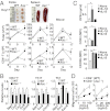
Asc−/− and Nlrp3−/− mice are resistant to EAE. (A) EAE development. Representative data from three independent experiments are shown. Disease scores were presented as mean ± SEM for each group (n = 5). (B) LFB and H&E staining of spinal cord sections from WT and Asc−/− mice at 17 d after EAE induction. Squares indicate representative regions shown at a high magnification on the right. Arrowheads indicate regions of demyelination. Representative data from three independent experiments are shown. (C and D) Numbers of total cells (C) and CD4+ T cells (D) obtained from spinal cords of WT, Asc−/−, and Nlrp3−/− mice at 17 d after EAE induction (n = 6–9). Horizontal lines denote mean values. (E) Intracellular staining of IL-17 and IFNγ and numbers of Th17 and Th1 cells in spinal cords of WT Asc and Nlrp3−/− mice at 17 d after EAE induction (n = 6–8). *P < 0.05.
Fig. 2.
Fig. 2.
Reduced Th17 response does not account for EAE resistance in Asc−/− and Nlrp3−/− mice. (A) Intracellular staining of IL-17 and IFNγ, and numbers of Th17 cells in DLNs at 9 d after EAE induction (n = 4–6). (B) In vitro Th17 cell generation. OT-2 CD4+ T cells were activated by splenic DCs from naïve mice with a Th17-polarizing condition. Flow cytometry plots show IL-17 and IFNγ intracellular staining in CD4+ T cells. Representative data from three independent experiments are shown. (C) IL-17 concentration in culture supernatants from experiments shown in B in triplicate wells. Representative data from three independent experiments are shown. (D) Schematic procedure for the experiment shown in E. IL-17+ cells were enriched by microbeads from WT, Asc−/−, or Nlrp3−/− mice at 9 d after immunization. IL-17+ cells (1 × 10⁶ cells per mouse) were adaptively transferred into Rag2−/− mice. (E) Passive EAE induced by IL-17+ cell transfer. Disease scores were presented as mean ± SEM for each group (n = 5).
Fig. 3.
Fig. 3.
Attenuated expression of genes encoding migration-related proteins impairs CD4+ T-cell migration in immunized Asc−/− mice. (A Top) DLNs and spleens from WT and Asc−/− mice at 17 d after EAE induction. (Middle and Bottom) Numbers of total cells (Middle) and CD4+ T cells (Bottom) in the DLNs, spleens, and peripheral blood in WT and Asc−/− mice on the indicated days after EAE induction (n = 6–11). (B) Gene expression determined by qPCR in CD4+ T cells, Th17 cells, and Th1 cells (n = 4). (C) Naïve CD4+ T cells were stimulated with CD3/CD28 antibodies with or without rIL-1β (10 ng/mL) or rIL-18 (100 ng/mL) in tissue culture. Protein levels at 24 h after stimulation were determined by ELISA (secreted OPN) and FACS (CCR2 and CXCR6) (n = 4). Representative FACS data are presented in Fig. S4E. (D) CD4+ T-cell chemotaxis toward rCCL2 or rCXCL16 of indicated concentrations evaluated by a Transwell assay of triplicate wells. (B and D) Cells were obtained from spleens of WT or Asc−/− mice at 9 d postimmunization. Representative data from two independent experiments are shown. *P < 0.05.
Fig. 4.
Fig. 4.
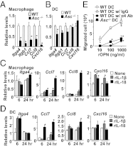
DCs and macrophages from immunized Asc−/− mice show attenuated expression of genes encoding chemokines or chemokine receptors. (AD) Gene expression in macrophage (A and C) and DCs (B and D). Bone marrow-derived macrophages (C) and DCs (D) were treated with or without rIL-1β (10 ng/mL) or rIL-18 (100 ng/mL) and harvested at the indicated time points. mRNA levels were determined by qPCR (n = 4). *P < 0.05. (E) DC chemotaxis toward rOPN of indicated concentrations (n = 4). Integrin α4 antibody or control IgG was incubated with DCs for 1 h, and then DCs were plated in upper chamber of a Transwell. (A, B, and E) Cells were obtained from spleens of WT or Asc−/− mice at 9 d after EAE induction. *P < 0.05 compared with WT DC data.
Fig. 5.
Fig. 5.
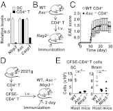
Presence of the NLRP3 inflammasome in APCs is sufficient to elicit T-cell migration. (A) Expression levels of genes encoding NLRP3 inflammasome components in DCs and CD4+ T cells were determined by qPCR. DCs and CD4+ T cells were obtained from spleen of WT mice at 9 d after immunization (n = 4). (B) Schematic procedure for the experiment shown in C. CD4+ T cells were isolated from spleens and lymph nodes of WT and Asc−/− naïve donor mice and transferred (1 × 10⁶ cells) into Rag2−/− recipients followed by MOG/complete Freund’s adjuvant (CFA) immunization. (C) EAE scores were presented as mean ± SEM for each group (n = 5). (D) Schematic procedure for the experiment shown in E. Naïve CD4+ 2D2 T cells were labeled with CFSE and transferred into WT, Asc−/−, or Nlrp3−/− mice that had been immunized at 2 d before the transfer. CD4+ T cells infiltrated into spinal cords and brains were enumerated at 4 d after the transfer. (E) Cell numbers of infiltrated CFSE-labeled CD4+ T cells into the spinal cord and brain (n = 5). *P < 0.05.
Fig. 6.
Fig. 6.
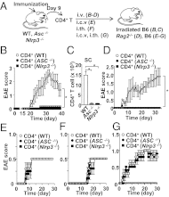
Bypassing the migration process to the CNS enables CD4+ T cells to induce EAE despite of priming in Asc−/− or Nlrp3−/− mice. (A) Schematic procedure for the experiments shown in BG. (BG) CD4+ T cells were obtained from spleens of WT, Asc−/−, or Nlrp3−/− mice at 9 d after immunization and transferred (3 × 10⁶ cells per mouse) into sublethally irradiated WT recipients (B and C) or Rag2−/− recipients (D). CD4+ T cells (1 × 10⁶ cells per mouse) were also transferred directly into the brains (E) or spinal cords (F) of WT recipients by i.c.v. or i.th. injection, respectively, or by the combination of both i.c.v. and i.th. injections (G). G also includes negative controls with splenic CD4+ T cells from naïve WT (△), Asc−/− (▲) or Nlrp3−/− (◆) mice transferred (1 × 10⁶ cells per mouse) to recipients (no EAE developed). (B and DG) EAE scores were presented as mean ± SEM for each group (n = 5). (C) Numbers of CD4+ T cells in spinal cords on day 44 after CD4+ T-cell transfer (n = 5). *P < 0.05.

"VSports app下载" References

    1. Fife BT, Huffnagle GB, Kuziel WA, Karpus WJ. CC chemokine receptor 2 is critical for induction of experimental autoimmune encephalomyelitis. J Exp Med. 2000;192:899–905. - "VSports" PMC - PubMed
    1. Brodmerkel CM, et al. Discovery and pharmacological characterization of a novel rodent-active CCR2 antagonist, INCB3344. J Immunol. 2005;175:5370–5378. - PubMed
    1. dos Santos AC, et al. CCL2 and CCL5 mediate leukocyte adhesion in experimental autoimmune encephalomyelitis—An intravital microscopy study. J Neuroimmunol. 2005;162:122–129. - "V体育2025版" PubMed
    1. Kataoka H, et al. FTY720, sphingosine 1-phosphate receptor modulator, ameliorates experimental autoimmune encephalomyelitis by inhibition of T cell infiltration. Cell Mol Immunol. 2005;2:439–448. - PubMed
    1. Yednock TA, et al. Prevention of experimental autoimmune encephalomyelitis by antibodies against α4β1 integrin. Nature. 1992;356:63–66. - PubMed

Publication types

VSports注册入口 - MeSH terms