<kbd dir="DUhZO0v"></kbd> Skip to main page content
U.S. flag

An official website of the United States government

Dot gov

The VSports app下载. gov means it’s official. Federal government websites often end in . gov or . mil. Before sharing sensitive information, make sure you’re on a federal government site. .

Https

The site is secure. The https:// ensures that you are connecting to the official website and that any information you provide is encrypted and transmitted securely V体育官网. .

. 2012 Dec;132(12):2691-9.
doi: 10.1038/jid.2012.201. Epub 2012 Jun 14.

Regulation of epithelial differentiation and proliferation by the stroma: a role for the retinoblastoma protein

Affiliations

Regulation of epithelial differentiation and proliferation by the stroma: a role for the retinoblastoma protein (V体育2025版)

Adam Pickard et al. J Invest Dermatol. 2012 Dec.

Abstract

Signaling between the epithelium and stromal cells is crucial for growth, differentiation, and repair of the epithelium. Although the retinoblastoma protein (Rb) is known to regulate the growth of keratinocytes in a cell-autonomous manner, here we describe a function of Rb in the stromal compartment. We find that Rb depletion in fibroblasts leads to inhibition of differentiation and enhanced proliferation of the epithelium. Analysis of conditioned medium identified that keratinocyte growth factor (KGF) levels were elevated following Rb depletion. These findings were also observed with organotypic co-cultures. Treatment of keratinocytes with KGF inhibited differentiation and enhanced keratinocyte proliferation, whereas reduction of KGF levels in Rb-depleted fibroblasts was able to restore expression of differentiation markers VSports手机版. Our findings suggest a crucial role for dermal fibroblasts in regulating the differentiation and proliferation of keratinocytes, and we demonstrate a role for stromal Rb in this cross-talk. .


Figures
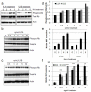
Figure 1
Figure 1. IL1A/B induce Rb phosphorylation in HFFs
A) Western blot analysis of Rb phosphorylation over a short time course of 10 ng/mL IL1A/B treatment. B and C) Induction of Rb phosphorylation following 24 hours treatment with various concentrations of IL1A and IL1B, respectively. Quantification of Rb phosphorylation is shown in D. E) Real time PCR analysis of KGF expression levels following IL1A/B treatment, for the indicated lengths of time. F) Induction of KGF expression by various concentrations of IL1A/B after 24h treatment at various concentrations. * p<0.05 and ** p<0.01 in a Students T test compared to untreated samples.
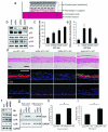
Figure 2
Figure 2. Rb-depletion in fibroblasts disrupts differentiation and proliferation of neighbouring epithelium
A) Schematic of organotypic cultures comprised of epithelial cells seeded on top of modified fibroblasts. B) Western blots confirming knockdown of Rb and p53 in HFFs compared to scrambled shRNA controls (scram). C) Proliferation in fibroblasts assessed by Brdu incorporation (See materials and methods). D) Immuno fluorescent detection of differentiation markers and Brdu incorporation in organotypic cultures indicate disruption of differentiation and enhanced proliferation in epithelium cultured with Rb depleted fibroblasts. Proliferation is quantified in E). Scale bars represent 100 Jm. F) KGF, IL1A and IL1B expression in Rb depleted fibroblasts detected by RT PCR. G) KGF expression in sub confluent and mitomycin C treated fibroblasts. H) ELISA detection of KGF secretion in monolayer co cultures and I) organotypic cultures.
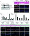
Figure 3
Figure 3. Rescue of Rb-depletion by adenoviral re-expression restores differentiation and proliferation defects
A) Restoration of Rb protein levels by adenoviral overexpression in shRb#2 fibroblasts. Re expression of Rb also reduced active AKT levels. B) Immuno fluorescent detection of keratin 1, filaggrin and Brdu identified that re expression of Rb restored differentiation and reduced proliferation in the epithelium of organotypic cultures. Brdu incorporation is quantified in C). Scale bars represent 100 Jm. D) Q PCR and E) ELISA detection of KGF following re expression of Rb. * represents p<0.05 in a Students T test. ** indicates GFP expression in fibroblasts.
Figure 4
Figure 4. Rb inactivation enhances KGF expression
A) HA tagged CDK6 expression in HFFs confirmed by Western blotting. B) Organotypic cultures grown with HA CDK6 expressing fibroblasts were stained for differentiation markers and Brdu incorporation which is quantified in C). D) Q PCR analysis of KGF, IL1A and IL1B in HA CDK6 expressing HFFs. * p<0.05 in a Students T test. E) Western blot detection of wild type Rb (WT Rb) and a non phosphorylatable mutant (Phos.Mut.Rb) expressed in HFFs. F) Q PCR assessment of the induction of KGF, IL1A and IL1B expression following 10 ng/mL IL1A (F), or IL1B (G) treatment of cells described in E. Induction of KGF/IL1A and IL1B expression in WT Rb HFFs was assigned as 100%, results are from triplicate experiments. * represents p<0.05 in a Students T test compared to WT Rb samples.
Figure 5
Figure 5. KGF inhibits differentiation of epithelial cells
A) Addition of KGF to keratinocyte cultures inhibited calcium induced differentiation as assessed by Western blotting of involucrin, keratin 1 and transglutaminase levels. B) Organotypic cultures grown in the presence of 2 or 10 ng/mL KGF showed disrupted differentiation. Representative cultures from triplicate analyses are shown. C) Knockdown of KGF levels in Rb depleted fibroblasts was assessed by Q PCR. D) Organotypic cultures using fibroblasts in C showed that KGF depletion did not significantly alter differentiation in control cultures (compare a c with d f and g i), however in cultures grown with Rb depleted fibroblasts, KGF depletion restored epithelial differentiation (compare j l with m o and p r). E) Brdu incorporation in the epithelium of cultures shown in D. Scale bars represent 100 Jm.

References

    1. Andreadis ST, Hamoen KE, Yarmush ML, Morgan JR. Keratinocyte growth factor induces hyperproliferation and delays differentiation in a skin equivalent model system. Faseb J. 2001;15:898–906. - VSports最新版本 - PubMed
    1. Berns K, Hijmans EM, Mullenders J, Brummelkamp TR, Velds A, Heimerikx M, et al. A large scale RNAi screen in human cells identifies new components of the p53 pathway. Nature. 2004;428:431–7. - PubMed
    1. Chang MW, Barr E, Seltzer J, Jiang YQ, Nabel GJ, Nabel EG, et al. Cytostatic gene therapy for vascular proliferative disorders with a constitutively active form of the retinoblastoma gene product. Science (New York, NY. 1995;267:518–22. - PubMed
    1. Chedid M, Rubin JS, Csaky KG, Aaronson SA. Regulation of keratinocyte growth factor gene expression by interleukin 1. The Journal of biological chemistry. 1994;269:10753–7. - "VSports" PubMed
    1. de Bruin A, Wu L, Saavedra HI, Wilson P, Yang Y, Rosol TJ, et al. Rb function in extraembryonic lineages suppresses apoptosis in the CNS of Rb deficient mice. Proceedings of the National Academy of Sciences of the United States of America. 2003;100:6546–51. - PMC - PubMed

"VSports在线直播" Publication types

MeSH terms

Substances