Skip to main page content
U.S. flag

An official website of the United States government

Dot gov

The . gov means it’s official. Federal government websites often end in . gov or VSports app下载. mil. Before sharing sensitive information, make sure you’re on a federal government site. .

Https

The site is secure. The https:// ensures that you are connecting to the official website and that any information you provide is encrypted and transmitted securely V体育官网. .

Review
. 2012 Jun 8;110(12):1628-45.
doi: 10.1161/CIRCRESAHA.111.259960.

Endocardial and epicardial epithelial to mesenchymal transitions in heart development and disease

Affiliations
Review

Endocardial and epicardial epithelial to mesenchymal transitions in heart development and disease

Alexander von Gise et al. Circ Res. .

Abstract

Epithelial to mesenchymal transition (EMT) converts epithelial cells to mobile and developmentally plastic mesenchymal cells. All cells in the heart arise from one or more EMTs. Endocardial and epicardial EMTs produce most of the noncardiomyocyte lineages of the mature heart VSports手机版. Endocardial EMT generates valve progenitor cells and is necessary for formation of the cardiac valves and for complete cardiac septation. Epicardial EMT is required for myocardial growth and coronary vessel formation, and it generates cardiac fibroblasts, vascular smooth muscle cells, a subset of coronary endothelial cells, and possibly a subset of cardiomyocytes. Emerging studies suggest that these developmental mechanisms are redeployed in adult heart valve disease, in cardiac fibrosis, and in myocardial responses to ischemic injury. Redirection and amplification of disease-related EMTs offer potential new therapeutic strategies and approaches for treatment of heart disease. Here, we review the role and molecular regulation of endocardial and epicardial EMT in fetal heart development, and we summarize key literature implicating reactivation of endocardial and epicardial EMT in adult heart disease. .


Figures
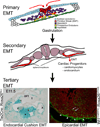
Fig. 1
Fig. 1. Formation of the heart through a series of EMTs
Myocardium and endocardium arise from two EMTs. Additional lineages present in the adult heart are formed by additional EMT events: neural ectoderm undergoes a primary EMT to yield cardiac neural crest (not shown); endocardium overlying the endocardial cushions undergoes a tertiary EMT to yield mesenchyme of the AV canal (*) and proximal OFT (†) cushions; and epicardium undergoes a tertiary EMT to yield cardiac fibroblasts, vascular smooth muscle, and potentially a subset of cardiomyocytes. Endothelium or epicardium-restricted Cre (Tie2Cre or Wt1CreERT2, respectively) were used to activate Cre-dependent reporters Rosa26fsLz or Rosa26mTmG, labeling descendants with LacZ (blue) or GFP (green), respectively. Distal OFT cushion (‡) was not labeled by Tie2Cre, reflecting mixed contributions from multiple precursor populations. Arrowheads, epicardium. Arrows, epicardium-derived cells. (Illustration credit: Cosmocyte/Ben Smith).
Fig. 2
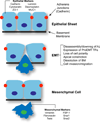
Fig. 2. Cellular changes during EMT
A typical epithelial sheet contains polarized epithelial cells atop a basement membrane (BM). Cells are joined to one another by adhesion junctions (AJs) containing cadherins. EMT requires dissassembly and downregulation of adhesion junction components, expression of pro-EMT transcription factors (TFs) like SNAI1 and SNAI2, and delamination of the cell through a combination of apical constriction and dissolution of the basement membrane. The resulting mesenchymal cell is mobile and developmentally plastic, and it expresses typical markers such as those indicated.
Fig. 3
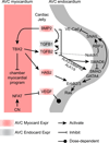
Fig. 3. Molecular regulation of endocardial cushion EMT
Endocardial EMT to form valve mesenchyme is regulated by signaling between specialized AVC myocardium and overlying cushion endocardium. Cardiac jelly, the extracellular matrix of the cushions, is also required for endocardial EMT. (Illustration credit: Cosmocyte/Ben Smith).
Fig. 4
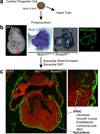
Fig. 4. Developmental plasticity of epicardial progenitor cells
(a) Progenitor cells expressing Isl1 and Nkx2–5 generate proepicardium (PE, red arrow) and the heart tube, containing the nascent atria (A) and ventricles (V). Thus PE cells are labeled by Nkx2–5IRES-Cre (b) and Isl1Cre (not shown), but do not actively express these markers. Whether precursors of proepicardium and heart tube are the same or merely express the same markers is presently unknown. Subsequently, PE cells migrate over the surface of the heart to form epicardium, which undergoes EMT to form EPDCs (c). EPDCs differentiate into interstitial and annulus fibrosis fibroblasts, smooth muscle cells, endothelial cells, cardiomyocytes, and mesenchymal stem cells. Cre-labeled cells expressed GFP (green), and unlabeled cells expressed RFP (red). (b) was modified from Zhou et al., BBRC, 2008, with permission.
Fig. 5
Fig. 5. Molecular regulation of epicardial EMT
Pathways that regulate EPDC fate, myocardial growth, and coronary vessel development without directly influencing epicardial EMT are not shown. Question marks indicate inconsistency in existing data.

References

    1. Perez-Pomares JM, Munoz-Chapuli R. Epithelial-mesenchymal transitions: a mesodermal cell strategy for evolutive innovation in Metazoans. Anat Rec. 2002;268:343–351. - PubMed
    1. Baum B, Settleman J, Quinlan MP. Transitions between epithelial and mesenchymal states in development and disease. Semin Cell Dev Biol. 2008;19:294–308. - PubMed (VSports在线直播)
    1. Thiery JP, Acloque H, Huang RY, Nieto MA. Epithelial-mesenchymal transitions in development and disease. Cell. 2009;139:871–890. - "V体育官网入口" PubMed
    1. Nieto MA. The ins and outs of the epithelial to mesenchymal transition in health and disease. Annu Rev Cell Dev Biol. 2011;27:347–376. - PubMed
    1. Acloque H, Adams MS, Fishwick K, Bronner-Fraser M, Nieto MA. Epithelial-mesenchymal transitions: the importance of changing cell state in development and disease. J Clin Invest. 2009;119:1438–1449. - PMC - PubMed

Publication types