Skip to main page content
U.S. flag

An official website of the United States government

Dot gov

The . gov means it’s official. Federal government websites often end in . gov or VSports app下载. mil. Before sharing sensitive information, make sure you’re on a federal government site. .

Https

The site is secure. The https:// ensures that you are connecting to the official website and that any information you provide is encrypted and transmitted securely V体育官网. .

. 2012 May 25;149(5):1098-111.
doi: 10.1016/j.cell.2012.02.065.

"V体育官网入口" The Skp2-SCF E3 ligase regulates Akt ubiquitination, glycolysis, herceptin sensitivity, and tumorigenesis

Affiliations

VSports - The Skp2-SCF E3 ligase regulates Akt ubiquitination, glycolysis, herceptin sensitivity, and tumorigenesis

Chia-Hsin Chan (VSports app下载) et al. Cell. .

Erratum in

V体育官网 - Abstract

Akt kinase plays a central role in cell growth, metabolism, and tumorigenesis. The TRAF6 E3 ligase orchestrates IGF-1-mediated Akt ubiquitination and activation. Here, we show that Akt ubiquitination is also induced by activation of ErbB receptors; unexpectedly, and in contrast to IGF-1 induced activation, the Skp2 SCF complex, not TRAF6, is a critical E3 ligase for ErbB-receptor-mediated Akt ubiquitination and membrane recruitment in response to EGF VSports手机版. Skp2 deficiency impairs Akt activation, Glut1 expression, glucose uptake and glycolysis, and breast cancer progression in various tumor models. Moreover, Skp2 overexpression correlates with Akt activation and breast cancer metastasis and serves as a marker for poor prognosis in Her2-positive patients. Finally, Skp2 silencing sensitizes Her2-overexpressing tumors to Herceptin treatment. Our study suggests that distinct E3 ligases are utilized by diverse growth factors for Akt activation and that targeting glycolysis sensitizes Her2-positive tumors to Herceptin treatment. .


Figures

Figure 1
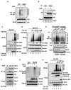
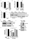
Figure 1. Skp2 is required for EGF-mediated Akt ubiquitination
(A) WT and Skp2-/- MEFs were serum-starved, treated with or without EGF, and harvested for in vivo ubiquitination assay. (B) 293T cells were serum-starved, treated with or without EGF and harvested for Co-immunoprecipitation (IP) assay followed by immunoblot (IB) analysis. (C) In vivo ubiquitination assay in 293T cells transfected with hemagglutinin (HA)-Akt1 and His-ubiquitin (His-Ub), along with Xpress-Skp2 (Xp-Skp2). Ni–nitrilotriacetic acid (NTA) indicates nickel bead precipitate; WCE indicates whole-cell extracts. * indicates non-specific signal. (D) In vivo ubiquitination assay in 293T cells transfected with various constructs. (E) Control- and Skp2-knockdown BT-474 cells were serum-starved, treated with or without EGF and harvested for in vivo ubiquitination assay. (F) 293T cells were transfected with Xp-Skp2, serum-starved, treated with or without EGF, and harvested for Co-IP assay followed by IB analysis. (G) Cos1 cells were serum-starved, treated with or without IGF-1 or EGF and harvested for in vivo ubiquitination assay. (H) In vivo ubiquitination assay in 293T cells transfected with HA-Akt1, His-Ub, along with WT Skp2 and E3-ligase dead mutants (Skp2-NES and Skp2-LRR). See also Figure S1.
Figure 2
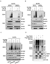
Figure 2. Skp2 SCF complex is a direct E3 ligase for Akt
(A) Luciferase or Cul-1-silenced 293 cells transfected with various constructs were harvested for in vivo ubiquitination assay. (B) In vivo ubiquitination assay in Luciferase or Skp1-silenced 293 cells transfected with various constructs. (C) In vivo ubiquitination assay in various 293 knockdown cells transfected with different plasmids. (D) GST-Akt proteins were incubated with adenosine triphosphate, E1, and E2 (both UbcH5c and Ubc13/Uev1) along with or without Flag-Skp2-SCF or Flag-TRAF6 for in vitro Akt ubiquitination assay. See also Figure S2.
Figure 3
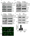
Figure 3. Skp2 SCF complex is required for Akt activation and membrane recruitment
(A) WT and Skp2-/- MEFs were serum-starved, treated with EGF for various time points and harvested for IB analysis. (B, C) BT-474 cells with control- and Skp2-silenced were serum-starved, treated with EGF (B) or HRG (C) for various time points and harvested for IB analysis. (D) WT and Skp2-/- MEFs were serum-starved, treated with EGF for various time points, and the membrane (mem) and cytosolic (cyt) fractions were isolated for IB analysis. The relative intensity of was quantified with ImageQuant software and normalized with the Akt levels in WT MEFs without EGF treatment. (E) WT and Skp2-/- MEFs were serum-starved, treated with EGF for 5 min and fixed for immunofluorescence assay. The arrow indicates the membrane localization of Akt. The quantification results were shown in Figure S3E. See also Figure S3.
Figure 4
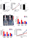
Figure 4. Skp2 regulates glucose uptake and glycolysis in vitro and in vivo
(A) Lactate production was measured in BT-474 cells with Luciferase and Skp2 knockdown. (B) Glucose uptake was measured in BT-474 cells with Luciferase and Skp2 knockdown. Cells treated with or without LY294002 were grown in the presence of the fluorescent analog NBDG for various time points, and glucose uptake was quantified using FACS analysis. (C) Lactate production was measured in BT-474 cells treated with or without LY294002. (D) Representative PET/CT images in nude mice bearing breast tumors with Luciferase- or Skp2-knockdown. White dotted lines indicate area of the breast tumors and black dotted lines indicate area of muscle tissues that were analyzed for in vivo glucose uptake. (E, F) Glucose uptake was expressed as Standard Uptake Value (SUV) ratio (E) or as percent of injected dose per gram (F) of labeled [18F] FDG-glucose incorporation in mice bearing Luciferase- or Skp2-silenced breast tumors. Glucose uptake in breast tumors was normalized with muscle, thymus or liver tissues in each mouse. The quantified results are presented as means ± s.d. (n = 5). (G) Breast tumor development in nude mice bearing breast tumors with Luciferase- or Skp2-knockdown (n=5). The arrow indicates the time point for in vivo glucose uptake analysis. **, p<0.01. See also Figure S4.
Figure 5
Figure 5. Skp2 regulates glycolysis through promoting Akt ubiquitination and activation
(A) Lactate production was determined in BT-474 cells with Luciferase, Skp2 knockdown, or Skp2 knockdown plus Myr-Akt overexpression upon EGF stimulation. (B) Real-Time PCR analysis of Glut1 mRNA levels in BT-474 cells or MDA-MB-468 cells with Luciferase or Skp2 knockdown. (C) Real-Time PCR analysis of Glut1 mRNA levels in WT and Skp2-/- MEFs. (D) Cos1 cells with Luciferase or Skp2 knockdown were serum-starved, treated with EGF for various time points and harvested for the isolation of membrane and cytosolic fractions, followed by IB analysis. (E) Cos1 cells with Luciferase or Skp2 knockdown were serum-starved, treated with EGF for various time points and harvested for IB analysis. (F) IB analysis of Glut1 protein expressions in whole cell extracts (WCE) or membrane fractions (Mem) of BT-474 cells with Luciferase, Skp2 knockdown or Skp2 knockdown plus Myr-Akt overexpression. (G) Lactate production was measured in Luciferase- and Skp2-silenced BT-474 cells transfected with various constructs as indicated upon EGF stimulation. See also Figure S5.
Figure 6
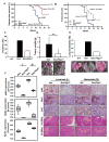
Figure 6. Skp2 deficiency restricts in vivo Akt activation and mammary tumor development upon Neu overexpression
(A) Kaplan–Meier plot analysis of cumulative disease-free survival of WT, MMTV-Neu and MMTV-Neu/Skp2-/- mice. (B) Kaplan–Meier plot analysis of tumor-free incidence of WT, MMTV-Neu and MMTV-Neu/Skp2-/- mice. (C) The percentage of mice that develop mammary tumor was analyzed from a cohort of WT, MMTV-Neu and MMTV-Neu/Skp2-/- mice at age around 10 months (WT, n=10; MMTV-Neu, n=13; MMTV-Neu/Skp2-/-, n=10). (D) Mammary tumors were obtained and weighed from MMTV-Neu and MMTV-Neu/Skp2-/- mice at the age around 10 months (MMTV-Neu, n=11; MMTV-Neu/Skp2-/-, n=10). Arrows indicate mammary tumors. **, p<0.01. (E) The percentage of mice that develop lung metastasis was analyzed from a cohort of WT, MMTV-Neu and MMTV-Neu/Skp2-/- mice at age around 12 months (WT, n=10; MMTV-Neu, n=11; MMTV-Neu/Skp2-/-, n=9). (F) Histological and quantification analysis of pAkt, Glut1 and Ki-67 protein expression in MMTV-Neu and MMTV-Neu/Skp2-/- mice. Scale bar indicates 200 μm. All p-value < 0.001 by using Mann-Whitney U Test, except the p-value for Glut1 expression between localized and metastatic MMTV-Neu/Skp2-/- (p=0.105).
Figure 7
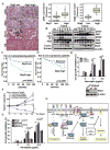
Figure 7. Skp2 deficiency prolongs survival of Her2-positve patients and confers Herceptin sensitivity in Her2-positive cells and tumors
(A) Histological and quantification analysis of pAkt expressions in Her2-positive patients with low or high expression of Skp2. Scale bar indicates 200 μm. All p-value < 0.001 by using Mann-Whitney U Test. (B) Breast tumors and normal breast tissues were extracted and subjected for IP assay, followed by IB analysis. (C) Kaplan–Meier plot analysis of metastasis-free survival of 80 cases of Her2-positive patients with low or high expression of Skp2. (D) Kaplan–Meier plot analysis of metastasis-free survival of 132 cases of Her2 low-expressing patients with low or high expression of Skp2. (E) Cell growth inhibition assay and IB analysis in BT-474 cells with Luciferase or Skp2 knockdown. BT-474 cells were treated with various doses of Herceptin for 6 days and cell numbers were counted using hemocytometer. (F) Tumor volume of Her2-positive tumors with or without Skp2 silencing upon treatment with IgG or Herceptin. Tumor volume at various time points of treatment is presented as percentage of original tumor size (~200 mm3) at day zero of treatment. (G) BT-474 cells with Luciferase or Skp2 knockdown were transfected with various plasmids, treated with various doses of Herceptin and viable cell numbers were counted using hemocytometer. (H) The working model of Skp2 in ErbB family-regulated Akt activation, glycolysis and tumorigenesis. See also Figure S6 and Tables S1-S4.

References

    1. Agus DB, Akita RW, Fox WD, Lewis GD, Higgins B, Pisacane PI, Lofgren JA, Tindell C, Evans DP, Maiese K, et al. Targeting ligand-activated ErbB2 signaling inhibits breast and prostate tumor growth. Cancer Cell. 2002;2:127–137. - PubMed
    1. Aragones J, Fraisl P, Baes M, Carmeliet P. Oxygen sensors at the crossroad of metabolism. Cell Metab. 2009;9:11–22. - V体育平台登录 - PubMed
    1. Barthel A, Okino ST, Liao J, Nakatani K, Li J, Whitlock JP, Jr, Roth RA. Regulation of GLUT1 gene transcription by the serine/threonine kinase Akt1. J Biol Chem. 1999;274:20281–20286. - "VSports手机版" PubMed
    1. Birnbaum MJ. On the InterAktion between hexokinase and the mitochondrion. Dev Cell. 2004;7:781–782. - PubMed
    1. Bornstein G, Bloom J, Sitry-Shevah D, Nakayama K, Pagano M, Hershko A. Role of the SCFSkp2 ubiquitin ligase in the degradation of p21Cip1 in S phase. J Biol Chem. 2003;278:25752–25757. - PubMed

Publication types

MeSH terms (V体育官网)